843 843 843 843 843 Mem Inst Oswaldo Cruz, Rio de Janeiro, Vol. 98(6): 843-848, September 2003
Cytotoxicity and Potential Antiviral Evaluation of Violacein
Produced by
Chromobacterium violaceum
CR Andrighetti-Fröhner, RV Antonio**, TB Creczynski-Pasa/***, CRM Barardi*,
CMO Simões/
+Laboratório de Virologia Aplicada, Departamento de Ciências Farmacêuticas *Departamento de Microbiologia e Parasitologia **Laboratório de Bioquímica, Departamento de Bioquímica ***Laboratório de Bioenergética e Comunicação Celular,
Universidade Federal de Santa Catarina, Campus Universitário Trindade, Florianópolis, SC, Brasil
Natural products are an inexhaustible source of compounds with promising pharmacological activities including antiviral action. Violacein, the major pigment produced by Chromobacterium violaceum, has been shown to have antibiotic, antitumoral and anti-Trypanosoma cruzi activities. The goal of the present work was to evaluate the cytotoxicity of violacein and also its potential antiviral properties.The cytotoxicity of violacein was investigated by three methods: cell morphology evaluation by inverted light microscopy and cell viability tests using the Trypan blue dye exclusion method and the MTT assay. The cytotoxic concentration values which cause destruction in 50% of the monolayer cells (CC50)were different depending on the sensitivity of the method. CC50 values were ≥ 2.07 ± 0.08 µM for FRhK-4 cells: ≥ 2.23 ± 0.11 µM for Vero cells; ≥ 2.54 ± 0.18 µM for MA104 cells; and ≥ 2.70 ± 0.20 µM for HEp-2 cells. Violacein showed no cytopathic inhibition of the following viruses: herpes simplex virus type 1 (HSV-1) strain 29-R/acyclovir resistant, hepatitis A virus (strains HM175 and HAF-203) and adenovirus type 5 nor did it show any antiviral activity in the MTT assay. However violacein did show a weak inhibition of viral replication: 1.42 ± 0.68%, 14.48 ± 5.06% and 21.47 ± 3.74% for HSV-1 (strain KOS); 5.96 ± 2.51%, 8.75 ± 3.08% and 17.75 ± 5.19% for HSV-1 (strain ATCC/VR-733); 5.13 ± 2.38 %, 8.18 ± 1.11% and 8.51 ± 1.94% for poliovirus type 2; 8.30 ± 4.24%; 13.33 ± 4.66% and 24.27 ± 2.18% for simian rotavirus SA11, at 0.312, 0.625 and 1.250 mM, respectively, when measured by the MTT assay.
Key words: violacein cytotoxicity antiviral MTT assay herpes simplex virus type 1 poliovirus rotavirus -hepatitis A virus - adenovirus
During the last few years efforts have been made to increase the number of substances with antiviral activity. Few substances are known which provide an effective treatment of viral infections in vivo (Balfour 1999). Also, the therapeutic potency of most of the antiviral agents encountered so far is counterbalanced by their severe side effects in humans (Glatthaar-Saalmüller et al. 2001) and the efficacy of these drugs is limited by increases in viral resistance (Pillay & Zambon 1998, De Logu et al. 2000). The search for antiviral substances with high efficacy, low toxicity, and minor side effects therefore must continue.
Natural products have been an abundant source of compounds which have proved useful in antiviral chemotherapy of infectious human diseases (Pujol et al. 1996, Bedoya et al. 2001) especially those originating from plant extracts and fermentation broths from soil bacteria, which provide compounds directly useful as drugs or as leads for the synthesis of new medicines (Nielsen 2002).
Chromobacterium violaceum is a Gram(-) bacteria found in water samples and soils from tropical and subtropical regions of the world. Due to its bio-technological potential, C. violaceum had its genome sequenced by the Brazilian National Genome Project. The most notable characteristic of C. violaceum is the production of the chemically well characterized pigment named violacein (Bromberg & Duran 2001). Previous studies indicated antibiotic and antichagasic (Duran & Menck 2001), antitumoral (Melo et al. 2000), and antileishmanial (Leon et al. 2001) activities of violacein.
The aim of this study was to assess the cytototoxicity and the potential antiviral activity of violacein against the viruses: Herpes Simplex Virus type 1 (HSV-1) strains KOS, 29-R/acyclovir resistant and VR733/ATCC; Poliovirus type 2 (PV-2); Simian rotavirus SA11 strain, Hepatite A virus (HAV) strains HAF203 and HM175 and Adenovirus type 5 (AdV-5), a respiratory strain.
MATERIALS AND METHODS
Compound-Violacein was isolated and purified from C. violaceum (CCT3496/JMC3496) as described by Duran et al. (1994). Violacein was dissolved in absolute ethanol (Merck) and 0.003% of dimethyl sulphoxide (DMSO, Merck) and stored at 4ºC protected from light until tested. The suitable dilutions for testing were made in cell culture medium as stated below and the stock solution was quantified by using a spctrophotometer (Pharmacia, Ultrospec 3000) at 577 nm.
Cell culture and viruses - The cell lines used were This work received financial support from CNPq/MCT/Brazil.
+Corresponding author. Fax: +55-48-331.9258. E-mail:
844 844 844 844
844 Antiviral Evaluation of Violacein • CR Andrighetti-Fröhner et al.
Vero cells (Adolpho Lutz Institute, Brazil), HEp-2 cells (Biological Science Institute, University of São Paulo, Brazil), MA104 cells (Biological Science Institute, University of São Paulo, Brazil) and FRhK-4 cells (Macquarie University, NSW, Australia). All the cell lines were grown in 199 Medium (Sigma) supplemented with 10% fetal bovine serum (FBS - Gibco BRL), penicillin G (100 U/ml), streptomycin (100 µg/ml) and amphotericin B (0.025 µg/ml) (Gibco BRL). The cell cultures were
maintained at 37ºC in a humidified 5% CO2 atmosphere.
The following viruses were used: Herpes Simplex Virus type 1 (HSV-1) strains KOS and 29-R/acyclovir resistant (Laboratory of Pharmacognosy, Faculty of Pharmacy, University of Rennes, France); HSV-1 strain VR733 (American Type Culture Collection, Rockville, MA, US); Poliovirus type 2 (PV-2) - a vaccinal strain Sabin II (Adolpho Lutz Institute, Brazil); Simian rotavirus SA11 strain (RV-SA11); Hepatitis A virus (HAV) strains HAF203 and HM175 (Federal University of Rio de Janeiro); and adenovirus type 5 (AdV-5) (Biological Science Institute, University of São Paulo, Brazil). HSV-1 strains and PV-2 were propagated in Vero cells; RV-SA11 was propagated in MA104 cells in the presence of trypsin (Sigma, 5 µg/ ml); AdV-5 was propagated in HEp-2 cells and HAV strains were propagated in FRhK-4 cells. Stock viruses were prepared as described previously (Barardi et al. 1998, Simões et al. 1999) and the supernatant fluids were harvested, titrated and stored at -80ºC until used. HSV-1 and AdV-5 titers were obtained by the limit-dilution method and expressed as 50% tissue culture infections
dose per ml (TCID50/ml) (Reed & Müench 1938); PV-2
titer was performed by the plaque method (Burlenson et al. 1992) and expressed as plaque forming units (pfu/ml); HAV and RV-SA11 titers were performed by immuno-fluorescence assay and expressed as focus forming units per ml (ffu/ml) (Barardi et al. 1998).
Cytotoxicity evaluation
Cell morphology evaluation by inverted light microscopy (Simões et al. 1999) - Vero or MA104 or
HEp-2 or FRhK-4 cell cultures (HEp-2x10⁵ cells/ml) were prepared in
96-well tissue culture plates (Corning, US). After a 24 h
period of incubation at 37ºC in a humidified 5% CO2
atmosphere cell monolayers were confluent, the medium was removed from each well and replenished with 200 µl of violacein dilutions per well (1:2) ranging from 5 to 0.078
µM prepared in 199 medium. For cellcontrols 200 µl of 199
medium without violacein was added. All cultures were
incubated at 37ºC in a humidified 5% CO2 atmosphere for
72 h. Cell morphology was observed daily for microscopically detectable morphological alterations, such as loss of confluency, cell rounding and shrinking, and cytoplasm granulation and vacuolization. Morphological
changes were scored and CC50 values (= cytotoxic effects
on 50% of cultured cells) were estimated from graphic plots (concentrations required to cause visible alterations in 50% of intact cells).
Cell viability test by Trypan blue dye exclusion method (Walum et al. 1990) - Vero or MA104 or HEp-2 or
FRhK-4 cell cultures (2x10⁵ cells/ml) were grown in
12-well tissue culture plates (Corning, US). After a 24 h period
of incubation, the same procedure for violacein cy-totoxicity assay described above was followed by using 1 ml of violacein dilutions per well. After 72 h the medium was removed, cells were trypsinized and an equal volume of 0.4% (w/v) Trypan blue dye aqueous solution was added to cell suspension. Viable cells were counted under
the phase contrast microscope. CC50 values were
estimated from graphic plots from data of % viable cells when compared to cell controls.
Cell viability test by 3-(4,5-dimethylthiazol-2-yl)-2,5-diphenyl tetrazolium bromide (MTT) method (Mosmmann 1983, Takeuchi et al. 1991, Sieuwerts et al. 1995 with minor modifications) - Vero or MA104 or HEp-2
or FRhK-4 cell cultures (2x10⁵ cells/ml) were prepared in
96-well tissue culture plates (Corning, US). After a 24 h period of incubation, the same procedure for violacein cytotoxicity assay described above was followed by using 200 µl of violacein dilutions per well. After 4 days, at 37ºC
in humidified 5% CO2 atmosphere, medium was removed
by suction from all wells and 50 µl of MTT (Sigma, 1mg/ ml) solution prepared in 199 medium was added to each well and the plates were incubated for 4 h at 37ºC. The MTT solution was removed without disturbing the cells and 100 µl of DMSO was added to each well to dissolve formazan crystals. After gently shaking the plates for 5 min, whereby crystals were completely dissolved, the absorbance was read on a multiwell spectrophotometer (Bio-Tek, Elx 800, US) at 540 nm. The percentage of cytotoxicity was calculated as [(A-B)/Ax100], where A and B are the absorbances of control and treated cells,
respectively. The CC50 was defined as the concentration
that reduced the absorbance of treated cells to 50% when compared to cell controls.
Antiviral assay
Cytopathogenicity inhibition - Vero or MA104 or
HEp-2 or FRhK-4 cell cultures (HEp-2x10⁵ cells/ml) were prepared in
96-well tissue culture plates (Corning, US). After a 24 h period of incubation, the same dilutions of violacein (100 µl per well) described above for cytotoxicity assays were added just before the inoculation of 100 µl of each one of the following viruses: HSV-1, strains KOS, ATCC/VR-733 and 29R; PV-2; HAV, strains HM175 and HAF203; RV-SA11 and AdV-5. RV-RV-SA11 was previously treated with trypsin (5 µg/ml) at 37ºC for 20 min (Estes et al. 1981). Respective controls of violacein toxicity, normal cells and viruses were run simultaneously. Plates were incubated for a period of time corresponding to four cycles of replication of each virus: 72 h for HSV-1, 24 h for PV-2, 48 h for rotavirus, 132 h for AdV-5 and HAV and were examined daily by inverted light microscopy for the appearance of cytopathic effect (CPE) on Vero, MA104, HEp-2 and FRhK-4 cells, respectively. The percentages of viral inhibition caused by violacein were calculated in relation to each virus control. Acyclovir [9-(2-hydroxyetho-xymethyl) guanosine, Sigma, 10, 5 and 2.5 µg/ml] was used as positive control for HSV-1 inhibition. MTT method - Vero or MA104 or HEp-2 or FRhK-4 cell
cultures (2x10⁵ cells/ml) were prepared in 96-well tissue
845 845845 845845 Mem Inst Oswaldo Cruz, Rio de Janeiro, Vol. 98(6), September 2003
of 100 µl of each one of the same viruses used before. Plates were incubated for 2 days for PV-2, 3 days for RV, 4 days for HSV-1 and 5 days for AdV-5 and HAV. The same method used to evaluate cell viability with MTT as described above was followed. The percentages of protection were calculated spectrophotometrically as [(A-B)/(C-B)x100], where A, B and C indicate the absorbances of violacein, virus and cell controls, respectively. Acy-clovir [9-(2-hydroxyethoxymethyl) guanosine, Sigma, 10, 5 and 2.5 µg/ml] was used as positive control for HSV-1 inhibition.
Data analysis - The 50% cytotoxic concentrations
(CC50) of violacein were estimated from
concentration-effect curves after linear regression analysis. The percentages of viral inhibition by violacein in relation to each tested virus represent mean ± standard error of the mean values of three different experiments. Student’s t-test (p < 0.05) was carried out as appropriate.
RESULTS AND DISCUSSION
Assessment of cytotoxicity is clearly an important part of the evaluation of a potential antiviral agent since a useful compound should show neither acute nor long-term toxicity against the host. Such a compound should be completely selective for virus specific processes with no or few effects on cellular metabolism (Simões et al. 1999).
Many methods have been developed for determining the antiviral activity of compounds in cell culture. Cytotoxicity evaluation in vitro is usually made by using cell viability assays, such as the uptake of a dye by dead cells after breakdown of the cellular permeability barrier
(ex.Trypan blue, eosin Y, etc.) or mitochondrial function
(ex. MTT or XTT assay), but other parameters, such as changes in cell morphology under microscopic examination have also been used as indicators of compound toxicity. These endpoints have been established for many years and in many cell types (Vlietinck et al. 1997, Eisenbrand et al. 2002).
The reduction of MTT ([3-(4,5-dimethylthiazol-2-yl)2,5-diphenyl tetrazolium bromide]) in the cell assesses the functional intactness of mitochondria based on the enzymatic reduction of the tetrazolium salt by the mitochondrial dehydrogenase in viable cells (Denizot & Lang 1986).
MTT and neutral red are probably the most commonly used colorimetric indicators of cell viability and they have been used to evaluate cytotoxicity in a quantitative way in contrast with cell morphology evaluation by inverted light microscopy which is qualitative and more subjective (Smee et al. 2002).
The toxicity of violacein to Vero, MA104, HEp-2 and FRhK-4 cells was investigated by three different methods: determination of effects on cell morphology (direct microscopic observation and scoring), and the cell viability tests by using the Trypan blue dye exclusion method and the colorimetric MTT assay.
Preliminary studies had already demonstrated that
violacein presents cytotoxicity to V79 fibroblasts (CC50 =
7 µM; MTT assay) (Melo et al. 2000).This cytotoxicity was also verified with the cell lines used in this study, and
Table I shows the different CC50 values obtained with
violacein, depending on the method used.
Violacein showed a concentration-response re-lationship since the cytotoxicity increased gradually with the increase of its concentration as shown by the tested methodologies (Figure).
The results of cytotoxicity evaluation were different
depending on the cell line used, and CC50 values obtained
by microscopic evaluation of cell morphology and by the Trypan blue exclusion method were significantly different
from CC50 values obtained by the MTT assay (p < 0.05 –
t-test) (Table I). It is important to note that if the cell viability tests were carried out under the same conditions, the results would probably have been closer. When microscopic evaluation of cell morphology and the Trypan blue method were used, the incubation time was 72 h. On the other hand, when MTT assay was made, the incubation time was higher (4 days). So, the differences found may well be a function of the time in which violacein was in contact with the cells.
According to Smee et al. (2002) the use of a dye is
TABLE I
Cytotoxicity of violacein on different cell lines expressed as CC50 values obtained from different methods
Method Cell line CC50 (µM) a
Vero 2.29 ± 0.23
Cell morphology evaluation MA104 2.69 ± 0.12 b by inverted light microscopy FRhK-4 2.42 ± 0.22
HEp-2 2.78 ± 0.13 b
Vero 2.23 ± 0.06 b
Cell viability test by Trypan MA104 2.54 ± 0.10 b blue dye exclusion method FRhK-4 2.07 ± 0.05 b HEp-2 2.70 ± 0.12 b
Vero 2.96 ± 0.12
Cell viability test by the MA104 3.55 ± 0.22
MTT assay FRhK-4 3.14 ± 0.32
HEp-2 3.42 ± 0.05
a: 50% cytotoxic concentration; b: CC50 values were significantly different from CC50 values obtained by MTT assay (p < 0.05 – t-test). Data represent the mean ± standard error of the mean values of three separate experiments.
very important to assess toxicity rather than solely relying on visual scoring. Cells may appear almost normal when observed microscopically, and yet be toxified by the test compound. In contrast, sometimes cells show mor-phological alterations and yet dye uptake methods can indicate values equivalent to untreated cultures.
846 846 846 846
846 Antiviral Evaluation of Violacein • CR Andrighetti-Fröhner et al.
order to determine if results obtained using the dye agree with those obtained by visual scoring.
Cell viability test by the Trypan blue dye exclusion method is more suitable to cells grown in suspension than to monolayers because dead cells can detach from monolayers and are therefore lost from the assay (Wilson
2000). This fact could justify the low CC50 values (Table
I) obtained by Trypan blue exclusion method when compared to the other cytotoxicity evaluation methods.
Various cell culture-based assays are available and can be successfully applied for the antiviral determination of synthetic or natural compounds (Vlietinck & Vanden Berghe 1998) which include visual quantitation of antiviral activity based upon the inhibition of the virus induced cytopathic effect (CPE) (Simões et al. 1999, Semple et al. 2001, Li et al. 2002,) or by less subjective measures, such
as the colorimetric MTT assay (Takeuchi et al. 1991, Kaneko et al. 2001, Betancur-Galvis et al. 2002) and fluorometric assays (Smee et al. 2002).
For viruses that cause microscopically discernable CPE in cells, visual scoring of CPE inhibition is performed more frequently because it is rapid, and allows a number of compounds to be evaluated together using 96-well microplates (Smee et al. 2002).
MTT was first applied to quantify cellular proliferation (Mosmann 1983, Denizot & Lang 1986) and is now widely used for screening antitumoral (Carmichael et al. 1987, Todryk et al. 2001, Betancur-Galvis et al. 2002, Lotfi et al. 2002) and antiviral (Bedard et al. 1999, Kaneko et al. 2001, Takahashi et al. 2001, Glatthaar-Saalmüller et al. 2001) activities of a large number of natural products. This assay has several advantages: it is easy to perform, the evaluations are objective, it can be automated using a personal computer, and the cytotoxicity evaluation can be made in parallel with antiviral activity evaluation (Takeuchi et al.1991).
The potential antiviral action of violacein (0.078 to 2.5 µM) against HSV-1 (strains KOS, ATCC/VR733 and 29-R/acyclovir resistant), PV-2, RV-SA11, HAV (strains HM175 and HAF-203) and AdV-5 was studied by the cyto-pathogenicity inhibition test and the MTT assay.
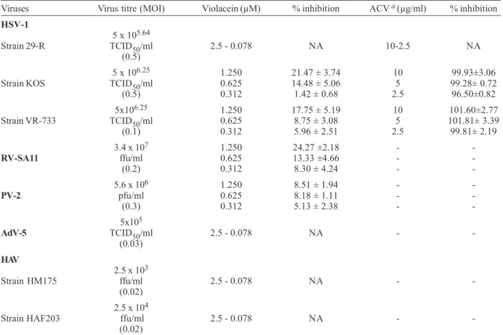
Violacein showed no cytopathic effect inhibition of HSV-1 (strain 29-R/acyclovir resistant), HAV (strains HM175 and HAF-203) and AdV-5 neither demonstrated antiviral activity through the MTT assay. However violacein showed a weak inhibition of HSV-1 (strains KOS and ATCC/VR733), PV-2 and RV-SA11 replication by MTT assay (Table II) which is more sensitive and accurate than cytopathogenicity inhibition evaluation. The obtained percentages of viral inhibition were < than
50%, therefore it was not possible to estimate the EC50
values (50% effective concentration) necessary to
calculate selectivity indices (SI = CC50 /EC50).
According to May et al. (1991) violacein (with 10% of deoxyviolacein) showed activity against herpes and polioviruses. Duran (1998) also registered a patent for a formulation of cyclodextrin/violacein for treating bacterial, viral, trypanocidal infections and for antitumoral activity. Duran and Menck (2001), citing the patent registered by May et al. (1991), stated that 0.25 µg/ml of violacein inhibited HSV replication by 62%, and 0.063 µg/ml of violacein inhibited poliovirus replication by 56% in HeLa cells. This information from patent documents does not inform the methodologies used nor the virus strains and types or experimental conditions under which the results were obtained. No comparison with present data was therefore possible.
The results obtained in the present study differ from the above cited information in that higher concentrations (0.312 to 1.25 µM) of violacein have shown only a weak inhibitory action on HSV-1 (strains KOS and ATCC/VR733) and PV-2 replication (Table II).
The violacein cytotoxicity data themselves have intrinsic value in defining toxic effects (e.g. as an indicator of acute toxic effects in vivo) and are also important for designing more in depth in vitro studies (Eisenbrand et al. 2002). These data will be useful for a better understanding Cytotoxicity of violacein towards different cell lines investigated
by different methods.
Cell m orphology evaluation by inverted light m icroscopy
0 2 4 6
0,08 0,31 1,25 5
Violacein (µM)
Score of
morphological
changes
VERO FRhK-4
HEp-2 MA104
Cell viability test by Trypan blue dye exclusion
0 50 100 150
0,08 0,31 1,25 5
Violacein (µM) % Cell viability in relation to cell
controls
VERO FRhK-4
HEp-2 MA104
Cell viability test by MTT assay
0 50 100 150
0,08 0,31 1,25 5
Violacein (µM) % Cell viability in relation to cell
controls
VERO FRhK-4
847 847847 847847 Mem Inst Oswaldo Cruz, Rio de Janeiro, Vol. 98(6), September 2003
of other biological activities attributed to violacein, such as antibacterial, antitumoral and anti- Trypanosoma cruzi activities (Melo et al. 2000, Duran & Menck 2001).
One strategy used to improve biological activity is the incorporation of active substances into liposomes which offer a substancial improvement in the therapeutic indices of the molecules entrapped in them (Gulati et al. 1998). Some antiviral drugs and their liposomal formulations have been subjected to in vitro and in vivo studies and have been shown to result in an increase of drug absorption, less toxicity than conventional drug formulations and a more prolonged effect (Law et al. 2000, Wutzler et al. 2002). In this way, violacein-containing liposomes have been prepared and preliminary results show that its antiviral activity against HSV-1 is increased (data not shown). Complementary studies are in course in our laboratory.
REFERENCES
Balfour HH 1999. Drug therapy: antiviral drugs. N Engl J Med 340: 1255-1268.
Barardi CRM, Emslie KR, Vesey G, Williams KL 1998. Development of a rapid and sensitive quantitative assay for rotavirus based on flow cytometry. J Virol Meth 74: 31-37.
Bedard J, May S, Barbeau D, Yuen L, Rando RF, Bowlin TL 1999. A high throughput colorimetric cell proliferation assay for the identification of human cytomegalovirus inhibitors. Antiviral Res 41: 35-43.
Bedoya LM, Palomino-Sanchez S, Abad MJ, Bermejo P, Alcami J 2001. Anti-HIV activity of medicinal plant extracts. J Ethnopharmacol77: 113-116.
Betancur-Galvis L, Morales GE, Forero JE, Roldan J 2002. Cytotoxic and antiviral activities of Colombian medicinal plant extracts of the Euphorbia genus. Mem Inst Oswaldo Cruz 97: 541-546.
Bromberg N, Duran N 2001. Violacein transformation by peroxidases and oxidases: implications on its biological properties. J Mol Catal B: Enzim 11: 463-467.
Burleson FG, Chamberts TM, Wiedbrauk DL 1992. Virology.A Laboratory Manual, Academic, San Diego, 250 pp. Carmichael J, Degraff WG, Gazdar AF, Minna JD, Mitchell JB
1987. Evaluation of a tetrazolium-based semiautomated colorimetric assay: assessment of chemosensitivity testing. Cancer Res 47: 936-942.
De Logu A, Loy G, Pellerano ML, Bonsignore L, Schivo ML 2000. Inactivation of HSV-1 and HSV-2 and prevention of cell-to-cell virus spread by Santolina insularies essential oil. Antiviral Res 48: 177-185.
Denizot F, Lang R 1986. Rapid colorimetric assay for cell growth and survival. J Immunol Meth 89: 271-277.
TABLE II
Antiviral activity of violacein expressed as the percentual inhibition of viral replication when compared to controls by using the MTT assay
Viruses Virus titre (MOI) Violacein (µM) % inhibition ACV a (µg/ml) % inhibition
HSV-1
5 x 105.64
Strain 29-R TCID50/ml 2.5 - 0.078 NA 10-2.5 NA
(0.5)
5 x 106.25 1.250 21.47 ± 3.74 10 99.93±3.06
Strain KOS TCID50/ml 0.625 14.48 ± 5.06 5 99.28± 0.72
(0.5) 0.312 1.42 ± 0.68 2.5 96.50±0.82
5x106.25 1.250 17.75 ± 5.19 10 101.60±2.77
Strain VR-733 TCID50/ml 0.625 8.75 ± 3.08 5 101.81± 3.39
(0.1) 0.312 5.96 ± 2.51 2.5 99.81± 2.19
3.4 x 10⁷ 1.250 24.27 ±2.18 -
-RV-SA11 ffu/ml 0.625 13.33 ±4.66 -
-(0.2) 0.312 8.30 ± 4.24 -
-5.6 x 10⁶ 1.250 8.51 ± 1.94 -
-PV-2 pfu/ml 0.625 8.18 ± 1.11 -
-(0.3) 0.312 5.13 ± 2.38 -
-5x10⁵
AdV-5 TCID
50/ml 2.5 - 0.078 NA -
-(0.03)
HAV
2.5 x 10³
Strain HM175 ffu/ml 2.5 - 0.078 NA -
-(0.02)
2.5 x 10⁴
Strain HAF203 ffu/ml 2.5 - 0.078 NA -
-(0.02)
848 848 848 848
848 Antiviral Evaluation of Violacein • CR Andrighetti-Fröhner et al.
Duran N 1998. Formulation of cyclodextrin/violacein based medicine – comprises enhancement of violacein solubility, with increase in versatility. BR PI 9801307-A, 4 Mar. Duran N, Menck CFM 2001. Chromobacterium violaceum: a
review of pharmacological and industrial perspective. Crit Rev Microbiol 27: 201-222.
Duran N, Antonio RV, Haun M, Pilli RA 1994. Biosynthesis of a trypanocide of Chromobacterium violaceum. World J Microbiol Biotechnol 10: 686-691.
Eisenbrand G, Pool-Zobel B, Baker V, Balls M, Blaauboer BJ, Boobis A, Carere A, Kevekordes S, Lhuguenot JC, Pieters R, Kleiner J 2002. Methods of in vitro toxicology. Food Chem Toxicol40: 193-236.
Estes MK, Graham DY, Mason BB 1981. Proteolytic enhan-cement of rotavirus infectivity molecular mechanism. J Virol 39: 879-888.
Glatthaar-Saalmüller B, Sacher F, Esperester A 2001. Antiviral activity of an extract derived from roots of Eleutherococcus senticosus. Antiviral Res 50: 223-228.
Gulati M, Grover M, Singh S, Singh M 1998. Lipophilic drug derivatives in liposomes. Int J Pharm 165: 129-168. Kaneko H, Keiichiro K, Mori S, Shigeta S 2001. Antiviral
activity of NMSO3 against adenovirus in vitro. Antiviral Res52: 281-288.
Law SL, Huang KJ, Chiang CH 2000. Acyclovir-containing liposomes for potencial ocular delivery corneal penetration and absorption. J Control Release63: 135-140.
Leon LL, Miranda CC, De Souza AO, Duran N 2001. Anti-leishmanial activity of the violacein extracted from Chromobacterium violaceum. Antimicrob Agents Chemoth 48: 449-450.
Li K, Ma SC, Yang YT, Ye SM, But PP 2002. Antiviral activities of flavonoids and organic acid from Trollius chinesis Bunge. J Ethnopharmacol79: 365-368.
Lotfi K, Zackrisson A, Peterson C 2002. Comparison of idarubicin and daunorubicin regarding intracellular uptake, induction of apoptosis and resistance. Cancer Lett 178: 141-149.
May G, Brummer B, Ott H 1991.Treatment of prophylaxis of polio and herpes virus infections – comprises admin. of 3-(1,2-dihydro-5-(5-hydroxy-1H- indol-3-yl)-2-oxo-3H-pyrrole-3-ylidene)-1,3-dihydro-2H-indol-2-one. Ger Offen DE 3935066, 25 April.
Melo PS, Maria SS, Vidal BC, Haun M, Duran N 2000. Violacein cytotoxicity and induction of apoptosis in V79 cells. In vitro Cell Dev Biol Anim36: 639-543.
Mossmann T 1983. Rapid colorimetric assay for cellular growth and survival: application to proliferation and cytotoxicity assays. J Immunol Meth 65: 55-63.
Nielsen J 2002. Combinatorial synthesis of natural products. Curr Opin Chem Biol 6: 297-305.
Pillay D, Zambon M 1998. Antiviral drug resistance. Brit Med
J 317: 660-662.
Pujol CA, Errea MI, Matulewicz MC, Damonte EB 1996. Antiherpetic activity of S1, an algal derived sulphated galactan. Phytoth Res 10: 410-413.
Reed LJ, Muench H 1938. A simple method of estimating fifty per cent endpoints. AmJ Hyg 27: 493-497.
Semple SJ, Pyke SM, Reynolds G, Flower RLP 2001. In vitro antiviral activity of the anthraquinone chrysophanic acid against poliovirus. Antiviral Res49: 169-178.
Sieuwerts A, Klijn JGM, Peters HA, Foekens JA 1995. The MTT tetrazolium salt assay scrutinized: how to use this assay reliably to measure metabolic activity of cell cultures in vitro for the assessment of growth characteristics, IC50 – values and cell survival. Eur J Clin Chem Clin Biochem 33: 813-823.
Simões CMO, Amoros M, Girre L 1999. Mechanism of antiviral activity of triterpenoid saponins. Phytoth Res 21: 317-325. Smee DF, Morrison AC, Barnard DL, Sidwell RW 2002. Comparison of colorimetric, fluorometric, and visual methods for determining anti-influenza (H1N1 and H3N2) virus activities and toxicities of compounds. J Virol Meth 106: 71-79.
Takahashi K, Matsuda M, Ohashi K, Taniguchi K, Nakagomi O, Abe Y, Mori S, Sato N, Okutani K, Shigeta S 2001. Analysis of anti-rotavirus activity of extract from Stevia rebaudiana. Antiviral Res 49: 15-24.
Takeuchi H, Baba M, Shigeta S 1991. An application of tetrazolium (MTT) colorimetric assay for the screening of anti-herpes simplex virus compounds. J Virol Meth 33: 61-71.
Todryk S, Melcher A, Bottley G, Gough M, Vile R 2001. Cell death associated with genetic prodrug activation therapy of colorectal cancer. Cancer Lett 174: 25-33.
Vlietinck AJ, De Bruyne T, Vanden Berghe DA 1997. Plant substances as antiviral agents. Curr Org Chem 1: 307-344. Vlietinck AJ, De Bruyne T, Pieters LA 1998. Plant-derived leading compounds for chemotherapy of human immunodeficiency virus (HIV) infection. Planta Med 64: 97-109.
Vlietinck AJ, Vanden Berghe DA 1998. Leads for antivirals from tradicional medicines. In HDV Prebdergastm, NL Etkin, DR Harris, PJ Houghton (eds), Plants for Food and Medicine, Royal Botanic Gardens, London, p. 333-344. Walum E, Strenberg K, Jenssen D 1990. Understanding Cell
Toxicology:Principles and Pratice, Ellis Howood, NewYork, p. 97-111.
Wilson AP 2000. Cytotoxicity and viability assays. In JRW Masters, Animal Cell Culture,3rd ed., Oxford University, Oxford, p. 175-219.