Biological activity of
Serratia
marcescens
cytotoxin
1Departamento de Microbiologia e Imunologia, Instituto de Biologia,
Universidade Estadual de Campinas, Campinas, SP, Brasil
2Departamento de Análises Clínicas, Toxicológicas e Bromatológicas,
Faculdade de Ciências Farmacêuticas de Ribeirão Preto, Universidade de São Paulo, Ribeirão Preto, SP, Brasil
3Departamento de Clínica Médica, Faculdade de Medicina de Ribeirão Preto,
Universidade de São Paulo, Ribeirão Preto, SP, Brasil G.V. Carbonell1,
C.R.N. Amorim1,
M.T. Furumura1,
A.L.C. Darini2,
B.A.L. Fonseca3
and T. Yano1
Abstract
Serratia marcescens cytotoxin was purified to homogeneity by ion-exchange chromatography on a DEAE Sepharose Fast Flow column, followed by gel filtration chromatography on a Sephadex G100 col-umn. The molecular mass of the cytotoxin was estimated to be about 50 kDa. Some biological properties of the cytotoxin were analyzed and compared with well-characterized toxins, such as VT1, VT2 and CNF from Escherichia coli and hemolysin produced by S. marces-cens. The sensitivity of the cell lines CHO, HeLa, HEp-2, Vero, BHK-21, MA 104 and J774 to the cytotoxin was determined by the cell viability assay using neutral red. CHO and HEp-2 were highly sensi-tive, with massive cellular death after 1 h of treatment, followed by BHK-21, HeLa, Vero and J774 cells, while MA 104 was insensitive to the toxin. Cytotoxin induced morphological changes such as cell rounding with cytoplasmic retraction and nuclear compactation which were evident 15 min after the addition of cytotoxin. The cytotoxic assays show that 15 min of treatment with the cytotoxin induced irreversible intoxication of the cells, determined by loss of cell viabil-ity. Concentrations of 2 CD50 (0.56 µg/ml) of purified cytotoxin did
not present any hemolytic activity, showing that the cytotoxin is distinct from S. marcescens hemolysin. Antisera prepared against S. marcescens cytotoxin did not neutralize the cytotoxic activity of VT1, VT2 or CNF toxin, indicating that these toxins do not share antigenic determinants with cytotoxin. Moreover, we did not detect gene se-quences for any of these toxins in S. marcescens by PCR assay. These results suggest that S. marcescens cytotoxin is not related to any of these toxins from E. coli.
Correspondence
G.V. Carbonell
Departamento de Microbiologia e Imunologia
Instituto de Biologia, UNICAMP 13083-970 Campinas, SP Brasil
Fax: +55-19-3788-7191 E-mail: [email protected]
Research supported by FAPESP.
Received July 2, 2001 Accepted October 21, 2002
Key words
·Cell culture ·Biological activity
·Serratia marcescens
·Cytotoxin ·Virulence factors
Introduction
Serratia marcescens has been considered to be an important nosocomial pathogen, re-sponsible for endemic and epidemic infec-tions, especially in newborns and patients submitted to invasive procedures (1-4).
serotype. Moreover, this cytotoxic activity is not mediated by plasmids. The S. marces-cens toxin is extracellular and heat labile, and optimal culture conditions were incuba-tion at temperatures ranging from 30 to 37ºC for 24 h under shaking in medium adjusted to pH 8.5 (6).
Recently, it was shown that the hemol-ysin of S. marcescens induces cytotoxic ef-fects on human epithelial cells, character-ized by vacuolization with subsequent cell lysis (7). The cytopathic effects of the S. marcescens cytotoxin correspond to cell rounding followed by gradual destruction of the monolayer, as observed by inverted mi-croscopy (5). This technique is routinely used to assess cytotoxicity in cell culture; however, it lacks sensitivity for detecting more details about early events of the cell injury process. Thus, it is still unclear whether the cytotoxin causes vacuolization and cell lysis. We report here the purification of the S. marcescens cytotoxin and compare its bio-logical characteristics with those of other well-characterized toxins.
Material and Methods
Cytotoxin purification
A 0.5-ml pre-culture of S. marcescens
(6) was inoculated into 500 ml Davis mini-mal medium (8) in a 1-liter Erlenmeyer flask with shaking at 150 rpm and incubated at 37ºC for 18-24 h. Six liters of culture from 12 such flasks was centrifuged at 10,000 g
for 15 min at 4ºC and ammonium sulfate was added slowly to the culture supernatant to 80% saturation. After centrifugation at 10,000
g for 15 min at 4ºC, the pellet collected was dissolved in 25 mM Tris-HCl, pH 7.0, fol-lowed by exhaustive dialysis against the same buffer. The preparation was applied to a DEAE Sepharose Fast Flow column (3.0 x 9.0 cm; Pharmacia LKB Biotechnology, Uppsala, Sweden) equilibrated with 25 mM Tris-HCl buffer, pH 7.0, and the cytotoxin
was eluted (5-ml fractions) with the same buffer containing 0.1 M NaCl. Fractions with toxicity were pooled, concentrated to 5 ml by ultrafiltration through a PM 10 membrane (Amicon Corp., Lexington, MA, USA) and applied to a Sephadex G100 column (2.5 x 68 cm; Pharmacia) equilibrated and eluted (2.5-ml fractions) with 25 mM Tris-HCl buf-fer containing 0.15 M NaCl, pH 7.0. Frac-tions with the highest cytotoxic activity were pooled and stored at -70ºC until use. The total protein concentration was determined by the method of Bradford using reagents from BioRad (Hercules, CA, USA) and se-rum albumin as standard (9).
Molecular mass estimation by gel filtration chromatography
The molecular mass of the cytotoxin was estimated by gel filtration on a Superdex 200 HR column (Pharmacia). A 0.1-ml portion of purified cytotoxin was applied to the col-umn equilibrated with 25 mM Tris-HCl buf-fer, pH 7.0, and eluted at 0.25 ml/min in an HPLC system (Shimadzu, Kyoto, Japan). Absorbance of the cytotoxin was measured at 280 nm and the material was collected and assayed for cytotoxicity on Chinese hamster ovary (CHO) cells. The molecular mass of the cytotoxin was estimated by comparing its elution volume with those of calibration standards such as ribonuclease A (13.7 kDa), chymotrypsinogen A (25 kDa), ovalbumin (43 kDa), albumin (67 kDa) and blue dex-tran 2000 (>100 kDa) (10).
Gel electrophoresis
The purified cytotoxin (30 µg/ml) was assessed for purity on 8% silver-stained so-dium dodecyl sulfate (SDS)-polyacrylamide gel (11).
Cell lines and cytotoxic assays
human epidermoid carcinoma (HEp-2), baby hamster kidney (BHK-21), CHO, mouse tu-mor macrophage (J774) and monkey kidney (MA 104) cells, all obtained from the Ameri-can Type Culture Collection (Rockville, MD, USA), were maintained in tissue culture flasks with Eagle’s modified essential medi-um (Gibco, BRL, São Paulo, SP, Brazil) supplemented with 10% fetal calf serum, and 0.75 mM L-glutamine. The J774 cells were cultivated in RPMI medium (Gibco) under the same conditions as described above. The cytotoxicity assays were per-formed as described in Ref. 6. Briefly, after formation of a cell monolayer, the medium was removed from each well and replaced with fresh medium without serum and con-taining the cytotoxin. The plates were incu-bated in the presence of 5% CO2 at 37ºC. The cell monolayer morphology was observed daily under an inverted microscope in order to detect the presence of any cytopathic ef-fect.
Determination of cell viability and CD50
In order to compare the sensitivities of the cell lines to S. marcescens cytotoxin, cell viability after the cytotoxic assay was quan-tified by the neutral red cytotoxicity assay (6). The concentration of toxin that killed 50% of CHO cells (CD50) was calculated as described in Ref. 12.
Morphological changes
The CHO cells were grown on coverslips placed inside 24-well plates using 1 ml of cell suspension (1 x 10⁴ cells/ml) per well and the plates were incubated at 37ºC in the presence of 5% CO2. After 24 h, the culture medium was aspirated and replaced with 1 ml of fresh medium and a 2 CD50 dose of cytotoxin was added to each well. At time-defined intervals, the coverslips were washed with phosphate-buffered saline (PBS) and fixed in 10% formaldehyde solution in PBS
for 1 h. The coverslips were then washed with distilled water and stained with 0.025% toluidine blue, pH 4.0, as described by Mello and Vidal (13). The coverslips were washed with distilled water, air dried, cleared in xylene and mounted on slides using Entellan (Merck, Darmstadt, Germany). The cellular alterations were observed with an Axioskop microscope (Nikon, Tokyo, Japan).
Antiserum production
Aliquots of 50 µl of a purified cytotoxin preparation (75 µg of protein per ml) were emulsified in an equal volume of Freund’s complete adjuvant and injected via the in-guinal node, with subsequent injections in incomplete adjuvant after 2 weeks. Animals were bled and the cytotoxicity-neutralizing capacity of the sera was tested.
Seroneutralization assays
In order to determine whether S. marces-cens cytotoxin antiserum could neutralize the cytotoxicity of VT1, VT2 or cytotoxic necrotizing factor (CNF) produced by E. coli, seroneutralization assays were carried out (14). Antisera produced in rabbits against purified cytotoxin of S. marcescens were serially diluted in Eagle’s medium and mixed with 1/40 dilution of the test toxins. The mixtures were incubated for 1 h at 37ºC, applied to Vero cells and incubated for 72 h at 37ºC. The plates were examined micro-scopically on a daily basis for the appear-ance of cytopathogenicity. Negative con-trols with preimmune serum were also in-cluded.
Hemolysis assays
dilu-tion were mixed with 1 ml of erythrocyte suspension and incubated for 30 min at 30ºC. The mixture was then centrifuged for 3 min at 2,500 rpm to remove unlysed erythrocytes and cell debris. The absorbance of the super-natants containing released hemoglobin was measured at 405 nm. Results were compared with total lysis (100%) caused by SDS used
as control.
Toxin gene sequence determination by genotypic analysis (PCR)
The S. marcescens strain was screened for the presence of gene sequences of toxins from E. coli using primers for VT1 (ST-I), VT2 (ST-II) (15) and CNF (16). Bacterial DNA to be amplified was released from the organism by boiling. The reactions were per-formed as previously described (17) using 2 µl dNTP solution containing 20 mM of each nucleotide, 1 µl Taq DNA polymerase (1 U/ µl), 3 µl 5 µM MgCl2, 5 µl PCR buffer, 5 µl DNA, 1 µl of each primer (50 pmol), and 35 µl sterile Milli-Q water. The solutions were pre-heated without the enzyme at 94ºC/10 min and submitted to 30 cycles (GeneAmp PCR System 9700, Perkin Elmer, Foster City, CA, USA) of 94ºC/2 min, 50ºC/2 min and 72ºC/1 min (VT2), 94ºC/2 min, 57ºC/2 min and 72ºC/1 min (ST-I and CNF); after these cycles they were submitted to 72ºC/7 min. PCR products were analyzed by 2% horizon-tal agarose gel electrophoresis with ethidium bromide staining under UV light.
Results
Cytotoxin purification
The crude cytotoxin preparations were initially subjected to ion-exchange chroma-tography on a DEAE Sepharose Fast Flow column. The column was eluted with 500 ml of 25 mM Tris-HCl buffer at a flow rate of 1 ml/min, and the fractions containing cyto-toxic activity were eluted with the same buffer containing 0.1 M NaCl (Figure 1A, horizontal bar). The cytotoxin was subse-quently chromatographed on a Sephadex G100 gel filtration column equilibrated with 25 mM Tris-HCl buffer containing 0.15 M NaCl. Cytotoxic activity was eluted in the third peak (Figure 1B, horizontal bar). Cyto-toxin obtained at this stage presented a single Figure 1. A, DEAE Sepharose Fast Flow column chromatography. A crude sample was
applied to a DEAE Sepharose Fast Flow column equilibrated with 25 mM Tris-HCl buffer, pH 7.0. Fractions with cytotoxic activity (5 ml) were eluted with a nonlinear gradient of 0.1 M NaCl in the same buffer. The horizontal bar indicates the fractions pooled for subsequent purification. B, Sephadex G100 column chromatography. The cytotoxic fractions (5 ml) obtained from the DEAE Sepharose column were applied to a Sephadex G100 column equilibrated with 25 mM Tris-HCl buffer, pH 7.0, containing 0.15 M NaCl. The horizontal bar indicates the fractions with cytotoxic activity.
Absorbance at 280 nm
1.2
1.0
0.8
0.6
0.4
0.2
0.0
2 4 6 8 10 12 14 16 18 20 22 24 26 28 30 32 34 36 Fraction number
2 4 6 8 10 12 14 16 18 20 22 24 26 28 30 32 34 36 38 40 Fraction number
Absorbance at 280 nm
0.5
0.4
0.3
0.2
0.1
0.0
NaCl (0.1 M)
A
protein band by SDS electrophoresis (Fig-ure 2). The cytotoxin peak was eluted be-tween the positions for bovine serum albu-min and ovalbualbu-min, corresponding to an apparent molecular mass of 47 to 50 kDa on the calibration curve.
Cytotoxicity and cell viability assays
Among the cell lines tested, CHO, HeLa, HEp-2, Vero, BHK-21 and J774 were sus-ceptible to S. marcescens cytotoxin and only MA 104 cells were resistant. Although the susceptible cell lines showed similar mor-phological changes when exposed to cyto-toxin, the cells were not affected to the same extent after exposure for a specific time. The highest sensitivity was observed with CHO and HEp-2 cells, with the cytopathic effect starting as early as 15 min after incubation with toxin and resulting in the destruction of the monolayer within 1-2 h (Figure 3). The BHK-21, HeLa, J774 and Vero cells showed monolayer destruction after 24 h, as deter-mined by the viability assays. Thus, the CHO cells were used in all experiments for the characterization of the cytotoxin. The CD50 of the cytotoxin is defined as the highest concentration of the sample that killed 50% of the CHO cells after 24 h. The purified cytotoxin had a CD50 of 0.28 µg/ml.
Morphological changes
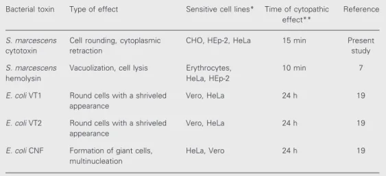
Morphological changes induced by the cytotoxin were studied in CHO cells stained with toluidine blue after different incubation periods. The first observable changes were evident about 15 min after the addition of the cytotoxin. The cytoplasm started to retract and the cells became rounded, with progres-sive nuclear compactation and extenprogres-sive sur-face blebbing being observed after 1 h (Fig-ure 4B). The morphological changes induced by the cytotoxin were then compared with S. marcescens hemolysin and VT1, VT2 and CNF from E. coli (Table 1).
Figure 2. Analysis of Serratia marcescens cytotoxin by SDS-PAGE using silver staining. Lane A, Cytotoxic fraction obtained from a Sephadex G100 column (30 µg/ml); lane B, molecular weight markers.
Cell viability (%)
100
Figure 3. Response of the CHO, HEp-2, HeLa, BHK-21, Vero, MA 104 and J774 cell lines after different incubation periods with a 2 CD50 dose (0.56 µg/ml) of purified Serratia marcescens cytotoxin. Cell viability was measured by neutral red assay.
12 12 12 12 12 12 12 12 12 12 12 12 12 12 1234 1234 1234 1234 1234 1234 1234 1234 1234 1234 1234 1234 1234 1234 1234 1234 1234 1234 1234 1234 1234 1234 1234 1234 1234 1234 1234 1234 1234 1234 1234 1234 1234 1234 1234 1234 1234 1234 123 123 123 123 123 123 123 123 123 123 123 123 123 123 123 123 123 123 123 123 123 123 123 12 12 12 12 12 12 12 12 12 12 12 12 12 12 12 12 12 12 12 1234 1234 1234 1234 1234 1234 1234 1234 1234 1234 1234 1234 1234 1234 1234 1234 1234 1234 1234 1234 1234 1234 1234 1234 1234 1234 1234 1234 1234 1234 1234 1234 1234 1234 1234 1234 1234 1234 12 12 12 12 12 12 12 12 12 12 12 12 123 123 123 123 123 123 123 123 123 123 123 123 123 123 123 123 123 123 123 123 123 123 123 123 123 123 123 123 123 123 123 123 123 123 123 123 123 123 123 123 123 123 123 123 123 123 123 123 12 12 12 12 12 12 12 12 12 12 12 123 123 123 123 123 123 123 123 123 123 123 123 123 123 123 123 123 123 123 123 123 123 123 123 123 123 123 123 123 123 123 123 123 123 123 123 123 123 123 123 123 123 123 123 123 123 123 75 50 25 0 12 12 12 12 12 CHO HEp-2 MA 104 J774 HeLa BHK-21 Vero
1 4 10 24
Incubation time (h)
Serum neutralization assays
At higher concentrations of antitoxin se-rum, i.e., lower dilution, the morphological changes induced by VT1, VT2 and CNF toxins from E. coli on Vero cells were clearly evident after 24 h. The results of neutraliza-tion studies showed that antiserum against S. marcescens cytotoxin did not neutralize the biological activity of these toxins (Figure 5).
Genotypic analysis (PCR)
The assays showed that S. marcescens
isolates do not amplify any detectable frag-ment, being characterized as negative for amplification by PCR gene sequence for VT1, VT2 and CNF toxins.
Discussion
The S. marcescens cytotoxin used in the present study was purified to homogeneity from a culture supernatant grown in Davis minimal medium (8) by ion-exchange chro-matography on a DEAE Sepharose Fast Flow column and submitted to gel filtration on a Figure 4. Morphological changes
induced by purified Serratia mar-cescens cytotoxin (2 CD50 dose)
in CHO cells stained with toluidin blue (100X). A, Typical morphol-ogy of the control cells; B, cyto-plasmic retraction, nuclear com-pactation and cytoplasmic bleb-bing (arrowheads) after 30 min of treatment.
Table 1. Characteristics of toxins that were compared with Serratia marcescens cytotoxin in this study.
Bacterial toxin Type of effect Sensitive cell lines* Time of cytopathic Reference effect**
S. marcescens Cell rounding, cytoplasmic CHO, HEp-2, HeLa 15 min Present
cytotoxin retraction study
S. marcescens Vacuolization, cell lysis Erythrocytes, 10 min 7
hemolysin HeLa, HEp-2
E. coli VT1 Round cells with a shriveled Vero, HeLa 24 h 19
appearance
E. coli VT2 Round cells with a shriveled Vero, HeLa 24 h 19
appearance
E. coli CNF Formation of giant cells, HeLa, Vero 24 h 19
multinucleation
*The most common cells used for the cytotoxicity assays.
**Approximate time for the appearance of a cytopathic effect after toxin addition. Hemolysis assays
S. marcescens cytotoxin did not show hemolytic activity in the liquid hemolytic assay, even when treated with crude cyto-toxin preparations (2 CD50 dose or 0.56 µg/ ml).
A
Sephadex G100 column. The fractions with cytotoxic activity were eluted in the third peak (Figure 1B, horizontal bar) and re-vealed a single protein band by SDS electro-phoresis (Figure 2). The molecular mass of the purified cytotoxin was estimated to be 47 to 50 kDa by gel filtration chromatography. This method is based on a calibration curve using proteins of known molecular masses as markers (10). The results show that the molecular mass of cytotoxin is apparently different from extracellular proteases pro-duced by this bacterium, which have molec-ular masses of 56, 60 and 73 kDa (18). However, this cytotoxin has not yet been characterized in terms of proteolytic activ-ity.
Figure 5. Effects of VT1 and CNF toxins on Vero cells in seroneu-tralization assays. A, Vero con-trol cells. B, Vero cells that re-ceived mixtures of antiserum against Serratia marcescens cy-totoxin incubated for 1 h at 37ºC with CNF toxin. C, Vero cells in-oculated with mixtures of anti-serum and VT toxin.
In order to choose a suitable target cell line, we compared the sensitivity of several cell lines to cytotoxin produced by clinical isolates of S. marcescens. This represents an important step related to the practical as-pects of characterization of the cytotoxin. CHO and HEp-2 cells appeared to be the most sensitive to the toxic effects of S. mar-cescens cytotoxin, followed by BHK-21, HeLa, J774 and Vero cells (Figure 3), while MA 104 cells were resistant to the toxin. Thus, CHO cells were used as a model to study the details of the mechanism of action of the cytotoxin.
The pattern of morphological changes gives a preliminary idea of the characteris-tics of the toxin and its mechanism of action
C
References
1. Archibald LK, Manning ML, Bell LM, Banerjee S & Jarvis WR (1997). Patient density, nurse-to-patient ratio and nosocomial infec-tion risk in a paediatric cardiac intensive care unit. Pediatric Infec-tious Disease Journal,16: 1045-1048.
2. Van Ogtrop ML, Van Zoeren-Grobben D, Verbakel-Salomons EMA & Van Boven CPA (1997). Serratia marcescens infections in neona-tal departments: Description of an outbreak and review of the literature. Journal of Hospital Infection, 36: 95-103.
3. Bosi C, Davin-Regli A, Charrel R, Rocca B, Monnet D & Bollet C (1996). Serratia marcescens nosocomial outbreak due to contami-nation of hexetidine solution. Journal of Hospital Infection, 33: 217-224.
4. Miranda G, Kelly C, Solorzano F, Leanos B, Coria R & Patterson JE (1996). Use of pulsed-field gel electrophoresis typing to study an outbreak of infection due to Serratia marcescens in a neonatal intensive care unit. Journal of Clinical Microbiology, 34: 3138-3141. 5. Carbonell GV, Alfieri AF, Alfieri AA, Vidotto MC, Levy CE, Darini ALC & Yanaguita RM (1997). Detection of cytotoxic activity on Vero cells in clinical isolates of Serratia marcescens. Brazilian Journal of Medical and Biological Research,30: 1291-1298.
6. Carbonell GV, Fonseca BAL, Figueiredo LTM, Darini ALC & Yanaguita RM (1996). Culture conditions affect cytotoxin produc-tion by Serratia marcescens. FEMS Immunology and Medical Mi-crobiology, 16: 299-307.
on the cells. The cells treated with S. marces-cens cytotoxin lost the typical morphology observed in the untreated cultures (Figure 4A) and showed cell rounding, cytoplasmic retraction, compactation of the nuclei and cytoplasmic blebbing (Figure 4B). The mor-phological changes induced in cell lines and the time of first observable effects are useful to distinguish the S. marcescens cytotoxin from other bacterial toxins, as shown in Table 1. The cell rounding seen in CHO cells in response to the S. marcescens cytotoxin is different from the cell rounding with shriv-eled appearance induced by VT1 and VT2 or from the giant, multinucleated cells induced by CNF from E. coli (19). The lack of neu-tralization of cytotoxic activity of VT1, VT2 and CNF by antisera prepared against S. marcescens cytotoxin indicates that these toxins do not possess an antigenic relation-ship. Moreover, cytotoxin-producing S. mar-cescens was negative when tested for the presence of gene sequences for VT1, VT2 and CNF toxins. The results of all compara-tive studies showed that cytotoxin is not
related to any of the toxins studied.
Almost all strains of S. marcescens se-crete a hemolysin that lyses all mammalian erythrocytes tested and is cytotoxic to hu-man epithelial cells. The cytopathic effects of hemolysin on HEp-2 cells were character-ized by rapid vacuolization (15 min) fol-lowed by lysis after 40 min (7). These mor-phological changes observed in cultured cells were clearly distinct from those of cytotoxin, as shown in Table 1. Therefore, the purified cytotoxin did not cause hemolytic activity in liquid assays. This is of particular impor-tance since this study provides clear evi-dence that the cytotoxin is distinct from S. marcescens hemolysin.
Acknowledgments
We are grateful to the staff of the Labora-tory of Microbiology, School of Medicine of Ribeirão Preto, USP, who provided the bac-terial strains, and to Ana Stella Menegon Degrossoli for technical assistance.
7. Hertle R, Hilger M, Weingardt-Kocher S & Walev I (1999). Cytotoxic action of Serratia marcescens hemolysin on human epithelial cells.
Infection and Immunity, 67: 817-825.
8. Davis BD & Minglioli ES (1950). Mutants of Escherichia coli requiring methionine or vitamin B12. Journal of Bacteriology, 60: 17-28. 9. Bradford MM (1976). A rapid method for the quantification of
micro-gram quantities of protein utilizing the principle of protein-dye bind-ing. Analytical Biochemistry, V: 248-254.
10. MacLeod DL, Gyles CL, Valdivieso-Garcia A & Clarke RC (1991). Physicochemical and biological properties of purified Escherichia coli
shiga-like toxin II variant. Infection and Immunity, 59: 1300-1306. 11. Laemmli UK (1970). Cleavage of structural proteins during assembly
of the head of bacteriophage T4. Nature, 227: 680-685.
12. Cleary TG, Mathewson JJ, Faris E & Pickering LK (1985). Shiga-like cytotoxin production by enteropathogenic Escherichia coli sero-groups. Infection and Immunity, 47: 335-337.
13. Mello ML & Vidal B (1980). Práticas em Biologia Celular. Edgard Blucher, Rio de Janeiro, RJ, Brazil, 71.
14. Parreira VR & Yano T (1998). Cytotoxin produced by Escherichia coli
isolated from chickens with swollen head syndrome (SHS). Veteri-nary Microbiology, 62: 111-119.
chain reaction and phenotypic assays. Journal of Veterinary Medi-cine. Series B: Infectious Diseases and Veterinary Public Health, 41: 49-59.
16. Blanco M, Blanco JE, Blanco J, Alonso MP, Balsanobre C, Mourino M, Madrid C & Juarez A (1996). Polymerase chain reaction for detection of Escherichia coli strains producing cytotoxic necrotizing factor type 1 and type 2 (CNF1 and CNF2). Journal of Microbiological Methods, 26: 95-101.
17. Blanco M, Blanco JE, Gonzalez EA, Mora A, Jansen W, Gomes T, Zerbini LF, Yano T, Pestana De Castro AF & Blanco J (1997). Genes
encoding for enterotoxins and verotoxins in porcine Escherichia coli
strains belonging to different O:K:H serotypes: relationship with toxic phenotypes. Journal of Clinical Microbiology, 35: 2958-2963. 18. Matsumoto K, Maeda H, Takata K, Kamata R & Okamura R (1984).
Purification and characterization of four proteases from a clinical isolate of Serratia marcescens kums 3958. Journal of Bacteriology, 157: 225-232.