Genotoxicity and antigenotoxicity assessment of shiitake (
Lentinula edodes
(Berkeley) Pegler) using the Comet assay
CK Miyaji
1, BQ Jordão
1, LR Ribeiro
2,3, AF Eira
2and IMS Cólus
1 1Universidade Estadual de Londrina (UEL), Londrina, Paraná, Brazil.
2Universidade Estadual Paulista (UNESP), Campus Botucatu, São Paulo, Brazil.
3Universidade Luterana do Brasil (ULBRA), Canoas, Rio Grande do Sul, Brazil.
Abstract
The mushroom shiitake (Lentinula edodes (Berkeley) Pegler) is been widely consumed in many countries, including Brazil, because of its pleasant flavor and reports of its therapeutic properties, although there is little available infor-mation on the genotoxicity and/or antigenotoxicity of this mushroom. We used the Comet assay and HEp-2 cells to evaluate thein vitro genotoxic and antigenotoxic activity of aqueous extracts of shiitake prepared in three different concentrations (0.5, 1.0 and 1.5 mg/mL) and three different temperatures (4, 22 and 60 °C), using methyl methanesulfonate (MMS) as a positive control and untreated cells as a negative control. Two concentrations (1.0 and 1.5 mg/mL) of extract prepared at 4 °C and all of the concentrations prepared at 22 ± 2 and 60 °C showed moder-ate genotoxic activity. To test the protective effect of the three concentrations of the extracts against the genotoxicity induced by methyl methanesulfonate, three protocols were used: pre-treatment, simultaneous-treatment and post-treatment. Treatments were repeated for all combinations of preparation temperature and concentration. Two extracts (22 ± 2 °C 1.0 mg/mL (simultaneous-treatment) and 4 °C 0.5 mg/mL (post-treatment)) showed antigenotoxic activity.
Key words: shiitake, Comet assay, HEp-2 cells; genotoxicity, antigenotoxicity.
Received: March 17, 2003; Accepted: September 15, 2003.
Introduction
The single cell gel electrophoresis assay (Comet) is a well-established highly sensitivity genotoxicity test that has been used to detect a broad spectrum of DNA damage (Fairbairnet al., 1995; Andersonet al., 1998). In the alka-line version of this test, DNA strand breaks and alkali-labile sites are detected and the extent of DNA migrations indi-cates the amount of DNA damage in the cell (Singh, 2000). According to Tice (1995) and Fairbairnet al. (1995), the al-kaline assay has been extensively used to assess the repair of single-strand breaks and the DNA damage induced by several agents and under a variety of experimental condi-tions (in vivoandin vitro). This assay can also be applied to examine the genotoxic and protective potential of several natural products.
The identification of antimutagenic agents present in the diet represents an effective strategy to influence or in-hibit pathological processes resulting from exposure to an increasing number of mutagenic and/or carcinogenic
sub-stances present in the environment (Wattemberg, 1983; Wattemberg, 1985; Ferguson, 1994).
Shiitake is an edible mushroom, highly appreciated due to its nutritional and medicinal properties, which has been reported to be of use in the reduction of arterial pres-sure and blood cholesterol levels as well as in the preven-tion of heart diseases and cancer (Wasser and Weis, 1999). However, few studies have been performed regarding the genotoxic and antigenotoxic potential of shiitake, and al-though Grüteret al. (1991) found that methanolic extracts of shiitake showed no mutagenic activity on bacteria the ef-fects of shiitake on eukaryotic organisms remains un-known. Using an in vivo micronucleus (MN) test and cyclophosphamide and N-ethyl-N-nitrosourea as muta-gens, Limaet al. (2001) found that shiitake strain LE 96/17 exhibited antimutagenic activity.
The objective of the study described in this paper was to investigate aqueous extracts of the shiitake mushroom (Lentinula edodes(Berkeley) Pegler), strain LE 96/17 for possible genotoxic or antigenotoxic effects on epidermoid carcinoma human larynx cells (HEp-2) exposed to the alkylant agent methyl methanesulfonate (MMS) using the alkaline version of the Comet assay.
Send correspondence to IMS Cólus. Universidade Estadual de Londrina, Departamento de Biologia Geral - CCB, Caixa Postal 6001, 86051-970, Londrina,PR, Brazil. E-mail address: colus@ sercomtel.com.br.
This study is part of a larger and more extensive re-search program to investigate distinctL. edodeslineages for genotoxic and antigenotoxic activity and identify those lineages having the most desirable characteristics for con-sumption; a further aim being to characterize and purify chemical components from these lineages and evaluate them in genotoxicity tests. Studies with crude aqueous ex-tracts are appropriate because they best represents the form in which shiitake is consumed. In addiction, with the ex-tracts it is possible to maintain the possible interactions among different chemical components, which effects could be not detected in studies that use only purified compo-nents.
Materials and Methods
Cell lines and culture conditions
Epidermoid carcinoma human larynx cells (HEp-2) were kindly provided by Prof. R.E.C. Linhares (Microbiology Department, State University of Londrina, Paraná -Brazil). Cells were grown at 37 °C in 10mL D-MEM/Ham F-10 (1:1) medium (Sigma) supplemented with 10% fetal calf serum (Cultilab), antibiotics (penicillin 0.06 g/L and streptomycin 0.10 g/L Sigma) and HEPES (2.38 g/L -Serva) in 25 cm2culture flasks (Nunc).
Chemicals
As recommended by the International Workshop on Genotoxicity Test Procedures (Ticeet al., 2000), for the positive control we used the mutagenic alkylating agent methyl methanesulfonate (MMS, Sigma; CAS: 66-27-3) dissolved and diluted in phosphate-buffered saline (PBS) to give a final concentration in the culture medium of 4x10⁻⁴M MMS in all cases.
Agarose LMP (low melting point, CAS: 9012-36-6, Gibco BRL) at 0.5% (w/v) and agarose NMP (normal melt-ing point, CAS: 9012-36-6, Gibco BRL) at 1.5% (w/v) were dissolved in Ca2+and Mg2+-free PBS.
Shiitake strains
For our experiments we used Lentinula edodes
(Berkeley) Pegler (shiitake) strain LE 96/17 produced and supplied by the Faculty of Agronomic Sciences, UNESP, Botucatu, SP, Brazil.
Preparation of aqueous extracts of shiitake and HEp-2 cell treatment protocol
Shiitake powder was produced from strain LE 96/17 by dehydrating the recently collected fruiting bodies (the edible portion of the fungus) to about 7% humidity and grinding them to a powder which was storing at room tem-perature protected from light until use.
To prepare aqueous suspensions we added 5 g of shiitake powder to 200 mL of deionized distilled water and agitated the suspension for 5 min, after which it was al-lowed to stand for different periods of time at different tem-peratures as follows: low temperature (LT), 4 °C for 1 h
before filtering; room temperature (RT), 22 ± 2 °C for 2 h before filtering and high temperature (HT), 60 °C for 5 min and filtered after cooling for 15 min at room temperature. The suspensions were filtered first through filter-paper and then through a 0.20µm bacteriological filter (Millipore) and the extracts stored at -20 °C for further use. The aque-ous extracts were applied to cell cultures at final concentra-tions equivalent to 0.5, 1.0 and 1.5 mg/mL of culture medium.
Three totally independent experiments were per-formed to determine the genotoxicity and antigenotoxicity of the different concentrations shiitake extracts. Positive (MMS) and negative (untreated cells) control groups were also included in the analysis. All experiments were carried out, in triplicate, using HEp-2 cells between the 3rdand 8th culture passage after thawing. For the experiments, 106 cells were seeded into tissue-culture flasks, incubated for one cycle (24 h) in complete D-MEM/Ham F-10 medium, washed with PBS and then submitted to one of the follow-ing treatments in serum-free medium: a) MMS for 2 h (pos-itive control); b) shiitake extract for 2 h (extract treatment); c) extract plus MMS for 2 h (simultaneous-treatment); d) shiitake extract for 2 h before washing the cells and adding MMS for 2 h (pre-treatment with extract); e) MMS for 2 h before washing the cells and adding shiitake extract for 2 h (post-treatment with extract). The cells were washed twice with PBS (pH 7.4) after each treatment. Treatments “b” to “e” were repeated for all combinations of preparation tem-perature (4, 22 ± 2 or 60 °C) and concentration (0.5, 1.0 or 1.5 mg/mL). Treatment “a” was the positive control, treat-ment “b” the genotoxicity experitreat-ment and treattreat-ments “c”, “d” and “e” the antigenotoxicity experiments. Cells from flasks that did not receive any of these treatments were used as the negative control.
Single cell gel electrophoresis
The procedures reported by Singhet al.(1988) were used with minor modifications as described by Speit and Hartmann (1999). Briefly, a base layer of 1.5% NMP agarose was placed on a microscope slide and 10µL of the HEp-2 test cells (which had been treated as described in “a” to “e” above), suspended in 120µL of 0.5% LMP agarose at 37 °C, were then spread on the base layer. A coverslip was added and the agarose was allowed to solidify at 4 °C for 15 min, after which the coverslip was gently removed and the slide immersed in freshly made lysing solution composed by 89 mL a stock solution (2.5 M NaCl, 100 mM EDTA, 10 mM Tris pH 10.0 and 1% sodium lauryl sarcosine) plus 10 mL of DMSO, 1 mL of Triton X-100; pH 10.0 at 4 °C for at least 1h, protected from light. At the end of the lysing period, slides were transferred to an electro-phoresis box containing a high pH (>13.0) buffer (300 mM NaOH, 1 mM EDTA) and incubated at 4 °C for 20 min to allow the DNA to unwind. A current of 25 V (1.0 V/cm, 300 mA) was applied for 20 min, after which, the slides
were submerged in a neutralization buffer (0.4 M Tris-HCl, pH 7.5) for 15 min, dried at room temperature and fixed in 100% ethanol for 10 min.
The slides were stored overnight, briefly rinsed in dis-tilled water, stained with 20 mg/mL ethidium bromide and covered with a coverslip. The stained nucleoids were im-mediately evaluated at 400X magnification using a Nikon fluorescence microscope fitted with a 515-560 nm excita-tion filter and a 590 nm barrier filter.
Scoring procedures
For each treatment, the extent and distribution of DNA damage indicated by the Comet assay were evaluated by examining 50 randomly selected and non-overlapping cells on the slides (i.e.150 cells per treatment).
On each slide, the cells were visually scored and allo-cated to one of four classes (0, 1, 2 and 3) according to the tail size as follows: class 0, undamaged, no tail; class 1, a short tail with a length smaller than the diameter of the head (nucleus); class 2, tail length between 1 and 2 times the di-ameter of the head; and class 3, maximally damaged, with a long tail more than twice the diameter of the head. The few comets observed with no head and those with almost all the DNA in the tail, or with a very wide tail, were excluded from the analysis since they could represent dead cells (Hartmann and Speit, 1997). The total score for 50 comets was obtained by multiplying the number of cells in each class by the damage class, according the formula: Total score = (0 x n0) + (1 x n1) + (2 x n2) + (3 x n3), where n =
number of cells in each class analyzed. Thus, the total score could range from 0 to 150.
The percentage reduction in the comet score in the treatments with shiitake extracts showing antigenotoxicity was calculated according to Manoharan and Banerjee (1985) and Waterset al. (1990) using the formula:
Reduction(%) mean score in A mean score in B mean s
= −
core in A mean score in C− ×100
where A is the group of cells treated with MMS (positive control), B the group of cells treated with the shiitake ex-tract plus MMS and C is the negative control.
Statistical analysis
The mean scores were calculated from the three inde-pendent experiments for each treatment. The Kruskal-Wallis Analysis of Variance on Ranks (p < 0.05) test fol-lowed by the Dunn’s Multiple Comparisons Test were used for comparing the means of each treatment with their nega-tive control in the genotoxicity assessment and posinega-tive control in the assessment of reduced genotoxicity.
Results
Genotoxicity
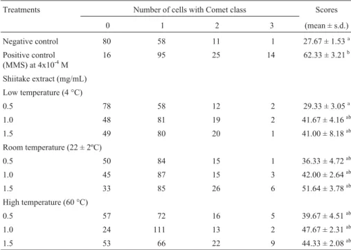
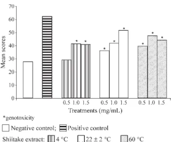
Table 1 and Figure 1 show the Comet assay results for the effects of shiitake aqueous extracts after 2 h treatment of HEp-2 cells. The 4 °C 0.5 mg/mL extract showed no genotoxic activity, since the mean score was not statisti-cally different from that of the negative control. However,
Table 1- Comet assay scores for HEp-2 cells treated with three different concentrations of shiitake aqueous extracts prepared at three different temperatures for evaluation of genotoxicity. Three replicates were made for each experiment and 50 nuclei were scored per repetition (n= 150 cells/group).
Treatments Number of cells with Comet class Scores
0 1 2 3 (mean ± s.d.)
Negative control 80 58 11 1 27.67 ± 1.53a
Positive control (MMS) at 4x10⁻⁴M
16 95 25 14 62.33 ± 3.21b
Shiitake extract (mg/mL)
Low temperature (4 °C)
0.5 78 58 12 2 29.33 ± 3.05a
1.0 48 81 19 2 41.67 ± 4.16ab
1.5 49 80 20 1 41.00 ± 8.18ab
Room temperature (22 ± 2ºC)
0.5 50 84 15 1 36.33 ± 4.72ab
1.0 45 87 15 3 42.00 ± 2.64ab
1.5 33 85 26 6 51.64 ± 3.78ab
High temperature (60 °C)
0.5 57 72 16 5 39.67 ± 4.51ab
1.0 24 111 13 2 47.67 ± 2.31ab
1.5 53 66 22 9 44.33 ± 2.08ab
the 4 °C 1.0 and 1.5 mg/mL extracts and all the extracts pre-pared at 22 ± 2 and 60 °C showed some genotoxic action, although the values were intermediate between those of the negative and positive controls.
Antigenotoxicity
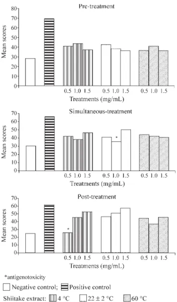
The antigenotoxic activity of the extracts was evalu-ated using three protocols (simultaneous-treatment, pre-treatment with extract and post-pre-treatment with extract), the comet scores of which were compared to the scores ob-tained for the DNA damaging agent MMS. Table 2 and Fig-ure 2 show the mean scores obtained in all protocols using three different preparation temperatures and concentrations of shiitake extracts (individual values for each repetition are not shown but were very similar).
A protective activity of the aqueous extracts was de-tected in the 22 ± 2 °C 1 mg/mL simultaneous-treatment and in the 4 °C 0.5 mg/mL post-treatment experiments. The values of the mean scores obtained after treatment of the cells with the other shiitake aqueous extracts were lower but not statistically different from that observed with MMS alone. For all the extracts, the percentage reduction in the comet score in the treatments with shiitake extracts show-ing antigenotoxic activity was calculated independently of the protective level (Table 2).
Discussion
Genotoxicity assessment
Research using the Ames test to assess the geno-toxicity of edible or medicinal mushrooms has produced conflicting results (Sterner et al., 1982a; Sterner et al., 1982b; Grüteret al., 1991).
Our results show that, except for the 4 °C 0.5 mg/mL extract, all of the shiitake extracts evaluated presented a
low level of genotoxicity, the values obtained in cells treated with shiitake extracts being intermediate between those obtained in the untreated control group and the group treated with the known mutagen MMS (Table 1; Figure 1).
The fact that the 4 °C 0.5 mg/mL extract showed no genotoxicity is not sufficient to indicate that this concentra-tion is not genotoxic, since the 0.5 mg/mL extracts prepared at 22 ± 2 and 60 °C showed a low-level of genotoxicity. In our study, we found no dose-response relationship regard-ing the genotoxic activity of shiitake in HEp-2 cells, which supports the findings of Moraleset al.(1990) who found no dose-response relationship when evaluating the mutagenic effects of some edible mushrooms on bacteria.
Analyzing the comet class distribution shown in Ta-ble 1 for the treatments with MMS and shiitake aqueous ex-tracts it is clear that cells with class 1 damage (minimal damage) were most prevalent and that cells with class 3 (maximum damage) were rare, that is, many cells presented few damage and few cells presented many damage. There-fore, the mushroom extracts caused moderated damage. This moderate genotoxicity appears to be related not only to the lower concentration of shiitake but also to effect of the preparation temperature on the active factors of Shiitake, since low comet scores were observed with those extracts prepared at the lowest temperatures.
Sterneret al. (1982a) reported that the heating and conservation conditions during the processing of mush-rooms can have a great influence on the biological activities of the mushrooms and Morales et al. (1990) detected slightly higher mutagenic activity in frozen mushrooms than in fresh ones, contrasting with the data presented in our present paper (Table 1; Figure 1). Chang (1996) considers that the mushrooms in general have several components with diverse biological activities and that these activities can vary according to how the mushrooms are prepared and consumed, while Grüter et al. (1991) suggest that a mutagen detectable in extracts may be unstable under nor-mal digestion conditions, not be absorbed by the gastroin-testinal tract or, alternatively, be activated as a toxic substance by the digestive processes.
Taken together, our findings show that the risks or benefits associated with the consumption of shiitake should be further investigated. The genotoxic activity of shiitake extracts detected in this study suggests the occurrence of different temperature-dependent pathways for the muta-genic agent present in the mushroom, and this could have practical implications for the preparation of shiitake.
Antigenotoxicity assessment
The interpretation of the results of the present study regarding the antigenotoxicity of the aqueous extract of shiitake is complex, since the extracts seem to have exerted their protective effects by more than one mechanism of ac-tion, which, according to Gebhart (1992), is not uncommon for mushrooms.
Miyajiet al. 111
Comet
assay
in
Lentinula
edodes
were scored per repetition (n= 150 cells/group).
Treatment with shiitake extract and MMS simultaneously (simultaneous-treatment)
Treatment with shiitake extract before exposure to MMS (pre-treatment with extract)
Treatment with shiitake extract after exposure to MMS (post-treatment with extract)
Treatments Mean
score
Number of cells with Comet class %R Mean score
Number of cells with Comet class %R Mean score
Number of cells with Comet class %R
0 1 2 3 0 1 2 3 0 1 2 3
Negative control 30.33a 73 64 12 1 28.33d 75 67 6 2 25.0g 81 63 6 0
Positive control (MMS) at 4x10⁻⁴M
66.00b 18 82 34 16 69.33e 12 83 40 15 62.0h 17 92 29 12
Shiitake extract (mg/mL)
Low temperature (4 °C)
0.5 42.33ab 47 81 20 2 66.36 41.00de 44 92 11 3 96.41 26.33g 81 60 8 1 69.10
1.0 38.33ab 56 75 17 2 77.57 43.67de 38 95 15 2 44.14 45.67gh 45 79 20 6 62.59
1.5 46.67ab 44 78 21 7 54.19 37.33de 43 98 7 2 24.32 53.0gh 31 84 30 5 78.05
Room temperature (22 ± 2ºC)
0.5 41.00ab 43 93 12 2 70.09 42.67de 37 101 9 3 41.43 46.67gh 39 87 19 5 65.02
1.0 35.67ac 65 66 16 3 85.03 38.33de 50 86 13 1 28.84 51.33gh 32 86 28 4 75.61
1.5 50.33bc 38 80 25 7 43.93 36.33de 51 90 8 1 11.70 57.67gh 20 90 31 9 80.49
High temperature (60 °C)
0.5 44.33ab 39 94 12 5 60.75 36.67de 49 94 5 2 46.84 44.67gh 39 91 17 3 79.66
1.0 42.33ab 50 78 17 5 66.36 40.67de 48 84 16 2 67.57 37.0gh 57 77 14 2 69.90
1.5 40.67ab 58 66 22 4 71.01 36.33de 52 88 9 1 44.14 45.67gh 44 78 21 7 80.49
MMS: Methyl methanesulfonate.
Pre-treatment of the HEp-2 cells with shiitake ex-tracts two hours before treatment with the mutagen MMS revealed no statistically significant protection attributable to the extracts (Table 2; Figure 2), although the percentage reduction in comet score (%R) was between 60 and 80%. Shiet al.(2002) also used a Comet assay and a similar pro-tocol to investigateL. edodesaqueous extracts prepared at 20 and 100 °C for genoprotector effects on hydrogen perox-ide damaged Raji cells, but found no significant protective effect.
According to Kurodaet al.(1992), the mechanism of bio-antimutagenesis is normally detected when the proto-col of post-treatment relative to the mutagen is used. In our study, except for the 4 °C 0.5 mg/mL extract, the majority of the extracts tested presented low efficiency in the post-treatment protocol (Table 2; Figure 2), even considering
that the 4 °C 0.5 mg/mL extract showed the lowest mean comet score and the highest percentage reduction in comet score of all the treatments used. It seems that the 4 °C 0.5 mg/mL extract would be the most appropriate to con-sume since it appeared to cause no genotoxic effects on the HEp-2 cells (Table 1).
In simultaneous-treatments antimutagenic agents may act as desmutagens by chemically or enzymatically in-activating mutagens, or inhibiting the activation of promutagens (Kurodaet al., 1992). Our experiments were performedin vitrowithout the addition of microsomal en-zymes (which can deactivate toxic chemicals), and the fact that the 22 ± 2 °C 1.0 mg/mL extract showed antigeno-toxicity action against MMS (Table 2; Figure 2) may indi-cate that this extract chemically inactivated MMS. The other extracts presented a moderate protector effect, al-though, statistically, the comet scores were not reduced to the levels of the untreated control (Table 2; Figure 2).
The results obtained in this study indicate that the ac-tive ingredient ofL. edodeswas present and effective in the extracts prepared at 4 and 22 ± 2 °C but at the temperature at which shiitake is frequently consumed (60 °C) it presented no protective effect. The active antimutagenic factor de-tected in the mushroom Lentinula edodes may be heat-labile (Shi et al., 2002 and Kada, 1981 (apud Grüter et al.,1990)), although the situation is unclear because Grüter
et al. (1990) state that several mushrooms have heat-resistant factors that reduce the mutagenicity of potent car-cinogenic agents such as aflatoxin B1and benzo[a]pyrene
inS. typhimurium.
The polysaccharide lentinan, one of the components obtained by the purification of shiitake, is very different from the whole mushroom, which leads to doubts whether consuming the whole mushroom has any preventative or therapeutic value, and if it does, what would be the ideal form in which to consume it and what would be the most ef-fective dose (Chang, 1996). Research has shown that there was considerable reduction in lentinan content when shiitake was stored for 7 days at 20 °C, whereas no alter-ation in lentinan content was observed when the mush-rooms were stored for 5 days at 5 °C or for 7 days at 1 °C (Minatoet al.,1999). We believe that the lentinan content remained constant in our study because the extract was pre-pared from recently collected fruit bodies which had been dried to about 7% humidity and powdered before storing at room temperature protected from light.
According to Lehmannet al.(2000), some modula-tors that exert protective effects in a certain situation can become deleterious when the genotoxic agent or parameter being analyzed is modified. The protective effects of the shiitake extracts observed in the present study in HEp-2 cells were obtained in relation to the damage caused by the monofunctional alkylant MMS (which acts directly on DNA bases and on the phosphate backbone causing a broad spectrum of DNA lesions). The alkylation followed by
Miyajiet al. 113
depurination can lead to single strand breaks (SSB) in the DNA which can be detected by Comet test (Sanderson and Shield, 1996; Helbig and Speit, 1997).
Ourin vitroresults are supported by the work of Lima
et al.(2001) who used the micronucleus test to evaluate the shiitake strain LE 96/17 in mice and observed an anti-clastogenic effect against cyclophosphamide and N-ethyl-N-nitrosourea. Taken together, these results indicate that antigenotoxic compounds can occur in shiitake strain LE 96/17 and can protect mammalian cells, bothin vitroandin vivo, from the effects of some alkylating agents, although ad-ditional studies are necessary with other types of genotoxic agents (preferably not alkylants) to elucidate for a better un-derstanding of the protective mechanism of shiitake extracts.
Acknowledgments
We thank Prof. José C. Soeiro for useful discussion regarding statistical analysis; the technical assistance of Mr. Dário P. Tormena and the excellent grammar review done by Dra. Angela Mehta. This study was supported by the Brazilian agencies Coordenação de Aperfeiçoamento de Pessoal de Nível Superior (CAPES), Fundação de Am-paro à Pesquisa do Estado de São Paulo (FAPESP) and by the Universidade Estadual de Londrina (UEL), Brazil.
References
Anderson D, Yu T-W and McGregor DB (1998) Comet assay as indicator of carcinogen exposure. Mutagenesis 13:539-555. Chang R (1996) Functional properties of edible mushrooms.
Nu-trition Reviews 54:S91-S93.
Fairbairn DW, Olive PL and O’Neill KL (1995) The Comet assay: a comprehensive review. Mutat Res 339:37-59
Ferguson LR (1994) Review antimutagens as cancer chemo-preventive agents in the diet. Mutat Res 307:395-410. Gebhart E (1992) Anticlastogenicity in cultured mammalian cells.
Mutat Res 267:211-220.
Grüter A, Friederich U and Würgler FE (1990) Antimutagenic ef-fects of mushrooms. Mutat Res 231:243-249.
Grüter A, Friederich U and Würgler FE (1991) The mutagenicity of edible mushroom in a histidine-independent bacterial test system. Food and Chem Toxicol 29:159-165.
Hartmann A and Speit G (1997) The contribution of citotoxicity to DNA effects in the single cell gel test (Comet assay). Toxicol Lett 10:183-188.
Helbig R and Speit G (1997) DNA effects in repair-deficient V79 Chinese hamster cells studied with the Comet assay. Mutat Res 377:279-286.
Kada T (1983) Environmental and biological factors suppressing induction of mutagens. Toxicology Forum 6:580-589. Kuroda Y, Jain AK, Tezuka H and Kada T (1992)
Antimutageni-city in cultured mammalian cells. Mutat Res 267:201-209. Lehmann M, Graf U, Reguly ML and Andrade HHR (2000)
Inter-ference of Tannic acid on the genotoxicity of mitomicyn C, methyl methanesulfonate and Nitrogen mustard in somatic cells of Drosophila melanogaster.Environ Mol Mutagen 36:195-200.
Lima, PLA, Delmanto RD, Sugui MM, Eira AF, Salvadori DMF, Speit G and Ribeiro LR (2001) Lentinula edodes(Berk.)
Pegler (shiitake) modulates genotoxic and mutagenic effects induced by alkylating agentsin vivo.Mutat Res 496:23-32. Manoharan K and Banerjee MR (1985)β-Carotene reduces sister chromatid exchange induce chemical carcinogens in mouse mammary cells in organ culture. Cell Biol Int Rep 9:783-789.
Minato K, Mizuno M, Terai H and Tsuchida H (1999) Autolysis of Lentinan, an antitumor polysaccharide, during storage of Lentinus edodes, shiitake mushroom. J Agric Food Chem 47:1530-1532.
Morales P, Bermúdez E, Sanz B and Hernández PE (1990) A study of the mutagenicity of some commercially canned Spanish mushrooms. Food Chem Toxicol 9:607-611. Sanderson BJ and Shield AJ (1996) Mutagenic damage to
mam-malian cells by therapeutic alkylating agents. Mutat Res 35:41-57.
Shi Y-I, James AE, Benzie IFF and Buswell JA (2002) Mush-room-derived preparation in the prevention of H2O2-induced oxidative damage to cellular DNA. Teratog, Carcinog Mutagen 22(2):103-111.
Singh NP (2000) Microgels for estimation of DNA strand breaks, DNA protein crosslinks and apoptosis. Mutat Res 455:111-127.
Singh NP, McCoy MT, Tice RR and Schneider EL (1988) A sim-ple technique for quantification of low levels of DNA dam-age in individual cells. Exp Cell Res 175:184-191. Speit G and Hartmann A (1999) The Comet assay (single cell gel
test) - a sensitive genotoxicity test for the detection of DNA damage and repair. In: Henderson DS (ed) Methods in mo-lecular biology, 113 DNA-repair protocols: eukaryotic sys-tems. Humana Press Inc., Totowa, pp 203-212.
Sterner O, Bergman R, Kihlberg E, Magnusson G, Nilsson L, Wickberg B and Zimerson E (1982a) Mutagens in larger fungi. I. 48 species screened for mutagenic activity in the Salmonella/ microsome assay. Mutat Res 101:269-281. Sterner O, Bergman R, Frenzén C, Kesler E and Nilsson L
(1982b) Mutagens in larger fungi. II. The mutagenicity of commercial pickledLactarius necatorin theSalmonella as-say. Mutat Res 104:233-237.
Tice RR (1995) The single cell gel: Comet assay: a microgel elec-trophoretic technique for detection of DNA damage and re-pair in individual cells. In: Phillips DH and Venitt S (eds) Environmental mutagenesis. Bios Scientific Publishers Ltd. Oxford, pp 315-339.
Tice RR, Agurell E, Anderson D, Burlinson B, Hartmann A, Kobayashi H, Miyamae Y, Rojas E, Ryu JC and Sasaki YF (2000) Single cell gel/Comet assay: guidelines forin vitro and in vivo genetic toxicology testing. Environ Mol Mutagen 35:206-221.
Wasser SP and Weis AL (1999) Therapeutic effects of substances occurring in higher basidiomycetes mushrooms: a modern perspective. Crit Rev Immunol 19:65-96.
Waters MD, Brady AL, Stack HF and Brockman HE (1990) Antimutagenicity profiles for some model compounds. Mutat Res 238:57-85.
Wattemberg LW (1983) Inhibition of neoplasia by dietary constit-uents. Cancer Res 43:2448s-2453s.
Wattemberg LW (1985) Chemoprevention of cancer. Cancer Res 45:1-8.