663 663663 663 663 Mem Inst Oswaldo Cruz, Rio de Janeiro, Vol. 99(7): 663-672, Novem ber 2004
Epidemiology of Leishmaniasis in Ecuador: Current Status of
Knowledge - A Review
M anuel Calvopina/* /+, Rodrigo X Armijos* /* * , Yoshihisa H ashiguchi
Department of Parasitology, Kochi Medical School, Kochi University, Kochi 783-8505, Japan *Unidad de Inmunologia y Medicina Tropical, Centro de Biomedicina, Universidad Central del Ecuador, Quito, Ecuador **Health Sciences Program,
College of Health Sciences, University of Texas at El Paso, El Paso, TX, US
Although leishmaniasis is regarded as a significant health problem in Ecuador by the Ministry of Health, and the incidence has increased over the last years, an official map on the geographic distribution of disease and sand fly vectors or a control strategy do not exist yet. This article reviews the current situation based on published informa-tion to improve our knowledge and understand the epidemiological situainforma-tion of leishmaniasis in Ecuador in order to help future research and to develop a national control strategy. The disease is endemic in most provinces throughout Pacific coastal region, Amazonian lowlands, and some inter-Andean valleys with a total 21,805 cases reported during 1990-2003. Whereas cutaneous leishmaniasis (CL) is found throughout Ecuador, mucocutaneous leishmaniasis (MCL) appears to be restricted to the Amazon region; one, parasitologically unconfirmed case of visceral form was reported in 1949. Most human infections are caused by Leishmania (Viannia) spp., which is distributed in the subtropical and tropical lowlands; infections due to L. (Leishmania) spp. are found in the Andean highlands and in the Pacific lowlands as well. The proven vectors are Lutzomyia trapidoi and Lu. ayacuchensis. Canis familiaris, Sciurus vulgaris, Potos flavus, and Tamandua tetradactyla have been found infected with Leishmania spp. It is estimated that around 3000-4500 people may be infected every year, and that 3.1 to 4.5 millions people are estimated to be at risk of contracting leishmaniasis.
Key words: leishmaniasis - Leishmania - epidemiology - Ecuador - review
Ecuador is located in north-west South America and straddles both the line of the Equator and the Andes moun-tain range (Fig. 1). The Andes cross the country from North to South and divide it into three different natural regions: the Pacific coast with subtropical and tropical lowlands, the Andean region with high mountains and valleys where temperatures range from 15 to 22oC and, in the East, a region which encompasses a section of Ama-zon lowlands covered by humid tropical rain forest. Eco-logically, Ecuador is an extremely diverse country with a total area of 283,560 km2, ranging from tropical to nival
(permanent snow) and from the rainforest to desert brush. The total population is 12,156,600 inhabitants, of which 6,053,987 live in the Pacific coast, 5,458,313 in the Andean region and 547,047 in the Amazon region; the remaining in the Galapagos Islands (Census 2001).
HISTORICAL BACKGROUND
Ancient representations of skin lesions and facial de-formities have been found on pre-Inca pottery from Peru, Colombia, and Ecuador, estimated to date from AD 400 to AD 900. According Ecuadorian ceramics, cutaneous (CL) and mucocutaneous (MCL) leishmaniasis has existed for hundreds or perhaps thousands of years before the ar-rival of the Spanish conquerors. Historians at the time of the conquerors wrote of skin lesions seen among the Inca
+Corresponding author. Fax: +81-88-8802617/+81-88-8802415.
E-mail: mcalvopina@hotmail.com Received 27 April 2004
Accepted 14 July 2004
Indians which resulted in mutilations similar to those of some human figures (huacos) (Ala-Vedra 1952). The first human case of CL in Ecuador was reported in 1920 (Rodriguez 1974). Hashiguchi and Gomez (1991) have pub-lished a detailed description of chronological events as-sociated with leishmaniasis up to 1987.
THE GEOGRAPHICAL DISTRIBUTION AND INCIDENCE RATES OF HUMAN INFECTIONS
Human leishmanial infections have been reported from 20/22 of the country’s provinces (Fig. 1). Cases are re-corded from sea level up to approximately 2700 m eleva-tion, mainly from rural areas. An endemic belt is formed along the western slopes of the Andes involving new settlements in Imbabura, Pichincha, Cotopaxi, Bolivar, and Cañar provinces. CL and MCL forms had been recorded from all 6 Amazonian provinces (Amunarriz 1991). Since 1986 Ecuadorian Andean leishmaniasis has been reported from valleys of Paute (2300-2500 m), Alausi (2300-2700 m), and Huigra (1200-1500 m).
Pa-664 664664
664664 Leishmaniasis in Ecuador • M anuel Calvopina et al.
Fig. 1:map of Ecuador. The distribution of leishmaniasis by regions and provinces are showed with the respective numbers of Leishmania
species identified (total 270 stocks). The disease is absent in Galapagos Islands and Carchi province. Shaded area indicates the Andean region with elevations > 1000 m above sea level. Provinces are identified by number: Esmeraldas (1), Manabi (2), Los Rios (3), Guayas (4), El Oro (5), Carchi (6), Imbabura (7), Pichincha (8), Cotopaxi (9), Tungurahua (10), Bolivar (11), Chimborazo (12), Cañar (13), Azuay (14), Loja (15), Sucumbios (16), Napo (17), Orellana (18), Pastaza (19), Morona Santiago (20), Zamora Chinchipe (21), and Galapagos (22)
665 665 665 665 665 Mem Inst Oswaldo Cruz, Rio de Janeiro, Vol. 99(7), N ovem ber 2004
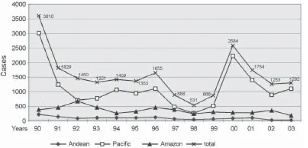
cific region. As in other leishmaniasis-endemic areas throughout Latin America, this increase may be attrib-uted to changes in land use, the construction of new dams and human activity patterns like internal migration, lead-ing to increase exposure of humans to the zoonotic Leish-mania life cycles (Desjeux 2001). The decline and the in-crease of numbers of cases could be due to the frequency of the El Niño Southern Oscillation in Ecuador, but there are no evidences (Davies et al. 2000). Despite, the official epidemiological data showing that the disease prevalence has been decreasing in the last decade, active case detec-tion studies have shown an increase in case numbers in several provinces (Alava et al. 1992, Barrera et al. 1994, Garcia et al. 1997, Nonaka et al. 1997). In fact, Armijos et al. (1997) and Hashiguchi et al. (1987) have reported that 14% (65/466) and 7.5% (7/93) of subjects surveyed in a sub-tropical rainforest area and Andean slope of Ecuador had CL, respectively. The incidence of leishmaniasis is ex-pected to remain stable or increase in the near future due to the large number of previously unexposed groups such as young children and persons who visit (e.g., tourists) or move from non-endemic areas (e.g., agriculturalist, lum-berjacks, workers for petrol exploitation, and subsequent colonization).
ATTITUDES OF THE LOCAL POPULATION TO LEISH-MANIASIS
Most adults from leishmaniasis-endemic areas of NW Ecuador are familiar with the disease and believe that it has a negative impact on the capacity to perform normal work duties and it needs some type of intervening treat-ment (Weigel et al. 1994, Weigel & Armijos 2001). How-ever, few knew about the transmission, etiology, ulcer healing, and conventional treatment. Women appeared to be more aware of the disease and scars because of the associated stigmatization of people affected. In the Ama-zon foci, because of tissue destruction and mutilation in the mucocutaneous form, patients tend to be isolated by the family and friends causing psychosocial impact with social and economic consequences (Amunarriz 1991). These local perceptions should be considered when plan-ning future control programs and would be helpful to es-tablish the communication channels that would be most effective with respect to control activities (e.g., identifica-tion of target groups). However, studies on people’s knowledge, attitudes, and practices towards leishmania-sis do not exist in the Andean and Amazon regions. Cer-tainly, the popular ideas and cultural practices in other foci might be different because of clinical presentation, cultural background, level of education, and environment between Amerindians in the Amazon and mestizo in the Andes foci. Further, plants and ethnomedical inventory used for the Amerindians in the Amazon foci do not exist in other regions (Amunarriz 1991).
The popular name for leishmaniasis in Ecuador differs from region to region. In the Pacific it is called sarna brava (angry sore), charra brava (angry ulcer) or charra co-lombiana (Colombian ulcer). In the Amazon region as milliai caracha or ulcera brava (angry ulcer) for CL and lepra de montaña (mountain leprosy) for MCL, whilst in the Andes people refer to it as nigua de raton (mice
chig-ger) or grano malo (bad sore). In all regions manta blanca (white blanket) is the popular name for the vector.
Traditional treatments are frequently used, especially by rural populations who often lack access to conven-tional treatment. For example, the use of tradiconven-tional meth-ods is reported to be as high as 70% in NW Ecuador (Weigel et al. 1994), and 100% in the Amazon region (Amunarriz 1991), with up to 150 different anti-leishmanial therapies used. The therapies described included various indigenous plants, chemicals or petroleum-based prod-ucts. Although widely used by the communities, their ef-ficacy is largely anecdotal and has never been proven in a controlled study. Other treatments methods such as cauterization with hot metal objects, strong acids, tobacco ash, and heated wax are also frequently used. These of-ten cause tissue burns and promote the development of secondary infections, lengthen the cure and leaving big-ger and permanent scars worse than those caused by the leishmaniasis itself (Weigel et al. 1994, Weigel & Armijos 2001).
CLINICAL FORMS AND THEIR CAUSATIVE PARASITES
The diseases occur in Ecuador in most of its recog-nized American tegumentary forms: CL, MCL, and diffuse cutaneous (DCL) leishmaniasis. The reported ratio of MCL:CL is low (1:13) (Hashiguchi & Gomez 1991). The unique cases reported as visceral leishmaniasis (VL) and DCL came from Esmeraldas province. DCL case was caused by L. (L.) mexicana (Reyna et al. 1994). To date, no other case of VL has been recorded, since the only case reported by Leon in 1949 was not confirmed by para-site isolation; it probably was a misdiagnosis. This is sup-ported by the apparent absence of L. chagasi/infantum parasites and their vectors in the country (Young & Rogers 1984, Alexander et al. 1992).
The clinical features of CL are different between high-land and lowhigh-land cases. “Andean type” occurs usually as single lesion, measuring less than 5 mm in diameter; most of them are crusted papules with plenty parasites, and reported to be caused only by L. (L.) mexicana and L. (L.) major-like (Armijos et al. 1990, Hashiguchi et al. 1991). The lesions here are clinically similar to “uta” described in the highlands of Peru; however the parasite species and the vector are different (Lainson 1983). Whereas “low-land type” is larger, usually wet with secondary bacterial infection and multiple lesions.
Commonly, CL lesions are ulcerative, but non-ulcer-ated such as papules, plaques, nodules, and erysipeloid forms are also seen (Chico & Guderian 1989, Nonaka et al. 1990, Armijos et al. 1997).
666 666666
666666 Leishmaniasis in Ecuador • M anuel Calvopina et al.
Sucumbios, Orellana, and Napo we encountered 13 active cases of MCL caused by L. (V.) braziliensis (Calvopina et al. 2001a), suggesting that this form and parasite could be prevalent in this humid tropical forest. MCL originating from the Pacific or Andean regions has never been re-corded (Hashiguchi et al. 1991, Barrera et al. 1994, Armijos et al. 1997). This is surprising, as Osorio et al. (1998) re-ported MCL among residents of the Colombian Pacific coast caused by the same species circulating in this Ecua-dorian region.
PARASITE DISTRIBUTIONS
Currently, six Leishmania spp. have been identified from humans, sand flies and non-human mammals (Table). The map (Fig. 1) shows the distribution of identified spe-cies by regions and provinces. Findings are that the ratio of L. (Viannia) to L. (Leishmania) infections is about 6:1. Clearly, L. (V.) panamensis and guyanensis are predomi-nant in the subtropical and tropical lowlands of the Pa-cific region (62.1% of all). Recently, Bañuls et al. (1997, 1999) doubted the separate taxonomic status of the latest two species. L. (L.) amazonensis has been only reported from subtropical Ecuador in human CL (Armijos et al. 1990, Furuya et al. 1997), but also infecting other mammals (Mimori et al. 1989). Notoriously, L. (V.) braziliensis is geographically the species most widespread in the coun-try, having been isolated from CL and MCL patients in nine of 15 surveyed provinces (Armijos et al. 1990, 1997, Bañuls et al. 1999, Furuya et al. 1997, Calvopina et al. 2001a). In the Amazon region only L. (V.) braziliensis was identified, but hybrid genotypes between L. (V.)
panamensis/guyanensis and L. (V.) braziliensis had also been reported (Bañuls et al. 1997). Furthermore, Pratlong et al. (2002) identified L. naiffi in a German patient with CL suspected to be infected in this region.
For Andean leishmaniasis the parasite is reported to belongs only to the subgenus Leishmania: L. mexicana for 82.4% (28/34) and L. major-like (Armijos et al. 1990, Hashiguchi et al. 1991), where lesions resembled to “uta” of Andean valleys of Peru; but the causative agent there is identified as L. (V.) peruviana (Lainson et al. 1983) hence, further studies with a standardized methodology have to be established in order to enable comparisons between both foci. L. major-like parasite had also been reported from Brazil, Paraguay, Venezuela, and Mexico (Cupolillo et al. 1994, Yamasaki et al. 1994) suggesting that some of these populations may have been imported into the Americas (Momen et al. 1993). The nucleotide sequences of cytochrome b of L. major-like isolated from the Ecuadorian Andes and the WHO reference strain L. major had a 99.9% homology (Luyo-Acero et al. 2004).
Parasite isolates from a sloth (Choloepus hoffmani) and a squirrel (Sciurus granatensis) caught in tropical forest of the Pacific region characterized before as L. (V.) equatorensis (Grimaldi et al. 1992), actually Uezato et al. (2001) and Katakura et al. (2003) using molecular methods identified that it belongs to the genus Endotrypanum spp. Since a small proportion of all leishmaniasis cases are ever diagnosed to species level, with a total of 270 cases from nine surveys, further studies are needed in order to isolate and identified the parasites circulating in most prov-inces reporting the disease.
TABLE
Distribution of Leishmania parasites in humans, vectors, and putative reservoir hosts by regions in Ecuador
Parasite Characterization Clinical Reservoir
identified methods forms Vectors hosts References
Pacific region
L. (V.) panamensis IE, mAb LCL, LRC,DL Lu. trapidoi Canis familiaris AR,MT,LeP,DE,CM,FM
L. (V.) guyanensis IE LCL Lu. trapidoi NI AR, BA
L. (V.) panam/guyan IE, RAPD LCL Lu. trapidoi Canis familiaris BA
L. (V.) braziliensis IE, mAb LCL NI NI AR, BA, FM
L. (L.) amazonensis IE, mAb, kDNA LCL NI Sciurus vulgaris, AR, MT, FM
Potos flavus &
Tam. tetradactyla L. (L.) major-like IE, kDNA LCL NI NI HY
L. (L.) mexicana IE, kDNA LCL, DCL NI NI RE Andean region
L. (L.) mexicana IE, mAb, kDNA LCL Lu. ayacuchensis Canis familiaris AR, HY, FM
L. (L.) major-like IE, mAb, kDNA LCL NI NI HY, FM Amazon region a
L. (V.) braziliensis IE, mAb, PCR LCL, MCL NI NI AR, CM, FM
L. (V.) brazil/panam-guyan IE, RAPD LCL Lu. serrana b NI LeP, BA
a: Lutzomyia spp. identified in Orellana province but negative for Leishmania: Lu. tortura, flaviscutellata, olmeca bicolor, gomezi, carrerai thula, geniculata and yuilli yuilli (Calvopina et al. 2001a). In Zumba-Zamora Chinchipe province: Lu. maranonensis,
667 667 667 667 667 Mem Inst Oswaldo Cruz, Rio de Janeiro, Vol. 99(7), N ovem ber 2004
AGE, GENDER, AND BODY DISTRIBUTION OF LESIONS
The age groups affected differ from region to region. In the Andean foci the most affected are children under two years, suggesting indoor-transmission and probably dogs as reservoirs (Hashiguchi et al. 1991). In the low-lands are children under the age of ten in tropical and five years in subtropical Pacific region (Alava et al. 1992, Armijos et al. 1997). The increased disease prevalence in children could be associated with no previous exposure to the parasite, the role of cellular immune status and/or with malnutrition (Weigel et al. 1995). Nevertheless, for the Amazon region in a passive hospital-based report the mean age of leishmaniasis diagnosis was 33.4 and 19.7 years for MCL and CL respectively (Amunarriz 1991), sug-gesting unknown reasons for delays in the diagnosis in this wide and disperse region. However, it is also evident that exposure to transmission at any age as do immigrants or tourists they will develop the disease.
No gender difference in the incidence is observed among children at any endemic regions surveyed (Hashiguchi et al. 1991, Weigel et al. 1994). However, the sex difference regarding disease prevalence in the adult population appears to be important. Thus, in the Amazon region a passive hospital-based reported 81% (22/27) of MCL and 83% (67/81) of CL cases to be male (Amunarriz 1991); Calvopina et al. (2001a) also encountered 85% (11/ 13) of MCL in male. Hence, the excess risk observed in this region is for male older than 14 years suggesting that it could be associated with their occupational exposure (i.e., hunting and agricultural work) characteristic of syl-vatic transmission cycle of Leishmania. In a community-based study in the subtropical of Pacific region adult male had a disease risk almost three times greater than of adult female (Weigel et al. 1994). But, several years after in the same study area, no differences were observed (Weigel & Armijos 2001) changes here appeared to be associated with change from sylvatic to domestic and peridomiciliary transmission.
Lesions are most frequent on the exposed parts of the body. The children face is more frequently affected (92%) in the Andean foci (Hashiguchi et al. 1990), probably be-cause mother’s cultural behavior in taking care small chil-dren in this cool region and where probably indoor-trans-mission occur. Whilst, in the lowlands, lesions or scars are mostly in the extremities (53.8 to 92%), it is hypoth-esized because of difference of biting behaviors of sand flies or clothing habits of inhabitants in hot climate (Amunarriz 1991, Alava et al. 1992, Armijos et al. 1997). Chiclero’s ulcer is not rare in lowlands foci. Other sites affected include the trunk and, less commonly the scalp, buttocks, eyelids, and genitalia. MCL affects the nasal mucosa, septum and turbinate, upper lip, pharynx and lar-ynx producing dyspnoea, and face deformities (Calvopina et al. 2001a).
VECTORS
The Table lists the sand fly species incriminated as vectors of Leishmania in Ecuador. More than 60 species of phlebotomine sand flies, including 55 species belong-ing to the genus Lutzomyia have been described (Young & Rogers 1984, Alexander et al. 1992). At least 15 Lutzomyia
spp. are known to be anthropophilic and are strongly sus-pected as vectors. Natural infection with promastigotes morphologically identical to Leishmania have been ob-served but not characterized in Lu. gomezi, hartmanni and panamensis. In the Pacific region, Lu. trapidoi, a proved vector here, is more abundant in the dry season whilst Lu. gomezi in the wet season (Le Pont et al. 1994a,b). Dujardin et al. (1996) demonstrated the presence of cryp-tic species in Lu. trapidoi. Studies carried out in 1982 (Hashiguchi et al. 1985) and in 1993 (Furuya et al. 1998) in this region, demonstrated that in 1982 the predominant species was Lu. trapidoi with 8.1% of flagellates in their hindgut, but 10 years later Lu. hartmanni predominate with 9.8% of infection. The latter species was recently confirmed to harbor Endotrypanum spp. (Uezato et al. 2001, Katakura et al. 2003), believed before to be L. (V.) equatorensis (Furuya et al. 1998).
For Andes foci, six anthropophilic Lutzomyia ( aya-cuchensis, hartmanni, nevesi, gomezi, serrana, and osornoi) have been identified so far (Gomez et al. 1994), with, only ayacuchensis shown to be naturally infected with L. (L.) mexicana (Hashiguchi et al. 1991). Lu. ayacuchensis abounds in the wet season and its infec-tion rate is found to be highest during the period from the early rainy season to the beginning of the dry season (Gomez & Hashiguchi 1991). It correlates with the appear-ance of new human cases. In these inter-Andean valleys, there is strong evidence that transmission of Leishmania occurs indoors and in urban settings (Hashiguchi et al. 1987, Armijos et al. 1990).
For the Amazon region, in the Zumba focus south wards of Ecuador bordering with Peru several man-biting species have been recorded, Lu. serrana abounds inside dwellings where it even bite during daytime (Alexander et al. 1992, Le Pont et al. 1994c), suggesting this species as a candidate vector in an indoor-transmission manner. In the inter-Andean Ecuadorian-Peruvian border Lu. robusta was also described (Galati et al. 1995). Both species are suspected vectors of human bartonellosis as well, since there is no probed vector for Bartonella bacilliformis endemic in those areas (Cooper et al. 1996). From Orellana province seven anthropophilic species captured in MCL focus were identified but all negatives for natural infec-tion (Calvopina et al. 2001a). In the whole country there are no reports on Lu. longipalpus and evansi a confirmed vectors of L. chagasi/infantum in the neighboring coun-tries.
PUTATIVE RESERVOIR HOSTS
668 668668
668668 Leishmaniasis in Ecuador • M anuel Calvopina et al.
but were not characterized (Hashiguchi et al. 1985). No positive parasite isolation or detection has been done from putative reservoir hosts in the Amazon region.
Serological studies to examine the role of dogs in the leishmaniasis transmission cycle carried out in the inter-Andean valley of Alausi found 19/58 (32.8%) of surveyed dogs positive for ELISA; no dog had positive liver-punc-ture aspirates (Mimori et al. 1992). Another study also showed high ELISA-rate positivity using antigens of L. (V.) panamensis and guyanensis in 12/17 dogs from Alausi and 12/20 from the Pacific region (Mori et al. 1994). These results showed high reactivity for ELISA but careful at-tention must be paid on the cross-reactivity (false-posi-tives) where Trypanosoma cruzi is endemic as is in low-lands of Ecuador (Aguilar et al. 1999). The positive samples should be confirmed parasitologically and characterized, even that, it does not distinguish whether dogs are acci-dental or reservoir hosts, dogs seems to be a victim-host as humans are. Due to low specificity and sensitivity of the methods employed, the PCR-based method could show better results if it is carried out properly (Reithinger et al. 2000).
DIAGNOSIS
In endemic areas of Ecuador the diagnosis of leishma-niasis is generally based on clinical criteria, because few laboratories have the capacity of confirming and most of them are far from rural endemic areas. Specialized labora-tories doing cultures, Montenegro skin test and/or PCR are rare and are restricted to research purposes. Micro-scopic examination of Giemsa-stained smears is the rec-ommended method by the public health authorities. The examination, however, has shown to have a lower sensi-tivity than culture and PCR (Aviles et al. 1999, Matsumoto et al. 1999, Calvopina et al. 2001b). The sensitivity of smears and cultures decreased if lesions were chronic and due to microbial contamination. The rate of parasite isola-tion in MCL patients was low as 23%, probably due to chronicity and scarcity of parasites in mucosal lesions (Calvopina et al. 2001a). Parasitological diagnosis is the “gold standard” in leishmaniasis, as it is highly specific (Davies et al. 2000). However, in laboratories from endemic areas of Ecuador there are many false positives on the microscopic examination of Giemsa-stained smears that might due to untrained staff. The direct immunofluores-cent antibody (DIFMA) test showed higher sensitivity and rapidity superior to scraping, culture and histology (Chico et al. 1995). The histology staining showed the lowest sensitivities (Aviles et al. 1999, Calvopina et al. 2001b); given the limited utility and dear of histology, this method should be reserved for selected patients.
Recently, several studies in Ecuador have found PCR-based method to be more sensitive (85.4% on average) than classical diagnostic techniques, named slit smears (45.4%), culture (57.2%), and histopathology (34.7%). In some way the sensitivity of PCR depended on the sample taken, from skin biopsies and exudates using cotton swabs it reached to 92% and 93.8%, respectively (Aviles et al. 1999, Calvopina et al. 2001b, Mimori et al. 2002), but using scrape/exudates or syringe-sucked fluid the sensitivities decrease to 70% (Matsumoto et al. 1999). The before
col-lection procedures should be validated on large samples. However, still a concern the false-negative rate showed by PCR that could be as high as 15.4% (Mat-sumoto et al. 1999). The employment of PCR allowed the identification of Leishmania at subgenus or complex level, but recently a study using polymorphism-specific primers yielded to identify species from clinical samples (Mimori et al. 1998). Other advantage of PCR is that diagnosis could be ob-tained on biopsy samples which are either in formalin or ethanol fixed and paraffin-embedded histological sections (Uezato et al. 1998, Mimori et al. 2002). Finding on PCR amplification of the Leishmania mini-exon gene and karyo-type characterization suggest that it would be used for diagnosis and for molecular epidemiology in Ecuador (Katakura et al. 1993, 1998). Nevertheless, in Ecuador, PCR system still expensive, personnel well trained and a refer-ence center to process the samples are still needed.
DIFFERENTIAL DIAGNOSIS
In Ecuador, the differential diagnosis of leishmaniasis includes a range of skin conditions, which depend on the prevailing disease pattern in the different leishmaniasis-endemic regions. Similar to other leishmaniasis-endemic countries in Latin America, clinical diagnosis of leishmaniasis can be over-estimated in rural areas where appropriate tests to confirm lesion etiology are absent (Lopez et al. 1993, De Brujin et al. 1993, Calvopina et al. 2001b). In a hospital-based survey Hosokawa et al. (2001) showed that 5/16 (31.2%) were misdiagnosed as having CL and MCL and had been receiving anti-leishmanial drugs. The five mis-diagnosed patients had basal cell carcinoma, chro-momycosis and ulcus cruris varicosum. Other skin condi-tions commonly misdiagnosed as leishmaniasis were fu-runcle, Bazin’s disease, liquen vulgaris, mycetoma, sporot-richosis, myasis, leprosy, and bacterial ulcers. MCL should be differentiated from paracoccidioidomycosis which is prevalent in the tropical lowlands of Ecuador (Fernandez 1990). Leprosy still endemic in rural Ecuador and because is overlapping leishmaniasis-endemic areas could make clinical confusions.
TREATMENT
669 669 669 669 669 Mem Inst Oswaldo Cruz, Rio de Janeiro, Vol. 99(7), N ovem ber 2004
after, the proportions of subjects who had used Glucantime remained low (20%) while the premature drug discontinu-ance appeared to have doubled (Weigel & Armijos 2001). Therefore, in order to monitor drug side-effects and re-sponsiveness, it is not unusual for MCL patients to be hospitalized for two or three times (Amunarriz 1991). There is no available data on unresponsiveness to Glucantime but is frequently observed mainly in MCL cases and CL acquired in Amazonian lowlands, no study has been done on parasite resistance. The intralesional infiltration with Glucantime alone or combined with intramuscular injec-tion is also popular in endemic areas and recommended for whom systemic chemotherapy is contraindicated. But, this method should not be applied in patients acquiring the infection in the Amazon region where L. (V.) braziliensis has the propensity to evolve into MCL. Sec-ond-line drugs (i.e. pentamidine and amphotericin B) are not available in the country.
Several clinical studies to test the efficacy of alterna-tive anti-leishmanial drugs have been carried out in Ecua-dor. Guderian et al. (1991) tested allopurinol plus pro-benecid against CL, but only 9 (41%) were healed, com-pared with 96% of Pentostam® group and 75% healing rate for the placebo group. Nonaka et al. (1992) applied paromomycin (PM) ointment as a monotherapy showing to be rather ineffective against CL in the Pacific region with clinical cure in 10-54% of patients. Krause and Kroeger (1994) using PM plus methylbenzethonium (MBCL) for CL in the Pacific region cured 85% with 9% healed spon-taneously in the placebo control group, but their follow-up was only 50 days. Armijos et al. (2004) using topical PM-MBCL and PM-urea reach clinical cure in 79.3% and 70% respectively, by 12 weeks of follow-up. Looking for an oral treatment Gomez et al. (1997) administered me-floquine (Mephaquin®) and artesunate (Plasmotrim®) to patients infected in the Pacific region, reporting cure rates of 100%. However, studies in the Pacific of Colombia (Hendrickx et al. 1998) and in the state of Bahia of Brazil (Laguna-Torres et al. 1999) showed that cure rates with mefloquine were similar than for the placebo controls and less than half observed in the Sb(v) group. Itraconazole (Sporanox®) was given orally at high dosage and for 90 days in 13 patients with MCL in the Amazon region, three patients were cured clinically and parasitologically, con-cluding that itraconazole is not longer effective in this clinical form (Calvopina et al. 2004). Nifurtimox (Lampit®) were administered in the Amazon region with apparently good results (Amunarriz 1991). Despite an uncontrolled study reporting cure rates of 100% using S-nitroso-N-acetylpenicillamine (SNAP), a compound that generates nitric-oxide (Lopez-Jaramillo et al. 1998), a subsequent study showed disappointing results in murine CL and in a clinical trial, showing low cure rate and a high rate of local reactions (Davidson et al. 2000).
The spontaneous healing of CL is common in patients infected in the Andean region, as well as in the subtropi-cal Pacific region that can be as high as 75% (Hashiguchi et al. 1991, Guderian et al. 1991). Hence, patients been infected in the Andes foci usually should not be treated systemically because the lesions are small and self-heal within months leaving a small and imperceptible scar
(Hashiguchi et al. 1991) and because there is no risk of metastasis. However, recently leishmaniasis recidivans has been reported in the subtropical Pacific region, such patients should be treated adequately. Patients with se-vere, sporotrichoid type, multiple lesions (> 3), diabetics and with lesions on the eyelid, the auricle of ear, face and nose which may cause disfiguring scars should also be treated promptly.
PREVENTION AND CONTROL MEASURES
Passive case finding and treatment is the only control measure followed in Ecuador with afore-mentioned limita-tions in sampling, diagnosis, drug availability, and train-ing of medical staff. A vaccination trial with killed cocktail of promastigotes was conducted in the subtropical-Ecua-dor (Armijos et al. 1998) showing significant protection during the first 12 months (72.9%). Protection declined until month 60 of follow-up, with no difference in protec-tion rate between the vaccinated and placebo groups (Armijos et al. 2003). Another study was conducted in a similar endemic area using Leishvaccin® plus BCG adju-vant, although two doses of the vaccine resulted in a significant increase in Montenegro skin test conversion, it did not appear to offer any significant protection against development of CL compared to the placebo group (Armijos et al. 2004). As outlined here, further region-spe-cific studies are required on the geographic distribution of disease, sandfly vectors, reservoirs hosts and risk fac-tors in order to identify and develop a leishmaniasis inter-vention strategy that is cost-effective.
CONCLUSIONS
Though the knowledge on the geographical distribu-tion and burden of leishmaniasis in Ecuador has increased over the last years, it is still incomplete and patchy. The situation on the structure of vector and reservoir system is limited. The national recording and reporting system even that is compulsory since 1985, is limited by gaps in information, usually quantitative, sometimes qualitative. As there is little accurate data on country specific preva-lence, estimates for the number of people infected and number at risk of infection must still be made based on extrapolations of passive case detection prevalence. Glucantime is still not reaching the majority of those who most need it; delivery of drug should be supported in a comprehensive reporting and actualized data. Most of the studies had been done by private initiatives. Health au-thorities must be able to translate the actual knowledge into better information, education, and research in accor-dance with the endemic level, principally in their periph-eral health system. In the future, would be important to design trials in order to clarify aspects like El Niño and its impact in annual fluctuation of the vector population as well as the impact of the global warming phenomenon. Imaginative research studies, adequate funds and infra-structure are urgently needed to define the disease epide-miology and to develop a national prevention and control strategy.
ACKNOWLEDGEMENT
670 670670
670670 Leishmaniasis in Ecuador • M anuel Calvopina et al.
REFERENCES
Aguilar VHM, Abad-Franch F, Racines VJ, Paucar AC 1999. Epidemiology of Chagas disease in Ecuador. A brief review.
Mem Inst Oswaldo Cruz94 (Suppl. 1): 387-393.
Ala-Vedra y Tama 1952. El boton de Oriente en Ecuador, ulcera tropical, leishmaniosis Americana. I Jorn Ped Ecuat. Guayaquil, Ecuador, p.1-23.
Alava JJ, Mora AE, Gomez EA, Hashiguchi Y 1992. Studies on leishmaniasis in an endemic focus of Leishmania on the Pacific coast of Ecuador. In: Studies on New World Leish-maniasis and its Transmission with Particular Reference to Ecuador, Kochi, Japan. Res Rep Series 3: 59-69.
Alexander JB, Takaoka H, Eshita Y, Gomez EA, Hashiguchi Y 1992. New records of phlebotomine sand flies (Diptera: Psychodidae) from Ecuador. Mem Inst Oswaldo Cruz87: 123-130.
Amunarriz M 1991. Leishmaniasis. In Estudios sobre Patologias Tropicales en la Amazonia Ecuatoriana, Cicame, Napo Ec-uador, Cap. 3, p. 41-64.
Armijos RX, Chico M, Cruz M, Guderian R, Kreutzer R, Berman J, Rogers M, Grogl M 1990. Human cutaneous leishmaniasis in Ecuador: Identification of parasites by en-zyme electrophoresis. Am J Trop Med Hyg42: 424-428. Armijos RX, Weigel MM, Aviles H, Maldonado R, Racines J
1998. Field trial of a vaccine against New World cutaneous leishmaniasis in an at-risk child population: Safety, immu-nogenicity, and efficacy during the first 12 month of fol-low-up. J Infect Dis177: 1352-1357.
Armijos RX, Weigel MM, Calvopina M, Hidalgo A, Cevallos W, Correa J 2004. Safety, Immunogenecity, and efficacy of an autoclaved Leishmania amazonensis vaccine plus BCG adjuvant against New World cutaneous leishmaniasis. Vac-cine22: 1320-1326.
Armijos RX, Weigel MM, Romero L, Garcia V, Salazar J 2003. Field trial of a vaccine against New World cutaneous leish-maniasis in an at-risk child population: How long does pro-tection last? J Infect Dis187: 1959-1961.
Armijos RX, Weigel MM, Calvopina M, Mancheno M, Rodriguez R 2004. Comparison of the effectiveness of two topical paromomycin treatments vs meglumine antimoniate for New World cutaneous leishmaniasis. Acta Trop91: 153-160.
Armijos RX, Weigel MM, Izurieta R, Racines J, Zurita C, Herrera W, Vega M 1997. The epidemiology of cutaneous leishmaniasis in subtropical Ecuador. Trop Med Int Health 2: 140-152.
Aviles H, Belli A, Armijos RX, Monroy F, Harris E 1999. PCR detection and identification of Leishmania parasites in clini-cal specimens in Ecuador: A comparison with classiclini-cal diag-nostic methods. J Parasitol85: 181-187.
Barrera C, Herrera M, Martinez F, Leon R, Richard A, Guderian RH, Mouchet J, Echeverria R, Le Pont F 1994. Leishmania-sis in Ecuador. 1. Incidence of cutaneous leishmaniaLeishmania-sis on the Pacific coast. Ann Soc Belg Med Trop74: 1-12. Bañuls AL, Guerrini F, Le Pont F, Barrera C, Espinel I, Guderian
R, Echeverria R, Tibayrenc MJ 1997. Evidence for hybrid-ization by multilocus enzyme electrophoresis and random amplified polymorphic DNA between Leishmania brasiliensis and L. panamensi/guyanensis in Ecuador. J Euk Microbiol44: 408-411.
Bañuls AL, Jonquieres R, Guerrini F, Le Pont F, Barrera C, Espinel I, Guderian R, Echeverria R, Tibayrenc M 1999. Genetic analysis of Leishmania parasites in Ecuador: Are
Leishmania (Viannia) panamensis and L. (V.) guyanensis
distinct taxa? Am J Trop Med Hyg6: 838-845.
Calvopina M, Guevara A, Gomez EA, Paredes W, Mimori T,
Guderian R, Nonaka S, Hashiguchi Y 2001b. Diagnosis of cutaneous leishmaniasis with PCR technique in comparision with conventional methods. In Studies on New World Leish-maniasis and its Transmission with Particular Reference to Ecuador. Kochi, Japan. Res Rep Series 6: 49-57.
Calvopina M, Guevara AG, Armijos RX, Hashiguchi Y, Davidson R, Cooper P 2004. Itraconazole in the treatment of New World mucocutaneous leishmaniasis. Inter J Dermatol
(in press).
Calvopina M, Guevara A, Armijos R, Gomez EA, Mimori T, Cooper P, Hashiguchi Y 2001a. Clinical features of muco-cutaneous leishmaniasis in the Amazonian region of Ecua-dor. In Studies on New World Leishmaniasis and its Trans-mission with Particular Reference to Ecuador. Kochi, Ja-pan. Res Rep Series 6: 82-89.
Calvopina M, Uezato H, Gomez E, Korenaga M, Nonaka S, Hashiguchi Y 2004. Leishmaniasis recidiva cutis due to Leish-mania (Viannia) panamensis in subtropical Ecuador. Inter J Dermatol43: 659-663.
Census 2001. VI Censo de Poblacion y V de Vivienda. Instituto Nacional de Estadisticas y Censos, Ecuador.
Chico M, Guderian RH 1989. Caracteristicas fotograficas de las leishmaniasis en el Ecuador. Rev Med Vozandes 3: 56-66.
Chico ME, Guderian RH, Cooper PJ, Armijos RX Grogl M 1995. Evaluation of a direct immunofluorescent antibody (DIFMA) test using Leishmania genus-specific monoclonal antibody in the routine diagnosis of cutaneous leishmania-sis. Rev Soc Bras Med Trop28: 99-103.
Cooper P, Guderian R, Paredes W, Daniels R, Perera D, Espinel M, Valdez M, Griffin G 1996. Bartonellosis in Zamora Chinchipe province in Ecuador. Trans R Soc Trop Med Hyg 90: 241-243.
Cupolillo E, Grimaldi Jr G, Momen H 1994. A general classifi-cation of New World Leishmania using numerical zymotaxonomy. Am J Trop Med Hyg50: 296-311. Davidson RN, Yardley V, Croft SL, Konecny P, Benjamin N
2000. A topical nitric oxide-generating therapy for cutane-ous leishmaniasis. Trans R Soc Trop Med Hyg94: 319-322. Davies CR, Reithinger R, Campbell-Lendrum D, Feliciangeli D, Borges R, Noris Rodriguez 2000. The epidemiology and control of leishmaniasis in Andean countries. Cad Saúde Pública 16: 925-950.
De Brujin MHL, Labrada LA, Smyth AJ, Santrich C, Barker DC 1993. A comparative study of diagnosis by the PCR and by current clinical methods using biopsies from Colom-bian patients with suspected leishmaniasis. Trop Med Parasitol44: 201-207.
Dereure J, Espinel I, Barrera C, Guerrini F, Martini A, Echeverria R, Guderian RH, Le Pont F 1994. Leishmaniasis in Ecua-dor. 4. Natural infestation of the dog by Leishmania panamensis. Ann Soc Belg Med Trop74: 29-33.
Desjeux P 2001. The increase in risk factors for leishmaniasis worldwide. Trans R Soc Trop Med Hyg95: 239-243. Dujardin JP, Le Pont F, Cruz M, Tarrieu LF, Guderian RH,
Echeverria R, Tibayrenc M 1996. Cryptic speciation in
Lutzomyia(Nyssomyia) trapidoi (Diptera: Psychodidae) detected by multilocus enzyme electrophoresis. Am J Trop Med Hyg54: 42-45.
Fernandez T 1990. Paracoccidiodiomicosis. In Texto de Medicina Tropical, Ed. Universidad de Guayaquil, Ecuador, p. 386-388.
671 671 671 671 671 Mem Inst Oswaldo Cruz, Rio de Janeiro, Vol. 99(7), N ovem ber 2004
Japan. Res Rep Series 5: 11-19.
Furuya M, Motonari S, Akimaru Y, Mimori T, Gomez EA, Hashiguchi Y 1998. Natural infection of Lutzomyia har-tmanni with Leishmania (V.) equatorensis in Ecuador.
Parasitol Internat47: 121-126.
Galati EA, Caceres AG, Le Pont F 1995. Description of Lut-zomyia (Pifanomyia) robusta n.sp. (Diptera, Psichodidae, Phlebotominae) from Peruvian Equadorean interandean ar-eas. Rev Saúde Pública29: 89-99.
Garcia W, Gomez EA, Hashiguchi Y 1997. Cutaneous leishma-niasis in El Carmen, province of Manabi, Ecuador. In Stud-ies on New World Leishmaniasis and its Transmission with Particular Reference to Ecuador. Kochi Japan. Res Rep Se-ries 5: 88-93.
Gomez EA, Hashiguchi Y 1991. Monthly variation in natural infection of the sandfly Lu. ayacuchensis with L. mexicana
in an endemic focus in the Ecuadorian Andes. Ann Trop Med Parasitol85: 407-411.
Gomez EA, Hosokawa A, Maruno M, Nonaka S, Suguri S, Hashiguchi Y 1997. Further studies on the oral treatment of cutaneous leishmaniasis with antimalarial drug mefloquine (Mephaquin®) in Ecuador. In Studies on New World Leish-maniasis and its Transmission with Particular Reference to Ecuador. Kochi, Japan. Res Rep Series 5: 102-107. Gomez EA, Katakura K, Matsumoto Y, Nonaka S, Mimori T,
Furuya M, Hashiguchi Y 1994. Further epidemiological studies of Andean leishmaniasis in Huigra. In Studies on New World Leishmaniasis and its Transmission with Par-ticular Reference to Ecuador. Kochi, Japan. Res Rep Ser 4: 71-84.
Grimaldi Jr G, Kreutzer R, Hashiguchi Y, Gomez E, Mimori T, Tesh R 1992. Description of Leishmania equatorensis sp. n. (Kinetoplastida:Trypanosomatidae), a new parasite in-fecting arboreal mammals in Ecuador. Mem Inst Oswaldo Cruz87: 221-228.
Guderian RH, Chico ME, Rogers MD, Pattishall KM, Grogl M, Berman JD 1991. Placebo controlled treatment of Ecua-dorian cutaneous leishmaniasis. Am J Trop Med Hyg45: 92-97.
Hashiguchi Y, Gomez EA 1991. Review of leishmaniasis in Ec-uador. Bull PAHO25: 64-76.
Hashiguchi Y, Gomez EA, de Coronel VV, Mimori T, Kawabata M 1985. Leishmania isolated from wild mammals caught in endemic areas of leishmaniasis in Ecuador. Trans R Soc Trop Med Hyg79: 120-121.
Hashiguchi Y, Gomez EA, de Coronel VV, Mimori T, Kawabata M 1985. Natural infections with promastigotes in man-biting species of sandflies in leishmaniasis-endemic areas of Ecuador. Am J Trop Med Hyg34: 440-446.
Hashiguchi Y, Gomez E, de Coronel VV, Mimori T, Kawabata M, Furuya M, Nonaka S, Takaoka H, Alexander JB, Quizhpe AM, Grimaldi Jr G, Kreutzer RD, Tesh RB 1991. Andean leishmaniasis in Ecuador caused by infection with
Leishmania mexicana and L. major-like parasites. Am J Trop Med Hyg44: 202-217.
Hashiguchi Y, Gomez EA, Quizhpe A 1990. Autochthonous Andean leishmaniasis from the Ecuadorian Andes. In Stud-ies on New World Leishmaniasis and its transmission, with particular reference to Ecuador. Kochi, Japan. Res Rep Se-ries 2: 126-139.
Hashiguchi Y, Gomez E, Vera V, Mimori T, Kawabata M 1987. Leishmaniasis in different altitudes on Andean slope of Ec-uador. Jap J Trop Med Hyg15: 7-15.
Hendrickx EP, Agudelo SP, Munoz DL, Puerta JA, Velez BID 1998. Lack of efficacy of mefloquine in the treatment of New World cutaneous leishmaniasis in Colombia. Am J Trop Med Hyg59: 889-892.
Hosokawa A, Maruno M, Takamiyagi A, Nonaka S, Gomez EA, Hashiguchi Y 2001. Differential diagnosis of cutaneous leishmaniasis in endemic areas of Ecuador. In Studies on New World leishmaniasis and its transmission with particu-lar reference to Ecuador. Kochi, Japan. Res Rep Ser 6: 58-68.
Hosokawa A, Paredes D, Nonaka S, Gomez E, Hashiguchi Y 1994. Dermatological survey in rural areas endemic for leish-maniasis and urban areas of Ecuador. In Studies on New World Leishmaniasis and its Transmission, with Particular Reference to Ecuador. Kochi, Japan. Res Rep Ser 4: 118-123.
Katakura K, Kawazu SI, Sanjyoba C, Naya T, Matsumoto Y, Ito M, Nagakura K, Aikawa M, Hashiguchi Y 1998. Leish-mania mini-exon gene for molecular epidemiology of leish-maniasis in China and Ecuador. Tokai J Exp Clin Med23: 393-399.
Katakura K, Matsumoto Y, Gomez EA, Furuya M, Hashiguchi Y 1993. Molecular karyotype characterization of Leish-mania panamensis, L. mexicana and L. major-like para-sites: agents of cutaneous leishmaniasis in Ecuador. Am J Trop Med Hyg48: 707-715.
Katakura K, Mimori T, Furuya M, Uezato H, Nonaka S, Gomez EA, Hashiguchi Y 2003. Identification of Endotrypanum
sp. from a sloth, a squirrel and Lutzomyia sandflies in Ecua-dor by PCR and sequencing of the mini-exon gene. J Vet Med Sci65: 649-653.
Krause G, Kroeger A 1994. Topical treatment of American cuta-neous leishmaniasis with paromomycin and methyl-benzethonium chloride: a clinical study under field condi-tions in Ecuador. Trans R Soc Trop Med Hyg88: 92-94. Laguna-Torres VA, Silva CA, Correia D, Carvalho EM,
Magalhaes AV, Macedo V de O 1999. Mefloquine in the treatment of cutaneous leishmaniasis in an endemic area of
L. (V.) braziliensis. Rev Soc Bras Med Trop32: 529-532. Lainson R 1983. The American leishmaniasis: some
observa-tions on their ecology and epidemiology. Trans R Soc Trop Med Hyg 77:569-596.
Lazo RF, Hashiguchi Y 1994. Generalized cutaneous leishma-niasis: parasitologically confirmed case in Ecuador. In Stud-ies on New World Leishmaniasis and its Transmission, with Particular Reference to Ecuador. Kochi, Japan. Res Rep Ser 4: 93-98.
Le Pont F, Barrera C, Caceres AL, Galati EA, Jarra O, Riofrio AR, Mouchet J, Echeverria R, Guderian R 1994c. Leishma-niasis in Ecuador. 6. Epidemiological and entomological note on the focus of leishmaniasis in Zumba. Ann Soc Belg Med Trop74: 43-49.
Le Pont F, Leon R, Guerrini F, Gantier JC, Mouchet J, Echeverria R, Guderian RH 1994b. Leishmaniasis in Ecuador. 3.
Lutzomyia trapidoi, vector of L. panamensis. Ann Soc Belg Med Trop 74: 23-28.
Le Pont F, Leon R, Mouchet J, Echeverria R, Guderian RH 1994a. Leishmaniasis in Ecuador. 2. Man/vector contacts in leishmaniasis: The case of Lu. trapidoi and Lu. gomezi. Ann Soc Belg Med Trop74: 13-21.
Leon LA 1957. Leishmanias y Leishmaniasis, Ed. Universitaria, Quito, Ecuador, 173 pp.
Lopez M, Inga R, Cangalaya M, Echevarria J, Llanos-Cuentas A, Orrego C, Arevalo J 1993. Diagnosis of Leishmania us-ing the polymerase chain reaction: a simplified procedure for field work. Am J Trop Med Hyg49: 348-356.
672 672672
672672 Leishmaniasis in Ecuador • M anuel Calvopina et al.
Luyo-Acero GE, Uezato H, Oshiro M, Takei K, Kariya K, Katakura K, Gomez-Landires E, Hashiguchi Y, Nonaka S 2004. Sequence variation of the Cytochrome b gene of vari-ous human infecting members of the genus Leishmania and their phylogeny. Parasitology128: 483-491.
Matsumoto T, Hashiguchi Y, Gomez EA, Calvopina MH, Nonaka S, Saya H, Mimori T 1999. Comparision of PCR results using scrape/exudates, syringe-sucked fluid and bi-opsy samples for diagnosis of cutaneous leishmaniasis in Ecuador. Trans R Soc Trop Med Hyg93: 606-607. Mimori T, Grimaldi Jr G, Kreutzer RD, Gomez EA,
McMahon-Pratt D, Tesh RB, Hashiguchi Y 1989. Identification using isoenzyme electrophoresis and monoclonal antibodies of
Leishmania isolated from humans and wild animals of Ec-uador. Am J Trop Med Hyg40: 154-158.
Mimori T, Matsumoto T, Calvopina MH, Gomez EA, Saya H, Katakura K, Nonaka S, Shamsuzzaman SM, Hashiguchi Y 2002. Usefulness of sampling with cotton swab for PCR-diagnosis of cutaneous leishmaniasis in the New World.
Acta Trop 81: 197-202.
Mimori T, Sasaki J, Nakata M, Gomez EA, Uezato H, Nonaka S, Hashiguchi Y, Furuya M, Saya H 1998. Rapid identifica-tion of Leishmania species from formalin-fixed biopsy samples by polymorphism-specific polymerase chain re-action. Gene210: 179-186.
Mimori T, Sud R, Gomez LE, Hashiguchi Y 1992. A seroepide-miological survey of canines in an area endemic for Andean leishmaniasis in Ecuador. In Studies on New World Leish-maniasis and its Transmission with Particular Reference to Ecuador. Res Rep Series 3: 45-48.
Momen H, Pacheco RS, Cupolillo E, Grimaldi G Jr 1993. Mo-lecular evidence for the importation of Old World Leishma-nia into the Americas. Biol Res26: 249-255.
Mori M, Asare C, Terabe M, Katakura K, Nanaka S, Gomez EA, Hashiguchi Y, Matsumoto Y 1994. Serological survey of the domestic dogs in leishmaniasis endemic areas of Ec-uador. In Studies on New World Leishmaniasis and its Trans-mission with Particular Reference to Ecuador. Kochi, Ja-pan. Res Rep Series 4: 64-70.
Nonaka S, Gomez EA, Hashiguchi Y 1990. A comparative study of cutaneous changes of leishmaniasis patients from high-land and lowhigh-land Ecuador. In Studies on New World Leish-maniasis and its Transmission with Particular Reference to Ecuador. Kochi, Japan. Res Rep Series 2: 150-161. Nonaka S, Gomez EA, Sud R, Alava JJ, Katakura K, Hashiguchi
Y 1992. Topical treatment for cutaneous leishmaniasis in Ecuador. In Studies on New World Leishmaniasis and its Transmission, with Particular Reference to Ecuador. Kochi, Japan. Res Rep Series 3: 115-124.
Nonaka S, Hosokawa A, Maruno M, Sud R, Gomez EA, Hashiguchi Y 1997. Clinical survey of cutaneous leishma-niasis in Ecuador for 5 years. In Studies on New World Leishmaniasis and its Transmission with Particular Refer-ence to Ecuador. Kochi, Japan. Res Rep Series 5: 82-87.
Osorio LE, Castillo CM, Ochoa MT 1998. Mucosal leishma-niasis due to Leishmania (Viannia) panamensis in Colom-bia. Clinical characteristics. Am J Trop Med Hyg59: 49-52. Pratlong F, Deniau M, Darie H, Eichenlaub S, Proll S, Garrabe E, le Guyadec T, Dedet JP 2002. Human cutaneous leish-maniasis caused by Leishmania naiffi is wide-spread in South America. Ann Trop Med Parasitol96: 781-785.
Reithinger R, Lambson B, Barker D, Davies C 2000. Use of PCR to detect Leishmania (Viannia) spp. in dog blood and bone marrow. J Clin Microbiol38: 748-751.
Reyna E, de Aroca M, Castillo A, Gomez E, Nonaka S, Katakura K, Furuya M, Hosokawa A, Hashiguchi Y 1994. Diffuse cutaneous leishmaniasis: The first report of a parasitologi-cally confirmed case in Ecuador. In Studies on New World Leishmaniasis and its Transmission with Particular Refer-ence to Ecuador. Kochi, Japan. Res Rep Series 4: 85-92. Rodriguez JD 1974. Lecciones de Parasitologia Humana:
Genero Leishmania, 5th ed., Universidad de Guayaquil, Ecuador, p. 170-185.
Takaoka H, Gomez E, Alexander Jr B, Hashiguchi Y 1990. Fur-ther studies on the natural infections of Ecuadorian sandflies. In Studies on New World Leishmaniasis and its Transmis-sion with Particular Reference to Ecuador. Kochi, Japan.
Res Rep Series 2: 40-48.
Uezato H, Hagiwara K, Hosokawa A, Maruno M, Nonaka S, Oshiro M, Nakashima Y, Furuya M, Hashiguchi Y 1998. Comparative studies of the detection rates of Leishmania
parasites from formalin, ethanol-fixed, frozen human skin specimens by PCR and Southern blotting. J Dermatol25: 623-631.
Uezato H, Maruno M, Khaskhely NM, Nonaka S, Oshiro M, Kariya K, Katakura K, Mimori T, Gomez EA, Sham-suzzaman SM, Hashiguchi Y 2001. Detection of
Endotrypanum using PCR and Southern blotting. In Studies on New World Leishmaniasis and its Transmission with Particular Reference to Ecuador. Kochi Japan. Res Rep Se-ries 6: 17-25.
Weigel MM, Armijos RX 2001. The traditional and conven-tional medical treatment of cutaneous leishmaniasis in rural Ecuador. Rev Panam Salud Publica10: 395-404.
Weigel MM, Armijos RX, Racines RJ, Zurita C, Izurieta R, Herrera E, Hinojosa E 1994. Cutaneous leishmaniasis in Subtropical Ecuador: popular perceptions, knowledge, and treatment. Bull PAHO28: 142-155.
Weigel MM, Armijos RX, Zurita C, Racines J, Reddy A, Mosquera J 1995. Nutritional status and cutaneous leish-maniasis in rural Ecuadorian children. J Trop Ped41: 22-28. Yamasaki H, Agatsuma T, Pavon B, Moran M, Furuya M, Aoki T 1994. Leishmania major-like parasite a pathogenic agent of cutaneous leishmaniasis in Paraguay. Am J Trop Med Hyg51: 749-757.