Influence of the Biotope on the Tick Infestation of Cattle
and on the Tick-Borne Pathogen Repertoire of Cattle
Ticks in Ethiopia
Sa´ndor Hornok1*, Getachew Abichu , Marina L. Meli , Bala2 3 ´ zs Tanczos , Kinga M. Sulyok ,´ 1 4 Miklo´s Gyuranecz4, Eniko˝ Go¨nczi3, Ro´bert Farkas1, Regina Hofmann-Lehmann3
1Department of Parasitology and Zoology, Faculty of Veterinary Science, Szent Istva´n University, Budapest, Hungary,2National Research Center, Department of Parasitology, Arachnoentomology Unit, Sebeta, Ethiopia,3Clinical Laboratory and Center for Clinical Studies, Vetsuisse Faculty, University of Zurich, Zurich, Switzerland, 4Institute for Veterinary Medical Research, Centre for Agricultural Research, Hungarian Academy of Sciences, Budapest, Hungary
Abstract
Background: The majority of vector-borne infections occur in the tropics, including Africa, but molecular eco-epidemiological studies are seldom reported from these regions. In particular, most previously published data on ticks in Ethiopia focus on species distribution, and only a few molecular studies on the occurrence of tick-borne pathogens or on ecological factors influencing these. The present study was undertaken to evaluate, if ticks collected from cattle in different Ethiopian biotopes harbour (had access to) different pathogens.
Methods:In South-Western Ethiopia 1032 hard ticks were removed from cattle grazing in three kinds of tick biotopes. DNA was individually extracted from one specimen of both sexes of each tick species per cattle. These samples were molecularly analysed for the presence of tick-borne pathogens.
Results:Amblyomma variegatumwas significantly more abundant on mid highland, than on moist highland.Rhipicephalus decoloratuswas absent from savannah lowland, where virtually onlyA. cohaerenswas found. In the ticksCoxiella burnetiihad the highest prevalence on savannah lowland. PCR positivity toTheileriaspp. did not appear to depend on the biotope, but some genotypes were unique to certain tick species. Significantly moreA. variegatumspecimens were rickettsia-positive, than those of other tick species. The presence of rickettsiae (R. africae) appeared to be associated with mid highland in case ofA. variegatumandA. cohaerens. The low level of haemoplasma positivity seemed to be equally distributed among the tick species, but was restricted to one biotope type.
Conclusions:The tick biotope, in which cattle are grazed, will influence not only the tick burden of these hosts, but also the spectrum of pathogens in their ticks. Thus, the presence of pathogens with alternative (non-tick-borne) transmission routes, with transstadial or with transovarial transmission by ticks appeared to be associated with the biotope type, with the tick species, or both, respectively.
Citation:Hornok S, Abichu G, Meli ML, Ta´nczos B, Sulyok KM, et al. (2014) Influence of the Biotope on the Tick Infestation of Cattle and on the Tick-Borne Pathogen Repertoire of Cattle Ticks in Ethiopia. PLoS ONE 9(9): e106452. doi:10.1371/journal.pone.0106452
Editor:Brian Stevenson, University of Kentucky College of Medicine, United States of America ReceivedJune 27, 2014;AcceptedJuly 29, 2014;PublishedSeptember 23, 2014
Copyright:ß2014 Hornok et al. This is an open-access article distributed under the terms of the Creative Commons Attribution License, which permits unrestricted use, distribution, and reproduction in any medium, provided the original author and source are credited.
Data Availability:The authors confirm that all data underlying the findings are fully available without restriction. All sequences have been deposited in the GenBank (accession numbers KJ941104-12).
Funding:MG, KMS and the study in part were supported by the Lendu¨let program (LP2012-22) of the Hungarian Academy of Sciences. Additional funding was provided by the Research Faculty budget of the SZIU-FVS (KK-UK-12006). The funders had no role in study design, data collection and analysis, decision to publish, or preparation of the manuscript.
Competing Interests:The authors have declared that no competing interests exist. * Email: [email protected]
Introduction
Due to the veterinary-medical importance of hard ticks (Acari: Ixodidae) and costs of their control, the transmission of tick-borne diseases remains a challenge for cattle industry in the tropical and subtropical areas of the world, and it is a priority concern for many countries in these regions [1]. On the other hand, while modern molecular biological methods allow more effective detection of pathogens in tick species, these are expensive and require sophisticated laboratory instruments. As a consequence, although vector-borne (tick-borne) infections occur more frequently in the
tropics [2], these are less frequently studied with up-to-date methods and relevant data are scarce in the literature.
Ethiopia has the largest livestock population in Africa [3], including 52 million cattle. Because ticks are widely distributed in the country [4], with more than 40 species of 10 genera [5], they severely affect cattle. Major tick-borne diseases in Ethiopia include anaplasmosis, babesiosisand theileriosis [6]. In such a scenario it is utterly important to know those epidemiological factors, which may increase or decrease tick burdens of animals and thus the risks of tick-borne pathogen transmission. However, most regional
studies report only the occurrence of tick species on Ethiopian cattle [7,8], and molecular investigations are very few [9].
The farming system, the local cattle breed and the human population show marked differences throughout the various landscapes of Ethiopia. Human and livestock settlements have concentrated in the moist highland areas, whereas dry lowlands allow conditions for traditional nomadic life [3,10]. In the face of such historical settings limited financial resources may explain why less attention was paid to compare epidemiological factors of tick-borne diseases between these highly divergent regions.
Thus the primary aim of the present study was to assess if (1) the tick infestation of cattle (i.e. tick species and/or their abundance), and (2) the presence of certain tick-borne pathogens/groups in ticks is influenced by the regional biotope used for grazing. In this context it was less important to consider here if PCR positivity implies a potential vector role for the relevant tick species in transmitting the detected pathogen. Instead, results focus on and are compared according to biotopes in which ticks could have acquired the evaluated pathogen(s), either from the current or one of their previous hosts.
Materials and Methods
Sample collection
The study area is situated in South-Western Ethiopia, along the Didessa valley (in the region between Nekemte and Jima, coordinates: 09u059N, 36u339E–7u409N, 36u50E). Three types of habitats (biotopes) were selected for tick collection. These biotopes have different altitude, rainfall, relative humidity, temperature, vegetation coverage, wildlife, cultivated crops and livestock animals. Ticks were collected from cattle between June-July of 2012 in the following biotopes: (A) moist highland (above 1500 m altitude, in excess of 900 mm rain annually, temperature 18– 20uC, with dense forest vegetation); (B) mid highland (less moist and cool, with mixed vegetation coverage showing altitudinal change); (C) savannah lowland (500–1500 m altitude, annual rainfall below 900 mm, temperature 18–24uC, with shrubs, gallery forests around rivers, woodland). Ticks were removed with strong pointed forceps from the skin of 109 cattle (35, 56 and 18 animals according to the above three biotope types, respectively) in 18 herds. Because this was part of the regular veterinary care and the field studies did not involve endangered or protected species, no specific permissions were required for these activities. All specimens were put into 70% ethanol in a separate vial according to host animal, and stored consequently at around room temperature.
DNA extraction
Amblyommaspecimens were prepared by mechanical removal of host tissues around their mouthparts, which are long and firmly cemented into the skin. All ticks were mechanically cleaned and their species identified according to [11,12]. DNA was individually extracted from one specimen of both sexes (and if available, one nymph) of each tick species per cattle. Prior to DNA extraction these 295 specimens were taken out from the 70% ethanol, air dried, and individually washed sequentially in detergent-contain-ing water, in tapwater and in distilled water. Air-dried ticks were then minced with pointed scissors at the bottom of Eppendorf-tubes, in 100ml of phosphate-buffered saline (PBS). DNA was extracted using the QIAamp DNA blood mini kit (QIAGEN, Hilden, Germany) following the manufacturer’s instructions and including an overnight digestion step (incubation at 56uC for at least 8 h) with tissue lysis buffer and Proteinase-K (QIAGEN,
Hilden, Germany). Extractions included an extraction control to monitor cross-contamination of samples.
Conventional PCR and sequencing for piroplasms
A 450 bp long portion of the18S rRNAgene of piroplasms was amplified with the primers PIRO-A1 [13] 59-AGG GAG CCT GAG AGA CGG CTA CC-39 and PIRO-B [14] 59-TTA AAT ACG AAT GCC CCC AAC-39. The reaction mixture contained 16concentration of Coralload PCR Buffer, 1.5 mM of MgCl2,
0.2 mM of each dNTP, 25 pmol of each primer and 1 U of HotStarTaq Plus DNA Polymerase (QIAgen GmbH, Hilden, Germany) in a final volume of 25ml. The reaction was run in a
T-personal thermocycler (Biometra GmbH, Go¨ttingen, Germany) according to the following program: initial denaturation for 5 min at 95uC was followed by a cycle 94uC for 30 s, 60uC for 30 s and 72uC for 40 s, repeated 35 times and finished with 72uC for 10 min. PCR products were visualized in 1.5% agarose gel prestained with ethidium-bromide. Strongly positive samples were selected for sequencing done by the Macrogen Inc. (Seoul, South Korea). Representative sequences were submitted to the GenBank (accession numbers KJ941104-12). In all PCR procedures positive (Babesia canisDNA) and negative controls (sterile deionized water) were included.
Real-time PCR forCoxiella burnetii
The samples were screened using a sensitive and specific TaqMan real-time PCR assay for the IS1111 element of C. burnetii[15]. The assay amplifies the superoxide dismutase gene and IS1111transposable element ofC. burnetiiwith the primers 59-CCG ATC ATT TGG GCG CT-39 (forward) and 59 -CGGCGGTGTTTAGGC-39 (reverse) and the probe 59 -6FAM-TTA ACA CGC CAA GAA ACG TAT CGC TGT G-MGB-39, at concentrations of 1600, 800 and 200 nM, respectively. The reaction was started with 95uC for 10 min, followed by 40 cycles at 95uC for 15 s and 60uC for 60 s.
PCRs and sequencing for rickettsiae
The presence of the members of spotted fever (SFG) and typhus groups (TG) rickettsiae was detected by using a previously published real-time TaqMan PCR assay specific for a 74-bp fragment of the citrate synthase (gltA) gene [16]. The PCR mixture contained a final concentration of 0.2mM of primers (forward: 59-TCG CAA ATG TTC ACG GTA CTT T-39and reverse: 59-TCG TGC ATT TCT TTC CAT TGT G-39) and probe (59-FAM-TGC AAT AGC AAG AAC CGT AGG CTG GAT G-BHQ-39), 12.5ml of 26qPCR MasterMix Plus Low ROX
(Eurogentec), and 5ml or 2.5ml of template in a final volume of
25ml. ThegltAassay was performed using 45 cycles on a ABI
7500 Fast Real-Time PCR system (Applied Biosystems), with an initial denaturation step of 20 s at 95uC, which was followed by 45 cycles of 95uC for 3 s and 60uC for 30 s [16]. In addition, from 21 samples with low threshold cycle (Ct) values the amplification of an approx. 750 bp fragment of thegltAgene and direct sequencing of the PCR product was attempted [17]. Representative sequences were submitted to the GenBank (accession numbers KJ941095-103). Sequences were edited and aligned with a consensus sequence using Geneious Version 7.1.5. For phylogenetic analysis, the sequences were aligned with known rickettsiae sequences from GenBank using ClustalW [18] and, if necessary, manually adjusted. Only the nucleotides available for all included sequences were used in the phylogenetic analysis. A bootstrap phylogenetic tree demonstrating the relationship between the isolates was created by the Neighbor-Joining method [19] using a distance matrix corrected for nucleotide substitutions based on the Eco-Epidemiology of Tick-Borne Pathogens in Ethiopia
Maximum Composite Likelihood model. The dataset was resampled 1,000 times to generate bootstrap values. Phylogenetic and molecular evolutionary analyses were conducted using MEGA version 6 [20].
Real-time PCR for haemotropic mycoplasmas (haemoplasmas)
The presence of haemotropic mycoplasmas or haemoplasmas was evaluated by using a universal screening assay based on the SYBR principle [21]. The primers were designed to fit the 16S rRNA gene of haemotropic Mycoplasma species with known sequences. The reaction volume was 25ml, consisting of 12.5ml of
KAPA SYBR FAST qPCR Kit Master Mix (2x) Universal (KAPA Biosystems), a final concentration of 200 nM of forward primer (59-AGC AAT RCC ATG TGA ACG ATG AA-39), and an equimolar mixture (200 nM) of two reverse primers (59-TGG CAC ATA GTT TGC TGT CAC TT-39 and 59-GCT GGC ACA TAG TTA GCT GTC ACT-39), and 5ml of template DNA.
Assays were performed using an ABI 7500 Fast Real-Time PCR system (Applied Biosystems). The SYBR green PCR protocol included an initial step at 95uC for 3 min, followed by 40 cycles of 95uC for 3 s and 60uC for 30 s. After the PCR run, dissociation was performed with the following thermal profile: 95uC for 15 s, 60uC for 1 min, a temperature increase from 60uC to 95uC with 1% gradient (for about 20 min), followed by 95uC for 15 s, and finally 60uC for 15 s.
Statistical analysis
Abundance rates were calculated from the number of individ-uals of one species, expressed as the percentage of the number of all ticks. Sample prevalence data were analysed by using Fisher’s exact test. Differences were regarded significant when P,0.05. Because of the large number of abundance/prevalence rates provided in the text below, exact confidence intervals are not shown.
Results
Abundance and habitat-dependent occurrence of ticks
Altogether 1032 ticks of seven species were collected: 1028 adults and four nymphs (Table 1). Concerning the adults, the most abundant wasAmblyomma variegatum(558 out of 1028: 54.3%), followed byA. cohaerens(307 out of 1028: 29.9%),Rhipicephalus decoloratus(135 out of 1028: 13.1%),Rh. evertsi(17 out of 1028: 1,7%),Rh. praetextatus(8 out of 1028: 0.8%),A. lepidum(2 out of 1028: 0.2%) andHyalomma rufipes(1 out of 1028: 0.1%). Among specimens of Amblyomma spp. the males predominated over females (752 vs. 115), whereas in case of Rh. decoloratus the contrary was true (male vs. female ratio was 9:126).
According to biotopes, A. variegatum was significantly more abundant on mid highland (445 out of 686 ticks: 64.9%), than on moist highland (111 out of 248 ticks: 44.8%) (P,0.001). On the other hand, A. cohaerens was significantly more abundant on savannah lowland (93 out of 98 ticks: 94.9%), than on either moist highland (82 out of 248 ticks: 33.1%) or mid highland (132 out of 686 ticks: 19.2%) (P,0.001). No Rh. decoloratus was found on savannah lowland. Pathogens detected in ticks are summarized in Table 1.
Piroplasms in ticks
Theileria mutans, T. veliferaand T. orientalis were detected with prevalence rates of 8.5% (25 out of 295), 5.4% (16 out of 295) and 5.8% (17 out of 295), respectively. In twoA. variegatummales
Babesia caballiwas also demonstrated. This strain had the highest,
but only 97% sequence similarity to aB. caballiisolate from South Africa (EU642514).
The presence and prevalence ofTheileriaspp. in ticks (and most likely in the cattle from which they had been removed) was apparently not related to the biotopes, as Rh. decoloratus was significantly (P,0.001) more frequentlyTheileria-positive, thanA. variegatum(orA. cohaerens) on both moist highland (12 out of 21 vs. 2 out of 35) and mid highland (16 out of 29 vs. 7 out of 81) (Table 1).
However, Theileria positivity seemed to be related to tick species, because the overall prevalence was significantly (P,0.001) higher inRh. decoloratus(28 out of 50: 56%), than inAmblyomma
specimens (25 out of 220: 11.4%). ConcerningRhipicephalusspp,
T. veliferawas identified only inRh. decoloratus. Apart from this tick species,T. orientalis was detected only inRh. praetextatus, andT. mutansin bothRh. praetextatusandRh. evertsi.
There was a considerable sequence variation of all three above
Theileriaspp, with altogether eight genotypes, with differences in up to 16 nucleotide positions (Tables 2–4). The highest degree of sequence polymorphism was observed in the case ofT. mutans, but T. velifera and T. orientalis had more genotypes in the samples. The numbers of ticks with the two genotypes (A and B) of
T. mutans were more equilibrated, than for T. veliferaand T. orientaliswith the dominance of genotype-A in comparison with B and C (Table 1, bottom).
In addition, it was noted, that if compared not on the species, but on the genotype level of the sameTheileriaspp, the B and C genotypes of T. orientalis were exclusively found in Rh. decoloratus (Table 1). Similarly, as B and C genotypes of T. velifera were unique to A. cohaerens (Table 1.), these could be detected only in association with savannah lowland. The genotypes ofT. mutanswere more evenly distributed among ticks species and biotopes.
Coxiella burnetiiin ticks
10.8% of ticks (32 out of 295) were positive for the Q fever agent (Table 1). PCR positivity was significantly (P,0.001) more frequently detected in ticks (collected from cattle) on savannah lowland (14 out of 18: 77.8%), than on mid highland (6 out of 173: 3.5%) or moist highland (12 out of 104: 11.5%). These data also imply, thatC. burnetii was significantly more prevalent in ticks from moist highland, than in those from mid highland (P = 0.011). The biotope-related occurrence ofC. burnetiiis confirmed by the comparison of the same tick species in different biotopes, i.e., there was significantly higher PCR positivity amongA. cohaerensticks in savannah lowland (12 out of 15: 80%), than in either mid highland (2 out of 51: 3.9%) or moist highland (2 out of 34: 5.9%).
Rickettsiae in ticks
The overall prevalence of rickettsiae in ticks of the present study was 64.1% (189 out of 295). In the rate of rickettsia prevalence there was a significant (P,0.001) difference between the tick species: more A. variegatum (114 out of 118: 96.6%) and A. lepidumspecimens (2 out of 2: 100%) were PCR positive, than those ofA. cohaerens(57 out of 100: 57%); but PCR positivity was also significantly more frequently detected among individuals of the latter species, than among those ofRh. decoloratus(11 out of 50: 22%) or otherRhipicephalusspp. (5 out of 25: 20%) (Table 1). The presence of rickettsiae in ticks was also associated with biotopes in case ofA. cohaerens: the prevalence was significantly lower on savannah lowland (4 out of 15: 26.7%), than on mid highland (33 out of 51: 64.7%) (P = 0.016) or on mid highland and moist highland taken together (53 out of 85: 62.4%) (P = 0.021). Similarly, Rh. decoloratus ticks significantly (P = 0.016) more Eco-Epidemiology of Tick-Borne Pathogens in Ethiopia
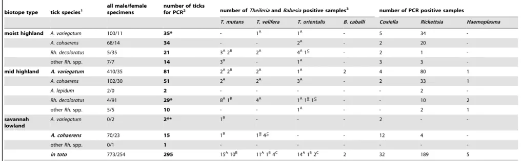
Table 1.Distribution of tick species collected from cattle in three biotopes, and results of their molecular analyses.
biotope type tick species1 all male/femalespecimens number of ticksfor PCR2 number ofTheileriaandBabesiapositive samples3 number of PCR positive samples
T. mutans T. velifera T. orientalis B. caballi Coxiella Rickettsia Haemoplasma
moist highland A. variegatum 100/11 35* - 1A 1A - 5 34
-A. cohaerens 68/14 34 - - 2A - 2 20
-Rh. decoloratus 5/35 21 3A2B 2A 4A1–C - 2 1
-otherRh.spp. 7/7 14 3B - 1A - 3 3
-mid highland A. variegatum 410/35 81 2A2B 2A 1A 2 4 80 1
A. cohaerens 102/30 51 2A 2A 3A - 2 33 1
A. lepidum 2/0 2 - - - 2
-Rh. decoloratus 4/91 29* 8A1B 4A 1A1–B1–C - - 10 2
otherRh.spp. 5/5 10 - - 1A - - 2 1
savannah lowland
A. variegatum 0/2 2** 1B - - - 2 -
-A. cohaerens 70/23 15 1B 1–B4–C - - 12 4
-otherRh.spp. 0/1 1 - - -
-in toto 773/254 295 15A10B 11A1B4C 14A1B2C 2 32 189 5
1Tick species significantly more abundant in a biotope type, than in other(s), are marked with bold character. Abbreviations are ‘‘A.’’ forAmblyomma, ‘‘Rh.’’ forRhipicephalus. Other
Rhipicephalusspp. implyRh. praetextatusandRh. evertsi.
2The number of asterisks (* or **) indicates the number of nymphs included in a sample number. 3Upper index capital letters (A, B, C) on the numbers of
Theileriapositive samples indicate genotypes (for legend see Tables 2–4).Theileriagenotypes unique to a tick species are marked with underlined superscript. TheHyalomma rufipesmale was PCR negative in all tests, therefore not shown.
doi:10.1371/journal.pone.0106452.t001
Eco-Epidemi
ology
of
Tick-Bor
ne
Pathogen
s
in
Ethiopia
PLOS
ONE
|
www.ploson
e.org
4
September
2014
|
Volume
9
|
Issue
9
|
Table 2.Sequence differences of18S rRNAgene ofTheileria mutansgenotypes identified in this study, compared to GenBank reference sequences.
designation nucleotid position in reference sequence (AF078815)
620 621 625 633 634 643 644 646 649 664 665 666 668 671 675 677
reference(AF078815) A T G T C A G G T A C T G T T T
genotype TM-A
N
N
N
N
N
N
N
–N
N
N
N
N
N
N
N
genotype TM-B C C C – – G A – C C G A – C C G
doi:10.1371/journal.pone.0106452.t002
Table 3.Sequence differences of18S rRNAgene ofTheileria veliferagenotypes identified in this study, compared to GenBank reference sequences.
designation nucleotid position in reference sequence (AF097993)
620 627 628 629 637 644 646 648 650 652 653 677–8
reference(AF097993) A C T A T T T G T T T –
genotype TV-A
N
N
N
N
N
N
N
N
N
N
N
Ggenotype TV-B G
N
C T A CN
N
– – C Ggenotype TV-C G T
N
C AN
– A – – C Gdoi:10.1371/journal.pone.0106452.t003
Eco-Epidemi
ology
of
Tick-Bor
ne
Pathogen
s
in
Ethiopia
PLOS
ONE
|
www.ploson
e.org
5
September
2014
|
Volume
9
|
Issue
9
|
frequently contained rickettsiae on mid highland (10 out of 29: 34.5%), than on moist highland (1 out of 21: 4.8%) (Table 1). In all 21 samples processed for sequencingR. africaewas identified, with one to eight nucleotide differences to a reference sequence (U59733). The phylogenetic relationships of these isolates with each other and with other rickettsia sequences from the GenBank are shown on Figure 1. In summary,R. africaewas detected inA. variegatum,A. cohaerensandA. lepidum.
Haemoplasmas in ticks
The low level of haemoplasma positivity seemed to be equally distributed among the tick species (Table 1). The haemotropic
Mycoplasmasp(p). in question could not be identified because of low bacterial loads (reflected by high Ct values). The presence of these pathogens was only detected in one type of tick biotope, i.e. on mid highland, but this association was not significant (P = 0.16) due to the small number of positive samples.
Discussion
Hard ticks (Acari: Ixodidae) feed on the blood of vertebrates. Although they adversely affect domestic animals in several ways, their economically most important effect on domestic animals is connected to their vector role, as they are able to transmit a broad spectrum of tick-borne pathogens. The productivity losses attributable to related diseases are estimated to be highest in the tropical parts of the world, including Africa [22].
In order to infect new hosts, all tick-borne agents need the availability of certain ixodid species, i.e. competent vectors, which thus determine their geographical distribution. At a local scale, the epidemiology of tick-borne infections is strongly interrelated with the ecology of relevant ticks. In this way tick-borne diseases are usually endemic, implying their focal occurrence according to the suitable habitats of competent vectors.
Results on the abundance of tick species on cattle in the present study are similar to those reported earlier from South-Western Ethiopia, i.e.A. variegatum,A. cohaerens,Rh. decoloratusandRh. evertsibeing the most important ixodid species [7,8]. The male-biased sex ratio ofAmblyommaspp. and female-biased sex ratio of
Rh. decoloratus is also consistent with previous findings [23]. However, here it was also demonstrated that the two most abundant tick species, A. variegatum and A. cohaerens are associated with mid-highland and savannah lowland, respectively, relevant to the endemicity and epidemiology of tick-borne pathogens they may carry.
Theileria spp. detected in ticks of cattle, i.e. T. mutans, T. veliferaandT. orientalisare widespread in Africa and are usually regarded as mildly pathogenic. However, they were also shown to cause severe anaemia, icterus, even deaths [24–27]. EspeciallyT. mutans have been associated with disease in cattle: invasion of brain capillaries by this piroplasm may result in a form of bovine theileriosis known as turning sickness [28].
In Africa these Theileria spp. are transmitted by ticks of the genus Amblyomma [28,29]. This is consistent with the present findings, i.e. the relatively high prevalence of these piroplasms in ticks of cattle, particularly inAmblyommaspp. However, the fact that according to the results of this study Rh. decoloratus
specimens also contained (had access to) all three above mentioned
Theileriaspp, it may deserve future attention to assess the vector competence of this tick species too. In addition, the prevalence of piroplasms (Theileria spp.) in cattle ticks was significantly higher (up to 56%) in the present study, than the 0.5–4% reported in Western Ethiopia recently [9], suggesting that their tick-borne transmission is more likely than previously thought.
Table 4. Sequence differences of 18S rRNA gene of Theileria orientalis genotypes identified in this study, compared to GenBank reference sequences. designation nucleotid position in reference sequence (AF236094) 626 630 637 639 654 676 – 7 reference (AF236094) AA G T T –– – genotype TO-A
NNNNNN
N
N
genotype TO-BNNNNN
TT A genotype TO-C T T – CC –– – doi:10.1371/journal.pone. 0106452.t004Eco-Epidemiology of Tick-Borne Pathogens in Ethiopia
A high degree of18S rRNAgene sequence polymorphism of these piroplasms was noted here, similarly to that reported in blood samples of the African buffalo (Syncerus caffer: the original hosts ofT. mutans,T. veliferaandT. orientalis) in South Africa [28]. Relevant to the epidemiological significance of the present findings, these genotypes are thought to circulate between some buffalo and cattle populations [28]. The present results also confirm that in comparison withT. mutansthe sequence variation is less evident in the18S rRNAgene ofT. velifera[28].
According to the data shown here the occurrence of (and risks associated with) theseTheileriaspp. and their genetic variants in South-Western Ethiopia are primarily dependent on the tick species (because they have specific competent vectors), and not on the type of tick-biotope where cattle are grazing. The latter may be
explained by the absence of transovarial maintenance ofTheileria
spp. by ticks in nature [29], unlike in case of babesiae.
Babesia caballiis the large babesia of the horse, causing mainly anaemia. This piroplasm was detected inA. variegatumticks, for the first in East Africa: a finding similar to the identification of the same piroplasm in cattle ticks in West Africa [30]. The sequence was new and highly (3%#)divergent from already reported ones. Since cattle were an unlikely source of this piroplasm in the present study, the potential vector role of A. variegatum in transmittingB. caballishould be further evaluated.
Coxiella burnetiiis a tick-borne zoonotic bacterium. Reports are scarce on its presence in ticks in East or West Africa. The Q fever agent is known to occur in Ethiopia for nearly half a century [31]. In a more recent study from West Africa [32] the infection rate
Figure 1. Phylogenetic comparison ofRickettsia africaeisolates identified in the present study and other rickettsiae based ongltA
gene sequences.Branch lengths correlate to the number of substitutions inferred according to the scale shown. doi:10.1371/journal.pone.0106452.g001
Eco-Epidemiology of Tick-Borne Pathogens in Ethiopia
withC. burnetiishowed significant variations in ticks, e.g. inA. variegatumit was 0% in one region, and 37.6% in another, which phenomenon could not be completely explained by the authors, and particularly not with differences between tick biotopes. Ticks usually carryC. burnetiias reservoirs, and may transfer it between stages (transstadially) and even may inherit it to the next generation (transovarially). Nevertheless, ticks play a subordinate, most likely reservoir role in the epidemiology of Q fever [32], the most significant sources of infection remaining environmental: contact with animal faeces or products, or inhalation of similar substances following aerosolisation. As conditions (prerequisites) for the latter (i.e. dry weather, open area, wind) appear to be more available in savannah woodland, these alternative transmission routes may explain why ticks significantly more often carried (or had access to)Coxiellain this kind of biotope (taking into account that cattle were the most likely source of infection of ticks in the present study). Taken together, results presented here may be the first indications that the reservoir role of ticks is related to their habitats, i.e. could be more significant in savannah woodland, than in mid highland or moist highland.
In another West African study [33], also evaluating ticks from cattle, the prevalence ofC. burnetiiwas higher (14% vs. 10.8%) and that of rickettsiae was significantly lower (12.5% vs. 64.1%) compared to findings in the present study. In an Ethiopian survey [34] the infection rate of ticks with rickettsiae was also considerably lower (4.1%). Therefore data obtained here, based on highly sensitive real-time PCR analysis, seem to attest for the first time, that rickettsiae may be present in the majority of A. variegatumandA. cohaerensticks. In all samples ofA. variegatum
processed for sequencingRickettsia africae, the causative agent of African tick-bite fever was found. This is in line with the known vectorial competence ofA. variegatumin the transmission of R. africae [35]. However, R. africae was also identified in a few specimens ofA. cohaerensandA. lepidum, justifying their future evaluation for vector role in the epidemiological cycle. Interest-ingly, the occurrence of rickettsiae in ticks of cattle, as shown here for the first time, may not only depend on the tick species (i.e. on their competent, specific vector), but also on the biotope type. This may be related to the transovarial maintenance of rickettsiae by ticks (bound to endemic foci) in nature [36].
Haemotropic mycoplasmas (haemoplasmas) are epi-erythrocytic bacteria that may cause anaemia and unthriftiness in various
domestic animals or even humans, and were recently reclassified (removed from the order Rickettsiales). Despite Eperythrozoon
(renamed asMycoplasma)wenyoniiwas reported for the first time in Africa [37], more recent data are not available on the occurrence of haemotropic mycoplasmas in cattle or in ticks in Africa/Ethiopia. The present results not only attest for the first time the occurrence of cattle-related haemoplasmas in ticks collected in Africa, but also show that ticks preferring a more humid environment (in association with mid highland biotopes) may be more exposed to these agents. This biotope-related occurrence may also be partly explained by certain features of haemoplasmas on the group level, because the majority of species have non-tick-borne and other, alternative routes of transmissions [38], thus rendering them more dependent on environmental factors.
In conclusion, the tick biotope, in which cattle were grazed in the evaluated period, influenced not only the tick burden of these hosts, but also the spectrum of pathogens in their ticks. The biotope appeared to be an important limiting factor in case of tick-borneCoxiella burnetiiand haemoplasmas, which are known to have alternative (non-tick-borne) transmission routes, but no specific tick vectors. The presence of rickettsiae in ticks was influenced by both the biotope type and the tick species, whereas that ofTheileria spp. only by the latter. This may reflect, that representatives of these two pathogen groups have specific tick vectors, and in these rickettsiae also have the means (i.e. transovarial transmission) of long-term maintenance in nature.
Acknowledgments
Molecular biology work was partially performed using the logistics of the Center for Clinical Studies at the Vetsuisse Faculty of the University of Zurich.
Author Contributions
Conceived and designed the experiments: SH GA MLM BT KMS MG EG RF RHL. Performed the experiments: SH GA MLM BT KMS MG EG RF RHL. Analyzed the data: SH GA MLM BT KMS MG EG RF RHL. Contributed reagents/materials/analysis tools: SH GA MLM MG RF RHL. Contributed to the writing of the manuscript: SH RHL.
References
1. Lodos J, Boue O, de la Fuente J (2000) A model to simulate the effect of vaccination againstBoophilusticks on cattle. Vet Parasitol 87: 315–326. 2. Levin BR, Svanborg Ede´n C (1990) Selection and evolution of virulence in
bacteria: an ecumenical excursion and modest suggestion. Parasitology 100: S103–115.
3. Benin S, Ehui S, Pender J (2006) Policies for livestock development in the Ethiopian highlands. In: Pender J, Place F, Ehui S (Eds.): Strategies for Sustainable Land Management in the East African Highlands. IFPRI, Washington DC.
4. Pegram G, Hoogsstraal H, Wassef HP (1981) Ticks (Acari Ixodidea) of Ethiopia Distribution, Ecology and Host relationship of species infecting livestock. Bull Entomol Res 71: 339–359.
5. Mekonnen S, Pegram RG, Gebre S, Mekonnen A, Jobre Y, et al. (2007) Zewdie S: A synthetic review of ixodid (Acari: Ixodidae) and argasid (Acari: Argasidae) ticks in Ethiopia and their possible roles in disease transmission. Ethiop Vet J 11: 1–24.
6. Mekonnen S, de Castro J, Gebre S, Hussein I, Regassa A (1992) Ticks, tick-borne diseases and their control in Western Ethiopia. Int J Trop Ins Sci 13: 661–664.
7. Abera M, Mohammed T, Abebe R, Aragaw K, Bekele J (2010) Survey of ixodid ticks in domestic ruminants in Bedelle district, Southwestern Ethiopia. Trop Anim Health Prod 42: 1677–1683.
8. Asrate S,Yalew A (2012) Prevalence of cattle tick infestation in and around Haramaya district, Eastern Ethiopia. J Vet Med Anim Health 4: 84–88.
9. Kumsa B, Signorini M, Teshale S, Tessarin C, Duguma R, et al. (2014) Molecular detection of piroplasms in ixodid ticks infesting cattle and sheep in western Oromia, Ethiopia. Trop Anim Health Prod 46: 27–31.
10. Mekonnen S (1994) Tick and tick-borne disease control in Ethiopia. In: Tick and tick-borne disease control in East, Central and Southern Africa, 1991–1994 (Eds. Musisi FL, Dolan TT). Proceedings of a joint OAU, FAO and ILRAD workshop held in Lilongwe, Malawi 25–28 April 1994. pp.19–21.
11. Hoogstraal H (1956) African Ixodoidea. 1. Ticks of the Sudan with special reference to Equatoria province and with preliminary review of genera Rhipicephalus,MargaropusandHyalomma. US Navy, Washington D.C. 12. Walker AR, Bouattour A, Camicas J-L, Estrada-Pen˜a A, Horak IG, et al. (2003)
Ticks of domestic animals in Africa: a guide to identification of species. Bioscience Reports, Edinburgh.
13. Muhlnickel CJ, Jefferies R, Morgan-Ryan UM, Irwin PJ (2002)Babesia gibsoni infection in three dogs in Victoria. Aust Vet J 80: 606–610.
14. Olmeda AS, Armstrong PM, Rosenthal BM, Valladares B, del Castillo A, et al. (1997) A subtropical case of human babesiosis. Acta Trop 67: 229–234. 15. Loftis AD, Reeves WK, Szumlas DE, Abbassy MM, Helmy IM, et al. (2006)
Rickettsial agents in Egyptian ticks collected from domestic animals. Exp Appl Acarol 40: 67–81.
16. Boretti FS, Perreten A, Meli ML, Cattori V, Willi B, et al. (2009) Molecular investigations ofRickettsia helveticainfection in dogs, foxes, humans andIxodes ticks. Appl Environ Microbiol 75: 3230–3237.
Eco-Epidemiology of Tick-Borne Pathogens in Ethiopia
17. Stenos J, Graves SR, Unsworth NB (2005) A highly sensetive and specific real-time PCR assay for the detection of spotted fever and typhus group Rickettsiae. Am J Trop Med Hyg 73: 1083–1085.
18. Thompson JD, Higgins DG, Gibson T J (1994) CLUSTAL W: improving the sensitivity of progressive multiple sequence alignment through sequence weighting, position-specific gap penalties and weight matrix choice. Nuc Acids Res 22: 4673–4680.
19. Saitou N, Nei M (1987) The neighbor-joining method: a new method for reconstructing phylogenetic trees. Mol Biol Evol 4: 406–425.
20. Tamura K, Stecher G, Peterson D, Filipski A, Kumar S (2013) MEGA6: Molecular Evolutionary Genetics Analysis Version 6.0. Mol Biol Evol 30: 2725– 2729.
21. Willi B, Meli ML, Lu¨thy R, Honegger H, Wengi N, et al. (2009) Development and application of universal Hemoplasma screening assay based on the SYBR green PCR principle. J Clin Microbiol 47: 4049–4054.
22. Jongejan F, Uilenberg G (2004) The global importance of ticks. Parasitology 129: S3–S14.
23. Mekonnen S, Hussein I, Bedane B (2001) The distribution of ixodid ticks (Acari: Ixodidae) in central Ethiopia. Onderstepoort J Vet Res 68: 243–251. 24. Rogers RJ, Callow LL (1996) Three fatal cases ofTheileria mutansinfection.
Aust Vet J 42: 42–46.
25. Ceci L, Kirvar E, Carelli G, Brown D, Sasanelli M, et al. (1997) Evidence of Theileria buffeliinfection in cattle in southern Italy. Vet Rec 140: 581–583. 26. Stockham SL, Kjemtrup AM, Conrad PA, Schmidt DA, Scott MA, et al. (2000)
Theileriosis in a Missouri beef herd caused byTheileria buffeli: case report, herd investigation, ultrastructure, phylogenetic analysis, and experimental transmis-sion. Vet Pathol 37: 11–21.
27. Kamau J, de Vos AJ, Playford M, Salim B, Kinyanjui P, et al. (2011) Emergence of new types ofTheileria orientalisin Australian cattle and possible cause of theileriosis outbreaks. Parasit Vectors 4: 22.
28. Chaisi ME, Collins NE, Potgieter FT, Oosthuizen MC (2013) Sequence variation identified in the 18S rRNA gene ofTheileria mutansandTheileria veliferafrom the African buffalo (Syncerus caffer). Vet Parasitol 191: 132–137. 29. Bishop R, Musoke A, Morzaria S, Gardner M, Nene V (2004) Theileria: intracellular protozoan parasites of wild and domestic ruminants transmitted by ixodid ticks. Parasitology 129: S271–83.
30. Tomassone L, Pagani P, De Meneghi D (2005) Detection ofBabesia caballiin Amblyomma variegatum ticks (Acari: Ixodidae) collected from cattle in the Republic of Guinea. Parassitologia 47: 247–251.
31. Philip CB, Hoogstraal H, Reiss-Gutfreund R, Clifford CM (1966) Evidence of rickettsial disease agents in ticks from Ethiopian cattle. Bull World Health Organ 35: 127–131.
32. Mediannikov O, Fenollar F, Socolovschi C, Diatta G, Bassene H, et al. (2010) Coxiella burnetiiin humans and ticks in rural Senegal. Plos Negl Trop Dis 4: e654.
33. Reye AL, Arinola OG, Hu¨bschen JM, Muller CP (2012) Pathogen prevalence in ticks collected from the vegetation and livestock in Nigeria. Appl Environ Microbiol 78: 2562–2568.
34. Mura A, Socolovschi C, Ginesta J, Lafrance B, Magnan S, et al. (2008) Molecular detection of spotted fever group rickettsiae in ticks from Ethiopia and Chad. Trans R Soc Trop Med Hyg 102: 945–949.
35. Socolovschi C, Huynh TP, Davoust B, Gomez J, Raoult D, et al. (2009) Transovarial and trans-stadial transmission ofRickettsiae africaeinAmblyomma variegatumticks. Clin Microbiol Infect 15 (Suppl. 2): 317–318.
36. Perlman SJ, Hunter MS, Zchori-Fein E (2006) The emerging diversity of Rickettsia. Proc Royal Soc B Biol Sci 273:2097–2106.
37. Neitz WO (1940) Eperythrozoonosis in cattle. Onderstepoort J Vet Sci 14: 9– 28.
38. Yang Z, Yan C, Yu F, Hua X (2007) Haemotrophic mycoplasma: Review of aetiology and prevalence. Rev Med Microbiol 18: 1–3.
Eco-Epidemiology of Tick-Borne Pathogens in Ethiopia