Functions in
Caenorhabditis elegans
Myriam Passannante1, Claude-Olivier Marti1, Catherine Pfefferli1, Paolo S. Moroni1, Ste´phanie Kaeser-Pebernard1, Alessandro Puoti1, Peter Hunziker2, Chantal Wicky1, Fritz Mu¨ller1*
1Department of Biology, University of Fribourg, Fribourg, Switzerland,2Functional Genomics Center Zu¨rich, University/ETH Zurich, Zu¨rich, Switzerland
Abstract
Biochemical purifications from mammalian cells and Xenopus oocytes revealed that vertebrate Mi-2 proteins reside in multisubunit NuRD (Nucleosome Remodeling and Deacetylase) complexes. Since all NuRD subunits are highly conserved in the genomes ofC. elegansandDrosophila, it was suggested that NuRD complexes also exist in invertebrates. Recently, a novel dMec complex, composed of dMi-2 and dMEP-1 was identified inDrosophila. The genome ofC. elegansencodes two highly homologous Mi-2 orthologues, LET-418 and CHD-3. Here we demonstrate that these proteins define at least three different protein complexes, two distinct NuRD complexes and one MEC complex. The two canonical NuRD complexes share the same core subunits HDA-1/HDAC, LIN-53/RbAp and LIN-40/MTA, but differ in their Mi-2 orthologues LET-418 or CHD-3. LET-418 but not CHD-3, interacts with the Kru¨ppel-like protein MEP-1 in a distinct complex, the MEC complex. Based on microarrays analyses, we propose that MEC constitutes an important LET-418 containing regulatory complex duringC. elegansembryonic and early larval development. It is required for the repression of germline potential in somatic cells and acts when blastomeres are still dividing and differentiating. The two NuRD complexes may not be important for the early development, but may act later during postembryonic development. Altogether, our data suggest a considerable complexity in the composition, the developmental function and the tissue-specificity of the different C. elegans Mi-2 complexes.
Citation:Passannante M, Marti C-O, Pfefferli C, Moroni PS, Kaeser-Pebernard S, et al. (2010) Different Mi-2 Complexes for Various Developmental Functions in
Caenorhabditis elegans. PLoS ONE 5(10): e13681. doi:10.1371/journal.pone.0013681
Editor:Mary Bryk, Texas A&M University, United States of America
ReceivedMay 12, 2010;AcceptedOctober 6, 2010;PublishedOctober 27, 2010
Copyright:ß2010 Passannante et al. This is an open-access article distributed under the terms of the Creative Commons Attribution License, which permits unrestricted use, distribution, and reproduction in any medium, provided the original author and source are credited.
Funding:This work is funded by the Swiss National Science Foundation (http://www.snf.ch/E/Pages/default.aspx) (SNF grants: 109938 and 31003A-125577). The funders had no role in study design, data collection and analysis, decision to publish, or preparation of the manuscript.
Competing Interests:The authors have declared that no competing interests exist.
* E-mail: [email protected]
Introduction
Epigenetics encompasses all inheritable changes capable of modulating gene expression that are not encoded by the DNA sequence itself. Such changes include modifications at the chromatin level, which can be achieved by four processes: DNA methylation, histone modifications, ATP-dependent chromatin remodeling and histone variant incorporation. The study of chromatin remodeling complexes has revealed a surprising complexity in the composition and the function of such complexes (reviewed in [1]). Based on sequence and structure, the ATPase subunits of these complexes are divided into four families: the SWI/SNF, ISWI, INO80 and CHD families. The CHD family is characterized by chromodomain containing proteins, including the Mi-2 proteins.
Mi-2 was first identified as an autoantigen in patients with dermatomyositis [2,3], and as a key component of the Nucleosome Remodeling and histone Deacetylase (NuRD, also called NURD or NRD) complex (reviewed in [4,5]). The vertebrate Mi-2/NuRD complex contains at least seven polypeptides. In addition to the Mi-2 protein, it also includes the class I histone deacetylases HDAC1 and HDAC2, the histone-binding proteins RbAp46/48, the methyl-binding MBD proteins and the metastasis-associated MTA proteins (reviewed in [5]). There is conflicting evidence regarding the exact composition of the NuRD complex because
the vertebrate genome encodes at least two homologues for most of the NuRD subunits, including the two Mi-2 isoforms Mi-2aand Mi-2b [5]. The existence of such isoforms suggests that the vertebrate NuRD complex might not be a single molecular species and that the subunit heterogeneity reflects a functional speciali-zation (reviewed in [5]).
In Drosophila, the existence of a NuRD complex has been strongly suggested by several interaction studies [6,7,8,9,10]. Recently, a new containing dMi-2 protein complex, dMec, was characterized in Drosophila. dMec is composed of dMi-2 and dMEP-1 [11] and is clearly distinct from the NuRD complex. dMec, which constitutes the major dMi-2 containing complex in
Drosophilacells, is strongly expressed in embryos but its role during embryogenesis is not known. It is also involved in the repression of proneural genes of the achaete-scute complex [11].
germline cytoplasm of most, if not all animals [13]. This suggested that the activity oflet-418is required to repress germline specific genes in somatic cells during development [14]. Furthermore, let-418 negatively regulates the expression of the Hox gene lin-39
[14]. In contrast tolet-418animals,chd-3mutants show no obvious phenotype. A role for chd-3, however, becomes visible in let-418;chd-3double mutants, which arrest as embryos in the absence of a maternal let-418 contribution [12]. Both, let-418 and chd-3
play a role during vulva formation. In the wild-type hermaphro-dite, the vulva is formed from the descendants of three of six equivalent vulval precursor cells (VPCs). The three cells are induced by multiple cell signaling pathways to adopt vulval cell fates. A large group of genes, the synthetic multivulva (SynMuv) genes, act redundantly to repress vulval differentiation. The SynMuv genes fall into two subgroups, termed A and B. While a single loss-of-function mutation in each subgroup does not result in an obvious vulval induction defect, a mutation in each of the two classes gives rise to a robust Muv phenotype (for a review see [15]). We found thatlet-418is a class B synMuv gene, whereaschd-3does not show a synMuv phenotype [12]. However, chd-3is required redundantly withlet-418for the proper execution of the 2ucell fate in vulval precursor cells and plays a role in the specification of the pharyngeal precursor cells [12].
To gain further insight into the different functions of the two Mi-2 paralogues inC. elegans, we have characterized the LET-418 and CHD-3 containing complexes. Here we show that C. elegans
harbours two distinct and previously undescribed Mi-2/NuRD complexes and an additional LET-418 containing complex, the MEC complex. The latter also contains the Kru¨ppel-like protein MEP-1 and represents the major LET-418 containing regulatory complex duringC. elegansembryonic and early larval development. Our data suggest that it acts before the bean stage to restrict the germline potential of somatic cells.
Results
LET-418 and CHD-3 are members of two distinct NuRD complexes
Because the putative null alleles let-418(s1617) and chd-3(eh4)
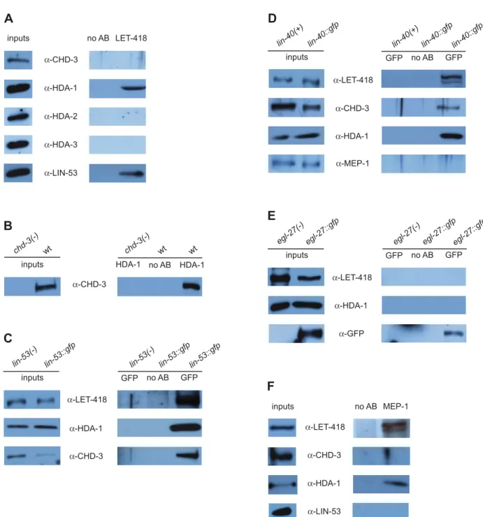
have a different phenotype we expected the two proteins LET-418 and CHD-3 to reside in separate complexes. As predicted, we found that anti-LET-418 antibodies failed to co-precipitate CHD-3 (Figure 1A). As a positive control we used anti-HDA-1 antibodies, since LET-418 interacts with HDA-1/HDAC (Figure 1A, second lane). These results show that LET-418 and CHD-3 do not co-precipitate, suggesting that they reside in different complexes.
Next we searched for interactions between LET-418 or CHD-3 with various orthologues of the core NuRD components encoded by the C. elegans genome. First we tested whether LET-418 interacts with the three class I histone deacetylases, 1, HDA-2 and HDA-3 [16]. However, LET-418 interacted only with HDA-1, but not with the two other HDACs HDA-2 or HDA-3 (Figure 1A). Similarly, we found that CHD-3 co-immunoprecip-itated with HDA-1 (Figure 1B).
RbAp46 and RbAp48 (pRB-associated proteins p46 and p48, also known as RBBP7 and RBBP4, respectively) are two other members of the mammalian NuRD complex (reviewed in [17]). TheC. elegansgenome encodes two RbAp homologues, LIN-53/ RBA-2 and RBA-1. To test a possible interaction of LIN-53/ RbAp with the twoC. elegansMi-2 orthologues, a protein extract was prepared from worms carrying a partially rescuinglin-53::gfp
fusion construct [18]. Anti-GFP antibodies against LIN-53::GFP co-immunoprecipitated both LET-418, CHD-3 and HDA-1
(Figure 1C). In a parallel approach we could also co-precipitate LIN-53 with specific anti-LET-418 antibodies (Figure 1A). Moreover, purification of LIN-53::TAP from a strain carrying a rescuing lin-53::tap construct yielded the same results (data not shown and see below). We also tested the second C. elegans
RbAp46/48 orthologue 1 using a strain expressing a RBA-1::TAP fusion protein. However, we found that RBA-RBA-1::TAP interacted neither with LET-418, nor with CHD-3 or HDA-1 (data not shown). Thus, our data suggested that theC. elegansMi-2 LET-418 and CHD-3 interact only with LIN-53/RbAp, but not with RBA-1/RbAp.
The MTA proteins are additional components of the vertebrate NuRD complexes (reviewed in [5]). The genome of C. elegans
encodes two proteins with homology to MTA1, namely LIN-40 (also called EGR-1) and EGL-27. LIN-40 is structurally more related to the vertebrate MTA family than EGL-27 [5]. Using protein extracts made from worms carrying a rescuinglin-40::gfp
fusion construct [19], we could co-immunoprecipitate LET-418, CHD-3 and HDA-1 with LIN-40::GFP (Figure 1D). We also tested the second MTA orthologue EGL-27. Immunoprecipitation of a rescuing EGL-27::GFP construct [20] yielded neither LET-418, nor CHD-3 or HDA-1 (Figure 1E and data not shown). Altogether, these results suggested that LET-418, CHD-3 and HDA-1 interact with LIN-40/MTA but not with EGL-27/MTA. To corroborate our co-immunoprecipitation data, we under-took a complex purification using the TAP (tandem affinity purification) method that was adapted toC. elegans. Since we could not obtain a full-length LET-418 tagged protein, we tagged the LIN-53/RbAp protein that interacts with both C. elegans Mi-2 orthologues (see above). The resultinglin-53::tapfusion construct was able to rescue the synMuv phenotype oflin-15A;lin-53(n833)
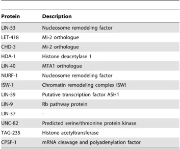
worms. Upon tandem affinity purification of LIN-53::TAP, we co-precipitated the NuRD subunits LET-418/Mi-2, CHD-3/Mi-2, HDA-1/HDAC and LIN-40/MTA, but not the orthologues EGL-27/MTA and RBA-1/RbAp (Table 1). Besides the NuRD components, we isolated additional proteins, among them several members of the DRM and the NURF-like complexes. This was expected since these two protein complexes also contain LIN-53 [21,22].
Altogether biochemical data suggests the presence of two different NuRD complexes inC. elegans. They differ in their Mi-2 orthologues LET-418 or CHD-3, but share the same core subunits HDA-1, LIN-53 and LIN-40. Their respective homologous proteins HDA-2/-3, RBA-1 and EGL-27 did not interact, although we cannot rule out that in some tissues or developmental stages they may reside in the same complex.
The LET-418/Mi-2 containing NuRD complex functions in the synMuv B pathway
Figure 1. Two NuRD complexes and a MEC complex are present inC. elegans.(A) LET-418 interacts with HDA-1 and LIN-53, but not with CHD-3 nor with HDA-2 and HDA-3. Extracts from wild-type mixed-stage worms were precipitated with eithera-LET-418 or no antibodies (negative control). Inputs and immunoprecipitates were subjected to Western analysis and immunoblotted with antibodies directed against proteins indicated next to each panel. (B) HDA-1 binds to CHD-3. Extracts from wild-type orchd-3(eh4)mixed-stage worms were precipitated with eithera-HDA-1 or no antibodies (negative control). Inputs and immunoprecipitates were subjected to Western analysis and immunoblotted with antibodies directed against CHD-3. (C) LIN-53::GFP interacts with LET-418, HDA-1 and CHD-3. Extracts fromlin-53::gfporlin-53(-)mixed-stage worms were precipitated with eithera-GFP or no antibodies (negative control). Inputs and immunoprecipitates were subjected to Western analysis and immunoblotted with antibodies directed against proteins indicated next to each panel. (D) LIN-40::GFP interacts with LET-418, HDA-1 and CHD-3, but not with MEP-1. Extracts from lin-40::gfpor lin-40(+) mixed-stage worms were precipitated with either a-GFP or no antibodies (negative control). Inputs and immunoprecipitates were subjected to Western analysis and immunoblotted with antibodies directed against proteins indicated next to each panel. (E) EGL-27::GFP does not interact with LET-418 nor with HDA-1. Extracts fromegl-27::gfporegl-27(-)mixed-stage worms were precipitated with either
a-GFP or no antibodies (negative control). Inputs and immunoprecipitates were subjected to Western analysis and immunoblotted with antibodies directed against proteins indicated next to each panel. The membrane was reprobed with anti-GFP as a positive control. (F) MEP-1 interacts with LET-418 and HDA-1, but not CHD-3 nor with LIN-53. Extracts from wild-type mixed-stage worms were precipitated with eithera-MEP-1 or no antibodies (negative control). Inputs and immunoprecipitates were subjected to Western analysis and immunoblotted with antibodies directed against proteins indicated next to each panel. Input: 5% of the immunoprecipitate; IP: 100% of the immunoprecipitate (1 mg of proteins). All co-immunoprecipitation experiments were reproducibly performed at least twice.
HDA-3, RBA-1 and EGL-27 do not act in the NuRD complex with LET-418.
MEP-1 and LET-418 reside in a complex distinct from NuRD
Previously, it was found that theC. elegansKru¨ppel-like protein MEP-1 interacts with LET-418 and HDA-1 ([27] and Figure 1F).
To further characterize this interaction and to see whether it occurs in the context of a NuRD complex, we tested if MEP-1 binds to the NuRD components LIN-53/RbAp and LIN-40/ MTA. However, we could not co-precipitate MEP-1 with LIN-53 (Figure 1F) nor with LIN-40::GFP (Figure 1D). We then asked whether MEP-1 was able to physically interact with the secondC. elegansMi-2 orthologue CHD-3. We found that CHD-3 did not co-precipitate with MEP-1 (Figure 1F). These results suggested that MEP-1 only interacts with LET-418 and HDA-1 but not with CHD-3 to form a complex that is distinct from the previously described NuRD complexes. By analogy to theDrosophila dMec, we named theC. elegansLET-418 and MEP-1 containing complex ‘‘MEC complex’’. However, in the dMec no histone deacetylase activity was detected [11].
Both, arrested let-418 and mep-1 L1 larvae show ectopic expression of the P granule component PGL-1 in their somatic cells [27], suggesting that a LET-418 and MEP-1 containing MEC complex is required to repress pgl-1 in somatic cells. To test whether other LET-418 interacting proteins may also be involved in the control ofpgl-1expression, we fed L1 larvae with dsRNA corresponding to all genes encoding NuRD complex components. Upon staining with anti-1 antibodies, we found ectopic PGL-1 expression only inlet-418(RNAi)andmep-1(RNAi), but not in lin-53/RbAp, lin-40/MTA, chd-3/Mi-2, hda-2/HDAC, hda-3/ HDAC, rba-1/RbAp and egl-27/MTA RNAi-depleted worms (Table 3). Due to the high percentage of dead embryos in hda-1(RNAi)worms [16], we could not analyze the effect of HDA-1 on PGL-1 expression.
In a parallel approach, we performed qRT-PCR experiments to determine thepgl-1mRNA levels in animals depleted forlet-418,
mep-1 and all genes encoding NuRD complex components. Consistent with the previous results, we found thatpgl-1mRNA levels were upregulated only inlet-418(RNAi)andmep-1(RNAi)L1 larvae, but not in animals that were RNAi-depleted forlin-53, lin-40,chd-3,hda-2,hda-3,rba-1andegl-27(Table 3). Altogether, the genetic data demonstrated thatpgl-1is jointly regulated by LET-418 and MEP-1, but not by NuRD subunits, thus supporting the notion that MEC and NuRD are distinct functional complexes.
The MEC and NuRD complexes differentially repress
lag-2::gfpexpression
Earlier, it was shown that LET-418 negatively regulates the expression of alag-2::gfpreporter gene in the gut [28]. In wild-type animalslag-2::gfpis expressed in the Distal Tip Cells (DTCs) and weakly along the ventral nerve cord (Figure 2A and [29]), whereas inlet-418mutants it shows an additional strong expression in the intestine (Figure 2B and [28]). A wild-type expression pattern was also observed inchd-3(RNAi)animals (Figure 2C), suggesting that CHD-3 is not involved in the regulation of lag-2::gfp. To determine, which of the LET-418 containing complexes might be responsible for the suppression of the ectopic expression, we analyzed the lag-2::gfpexpression in L3 larvae that were RNAi-depleted formep-1, hda-1, lin-53and lin-40. To our surprise, we reproducibly found three characteristic types oflag-2::gfp expres-sion patterns. Strong intestinal lag-2::gfp expression was only observed in let-418(RNAi), mep-1(RNAi) and hda-1(RNAi) worms (Figures 2B, 2D–2E) and [30]), whereas inlin-53(RNAi) lag-2::gfp
was expressed mainly in the epidermis but clearly not in the intestine (Figure 2F). Finally, inlin-40(RNAi)worms,lag-2::gfpwas strongly expressed only in the two most anterior cells of the intestine and in a weaker manner in a few posterior intestinal cells (Figure 2G). Worms that were RNAi-depleted for the non-NuRD proteins encoding geneshda-2,hda-3,rba-1,egl-27andmbd-2, had a wild-type lag-2::gfp expression pattern (data not shown). These
Table 1.LIN-53::TAP co-precipitates NuRD complex core components.
Protein Description
LIN-53 Nucleosome remodeling factor LET-418 Mi-2 orthologue
CHD-3 Mi-2 orthologue
HDA-1 Histone deacetylase 1 LIN-40 MTA1 orthologue
NURF-1 Nucleosome remodeling factor
ISW-1 Chromatin remodeling complex ISWI LIN-59 Putative transcription factor ASH1
LIN-9 Rb pathway protein
LIN-37
-UNC-82 Predicted serine/threonine protein kinase
TAG-235 Histone acetyltransferase
CPSF-1 mRNA cleavage and polyadenylation factor
LIN-53::TAP containing complexes were subjected to tandem affinity purification. Proteins co-purified with LIN-53::TAP were identified by liquid chromatography tandem mass spectrometry. Proteins were identified using a ProteinLynx Global server and Mascot Search engines.
doi:10.1371/journal.pone.0013681.t001
Table 2.rba-1 and egl-27are not synMuv B genes.
Genotype % Muv (n)
N2;rba-1(RNAi) 0 (132)
lin-15A(n767); rba-1(RNAi) 0 (365)
lin-15B(n744); rba-1(RNAi) 0 (9)*
lin-35(n745); rba-1(RNAi) 0 (9)*
lin-15A(n767); lin-53(RNAi) 74 (243)
N2;egl-27(RNAi) 0 (798)
lin-15A(n767); egl-27(RNAi) 0 (899)
lin-15B(n744); egl-27(RNAi) 0 (9)*
lin-35(n745); egl-27(RNAi) 0 (40)
lin-15A(n767); lin-40(RNAi) 0u
egl-27(n170);HT115 0 (172)
egl-27(n170); lin-15A(RNAi) 0 (253)
egl-27(n170); lin15B(RNAi) 0 (450)
egl-27(n170); lin-35(RNAi) 0 (395)
lin-40(ku285); lin-15A(RNAi) 22 (11)
*no adults or only few adults were obtained because most of the worms arrested as larvae.
usynMuv only withlin-40(ku285)allele (Chenet al., 2001).
The genetic background, the percentage of synMuv and the number of animals counted (n) are indicated.
results suggested that the MEC complex might negatively regulate
lag-2::gfpexpression in the intestine.lin-53(RNAi)andlin-40(RNAi)
produced different lag-2::gfp expression patterns (see Figure 2F– 2G), suggesting that NuRD and/or other LET-418 containing complexes may play additional, tissue-specific roles in the regulation of thelag-2::gfpreporter outside of the gut.
LET-418 and MEP-1 regulate common target genes
The repression of the germline genepgl-1and the transgene lag-2::gfpin the gut by LET-418 and MEP-1 implies a common role for the two proteins in regulating tissue-specific gene expression throughout development. To identify potential target genes, we performed a genome wide gene profile analysis inlet-418and mep-1 depleted animals. For our experiments we chose arrested L1 larvae, which also show ectopic P granule expression [12]. Since
mep-1(q660)mutants are sterile [31], we used RNA interference to generate mep-1 and let-418 depleted worms. Animals fed with bacteria expressinggfpdsRNA (pPE128.110 in HT115) were used as reference sample. Deregulated genes with a p-value of #0.01 and fold change $ 62 were further analyzed. The microarray results were validated by qRT-PCRs on ten randomly selected genes deregulated in bothlet-418(RNAi)andmep-1(RNAi)(five up-and five downregulated genes). The results confirmed the microarray data for all ten genes (Figure S1).
A total of 1113 genes showed changed expression levels in let-418(RNAi) L1, whereas 1104 genes were deregulated in mep-1(RNAi)worms. Given the similar phenotype oflet-418(RNAi)and
mep-1(RNAi)animals and the physical interaction of LET-418 and MEP-1, we expected a comparable gene expression profile. Indeed, we found that 914 (82%) of the deregulated genes were common betweenlet-418(RNAi)andmep-1(RNAi)animals (hence-forth referred to as ‘‘common genes’’). The majority of them (70%) were upregulated. Analyses using the statistical software MAGMA with R-scripts revealed a very strong correlation between their
deregulation pattern of these genes. A standard correlation factor of R = 0.98 was calculated according to the linear regression (R = 1 means that the relation is linear), demonstrating that gene expression was deregulated very similarly in let-418(RNAi) and
mep-1(RNAi)depleted L1 larvae (Figure 3). The high degree of correlation between the expression profiles is consistent with the idea that most, if not all, of those genes are controlled by the same MEC complex.
To test whether LET-418 and MEP-1 can also function independently from each other at this stage of development, we focused on the 18% of genes that were deregulated exclusively on either the let-418(RNAi) or the mep-1(RNAi) microarray. The expression of most of these genes was only moderately affected with a fold change around62. We tested the expression levels of 12 randomly selected genes by qRT-PCR analysis (six specifically deregulated genes were chosen on each microarray list). We found that the expression of six of them was not affected, neither in let-418(RNAi)nor inmep-1(RNAi)animals, whereas the six remaining genes were deregulated in both,let-418(RNAi)and inmep-1(RNAi)
L1 larvae (Figure S2A–S2B). Thus, the 12 tested genes corresponded either to false negative or false positive signals on their microarrays and we could find no evidence for LET-418 and MEP-1 acting independently from each other. Therefore, we can conclude that MEC represents the major LET-418/Mi-2 containing gene regulatory complex acting during early larval development inC. elegans.
LET-418 and MEP-1 regulate germline specific and early embryonic genes
Sincelet-418andmep-1 were proposed to repress the germline potential in somatic cells during embryonic and early larval development [27] and our own observation), we were interested to determine how many germline expressed genes were deregulated inlet-418(RNAi) and mep-1(RNAi) depleted L1 larvae. Recently,
Table 3.The MEC complex regulatespgl-1expression in L1 larvae.
Gene inhibited by RNAia ectopic PGL-1 staining pgl-1mRNA fold change
control RNAi no 1
MEC complex let-418 yesb 54.2
mep-1 yesb 62.7
hda-1 n.d.d n.d.d
LET-418 NuRD complex let-418 yesb 54.2
hda-1 n.d.d n.d.c
lin-53 noc 0.5
lin-40 no 0.5
CHD-3 NuRD complex chd-3 no 0.5
hda-1 n.d.d n.d.d
lin-53 nob 0.5
lin-40 no 0.5
non NuRD homologues hda-2 no 0.03
hda-3 no 0.1
rba-1 no 1.8
egl-27 no 0.5
aEfficiency of all dsRNAs were tested by looking for the expected phenotype. bUnhavaithayaet al.(2002).
cWanget al.(2005).
dcould not be determined due to the very high percentage of dead embryos in
hda-1(RNAi)worms.
Wang et al. [32] have identified 4699 germline expressed genes from a SAGE library constructed from dissected C. elegans
hermaphrodite gonads from young adults. They correspond to about 21% of all predictedC. elegansgenes, and henceforth will be referred to as ‘‘germline genes’’. We compared them with our 914 common genes co-regulated by LET-418 and MEP-1 and identified 222 (24.3%) putative germline genes among them. Although the germline genes were statistically not overrepresented
among the deregulated genes, the fact that most of them (187 genes) were upregulated indicated a possible shift of the gene expression pattern towards germline specificity in let-418(RNAi)
andmep-1(RNAi)depleted L1 larvae,
To further investigate this issue, we have searched for genes which expression is enriched in, or specific for germ cells. Such genes are suggested to have roles specific to germline functions. Since in C. elegans L1 larvae germline proliferation has not yet
Figure 2. The MEC complex negatively regulateslag-2::gfpexpression in the gut.Thelag-2::gfptransgene is expressed in the gut of let-418(RNAi),mep-1(RNAi)andhda-1(RNAi)L3 larvae. (A) L3 worms carryinglag-2::gfptransgene and fed bacteria containing empty vector (RNAi control) (A) show expression in the Distal Tip Cells (DTC) and in the ventral nerve cord. No ectopic expression is observed. (B, D–E)lag-2::gfpis ectopically expressed in the gut oflet-418(RNAi)(B),mep-1(RNAi)(D) andhda-1(RNAi)(E) L3 larvae. (C) No ectopic expression is observed inchd-3(RNAi)L3 larvae. (F)lag-2::gfpis mainly expressed in the epidermis inlin-53(RNAi)L3 larvae. (G)lag-2::gfpis ectopically expressed only in two cells in the most anterior part of the intestine inlin-40(RNAi)L3 larvae. The asterisk marks the DTC.
begun, the larvae only contain two germ cells (Z2 and Z3). Therefore, we expected germline-specific genes to be underrep-resented or even absent from L1 larvae. Among the 4699 germline genes, Wang et al. identified 733 (15.6%) germline-enriched and 330 (7%) germline-specific genes. We have compared their data with the list of our common genes and found that they include 19 (8.5%) germline-enriched and 40 (18%) germline-specific genes. With the exception of two enriched and one germline-specific genes they are all overexpressed and, particularly, the germline-specific genes are statistically overrepresented among the common genes. The upregulation of germline-enriched and germline-specific genes suggests that they may be ectopically expressed in the soma, an assumption that is also supported by the finding of P granules in intestinal and some hypodermal cells of let-418(RNAi)andmep-1(RNAi)depleted L1 animals [27]. We found that 17 of the currently 41 known genes encoding C. elegans P granule components [13] are derepressed in let-418(RNAi) and
mep-1(RNAi)depleted L1 larvae, including gld-1, gld-3, glh-1and
glh-2, pgl-1, pos-1, deps-1 and others. Further examples of upregulated germline-specific genes are him-3, htp-1, htp-2 and
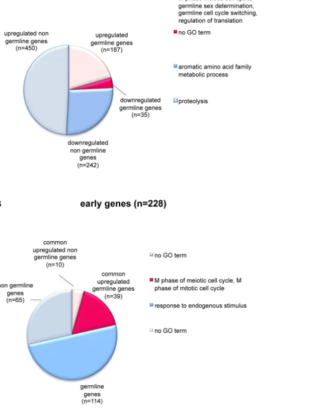
htp-3, which encode meiosis-specific HORMA domain-containing proteins involved in synaptonemal complex formation and meiotic chromosome segregation [33,34]. Altogether, our expression data support the idea that let-418 and mep-1 are involved in the repression of the germline potential of somatic cells in L1 larvae. To gain further insight into the biological roles of the common genes, we analyzed the gene ontology (GO) terms associated with them by using the FatiGO+software [35]. We found that the 187 upregulated germline genes are implicated in biological processes associated to hermaphrodite germline sex determination, M phase of meiotic cell cycle, germline cell cycle switching (the switch from mitotic to meiotic cell cycle) and regulation of translation (Figure 4A). They also comprise various genes encoding P granule
components, such asgld-1and gld-3,glh-1 andglh-2, pgl-1,pos-1
anddeps-1. The remaining 450 upregulated common genes, that could not be classified among the germline genes, were annotated as genes involved in proteolysis (Figure 4A). The 242 downreg-ulated non germline genes, finally, are implicated mainly in aromatic amino acid family metabolic processes. No GO terms could be associated with the 35 downregulated germline genes.
Among the common deregulated genes we found also 49 early embryonic genes (Figure 4B). They belong to a group of 228 genes, which are expressed at the beginning of embryogenesis and then downregulated to background levels by the onset of gastrulation [36]. Their expression temporally correlates with the developmental plasticity observed inC. elegansembryonic blasto-meres, which is lost during gastrulation [36]. Among the 49 genes, 39 belong to the class of germline genes and are significantly enriched in the GO terms associated with M phase of meiotic cell cycle and M phase of mitotic cell cycle (Figure 4B). Interestingly, the 114 remaining germline genes among the early genes which downregulation in L1 larvae does not depend on the MEC complex, are associated with response to endogenous stimulus. The 65 early genes that do not belong to germline genes did not show any significant GO term (Figure 4B). Altogether these data suggested that LET-418 and MEP-1 are specifically required during early embryogenesis to downregulate the early genes with mitotic and meiotic functions.
Ectopic P granules inmep-1(RNAi)animals can first be seen at or shortly after the two-fold stage of embryogenesis ([27] and own observations). This suggested thatmep-1andlet-418must act prior to this stage to ensure normal development. To further identify the time laps in which let-418 activity is required during early development, we shifted let-418 temperature-sensitive embryos at different embryonic stages from the permissive temperature (15uC) to the restrictive (25uC) temperature and followed their
develop-Figure 3. LET-418 and MEP-1 regulate common target genes. A strong correlation is observed between the genes deregulated in let-418(RNAi)andmep-1(RNAi)L1 larvae. The fold change of each of the common genes was plotted on the graph (X-axis: fold change oflet-418(RNAi)
genes, Y-axis: fold change of themep-1(RNAi)). Each circle represents a single gene. A standard correlation factor of R = 0.98 was calculated according to the linear regression.
Figure 4. LET-418 and MEP-1 regulate germline and early embryonic genes.Pie charts show repartition and functional annotation of the common deregulated genes (A) and the early genes (B) according to gene ontology (GO) annotations. The GO terms are indicated on the right of each pie chart. (A) The pie chart shows the repartition of the 914 common deregulated genes compared to the list of 4699 germline genes. The common genes are divided into four groups: upregulated germline (rose slice) and non germline (gray slice) genes; and downregulated germline (pink slice) and non germline (blue slice) genes. (B) The pie chart shows the repartition of the 228 early genes compared to the common upregulated genes and to the list of 4699 germline genes. 49 common upregulated genes are also early genes. They are subdivided in germline (pink slice) and non germline (rose slice) genes. The remaining early genes, that are not targets of MEC, are subdivided in germline (blue slice) and non germline (gray slice) genes.
ment. Embryos shifted to 25uC prior to the lima bean stage arrested at the L1 stage (Table 4). By contrast, embryos shifted to 25uC after the lima bean stage did not arrest and developed beyond L1 into mixed-stage larvae and fertile adults. Taken together, our results indicated that, in order to bypass the L1 larval arrest, thelet-418activity is required prior to the lima bean stage, which corresponds to the time point when embryonic cell proliferation largely ceases and morphogenesis starts [37].
LET-418 and CHD-3 have different functions and expression patterns
We were also interested to learn more about the function of chd-3, the secondC. elegansMi-2 orthologue. Therefore, we analyzed the gene expression profile of chd-3 L1 larvae by using transcriptional microarrays. At this stage,chd-3animals have no obvious phenotype. Consistently, we found that the expression of only a few genes was affected. For that reason we set the minimal median fold change limit at 1.5 instead of 2 (with ap-value#0.01). After validation of the candidates by qRT-PCR, we ended up with only 9 deregulated genes; the expression of 7 of them was moderately downregulated whereas the remaining two target genes were upregulated (data not shown). No GO term could be attributed to this limited number of genes. Only one upregulated gene, cyp-14A5, was also deregulated in let-418(RNAi) and mep-1(RNAi) animals, however it was downregulated in the latter. Obviously,chd-3does not play much of a regulatory role at this stage.
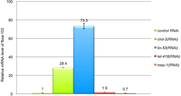
To test whether CHD-3 may act as a component of a NuRD complex, we analyzed the expression level of one of its target genes, fbxa-103, in worms RNAi-depleted for diverse NuRD members. fbxa-103 encodes a protein with an F-box motif predicted to mediate protein-protein interactions. We found that
fbxa-103 was upregulated inchd-3(RNAi)andlin-53(RNAi)animals, but not in let-418(RNAi) nor in mep-1(RNAi) worms (Figure 5). These data are consistent with the idea that CHD-3 negatively regulates the expression of fbxa-103 in the context of a NuRD complex.
Next we have analyzed the developmental expression patterns oflet-418andchd-3. Previously it was shown thatlet-418transcripts are present in the germline and maternally delivered to the early embryos, whereaschd-3mRNA is absent from the germline and appears first around the 20 cell stage after the onset of zygotic transcription ([12] and data not shown). To compare the expression patterns of the two genes during later larval development and in adults, we constructed transcriptional reporter genes by fusing the let-418 promoter region with the Venus
fluorescent reporter and the chd-3 promoter region with the
DsRed2reporter. For each reporter construct, three independent transgenic lines were analyzed. Both reporters were strongly expressed in most if not all cells of the embryo (data not shown). In young adults, both thelet-418andchd-3transgenes were primarily expressed in the head, the vulva, the tail, the ventral nerve cord
and the distal tip cells (Figure 6A–B and data not shown). The strong co-expression of let-418 and chd-3 in the vulva is in agreement with the finding that both genes are redundantly required for the specification of the 2ufate of P.5p and P.7p [12]. The let-418p::Venus construct was expressed in cells surrounding the pharynx, whereas the chd-3p::DsRed2 construct was mainly located in the pharynx itself (Figure 6A). The specific pharyngeal expression ofchd-3is in agreement with the finding thatchd-3, but notlet-418normep-1, plays a role in the pharyngeal precursor cells specification [38]. Another interesting difference was found in the somatic gonad, where only thelet-418reporter, but not thechd-3
reporter was strongly expressed (Figure 6B). These data suggested that let-418 and chd-3may have distinct tissue-specific functions during the postembryonic development and in adults.
Discussion
This study brings an important contribution towards the understanding of the developmental roles of the C. elegans Mi-2 orthologues LET-418 and CHD-3. We demonstrate that the two
C. elegans Mi-2 proteins are members of at least three different protein complexes, two distinct NuRD complexes and a MEC complex comprising LET-418/Mi-2, the Kru¨ppel-like protein MEP-1 and the histone deacetylase HDA-1 (Figure 7). Our data suggest that MEC constitutes an important LET-418 containing gene regulatory complex throughout embryonic and early L1 development, whereas the two NuRD complexes rather function during later larval development, for instance, during vulval development.
Previous studies have revealed a physical interaction between LET-418, MEP-1 and HDA-1 ([27] and own data), suggesting that the three proteins reside in a common regulatory complex. Consistent with this idea,let-418andmep-1mutants share the same phenotype that includes an L1 larval arrest and derepression of germline specific genes in somatic cells of the arrested larvae. Furthermore,mep-1(RNAi)depletedlet-418mutants also arrest at the L1 stage and show no additional synthetic phenotype, suggesting that the two proteins act together in the same biological process. Because of its embryonic lethal phenotype, we could not test whether depletion of hda-1 causes a similar L1 larval phenotype, as would be expected if HDA-1 was also a member of this complex during early development. However, depletion of
let-418, mep-1and hda-1 resulted in an identically strong ectopic expression of alag-2::gfpreporter gene in the intestine of L3 larvae. This suggested that a MEC complex containing LET-418, MEP-1 and HDA-1 is required for the negative control of a lag-2::gfp
reporter in the gut during later larval development. This is in contrast to the situation inDrosophila, where dRPD3/HDAC did not physically associate with dMi-2 and dMEP-1 [11]. However, we cannot rule out that inC. elegansthe composition of the MEC complex shows stage-specific differences. Moreover, theC. elegans
MEC complex probably contains additional subunits, since
Table 4.let-418activity is required prior to the bean stage to prevent the L1 larval arrest.
1–8 cells (17) 8–24 cells (20) 24–50 cells (19) 50- bean stage (21) bean stage (23) comma stage (23) 1.5x-stage (20) 2x-stage (20) 3x-stage (21) newly hatched (30)
.L1 0% 0% 0% 0% 0% 90% 100% 100% 100% 100%
L1 100% 100% 100% 100% 100% 10% 0% 0% 0% 0%
LET-418 activity is required prior the bean stage to allow normal L1-L2 transition.let-418(n3536)tsembryos grown at 15uC were transferred at 25uC. Animals were scored according to their body length and morphological structures three days after hatching at 25uC. In parentheses: number of scored animals.
fractionation of protein extracts from mixed-stage worms resulted in the co-elution of LET-418 and MEP-1 at an estimated complex size ranging from 0.8 to 1.5 MDa (Figure S3 and Materials and Methods S1).
Gene expression profiling experiments revealed that at least 82% of the deregulated genes in let-418(RNAi) and mep-1(RNAi)
worms are tightly co-regulated, thus supporting the association of LET-418 and MEP-1 in a common regulatory MEC complex. Furthermore, our data demonstrate that MEC represents the major LET-418/Mi-2 containing gene regulatory complex during earlyC. elegansdevelopment. Most of the deregulated genes (about 70%) are upregulated inlet-418(RNAi) andmep-1(RNAi)animals,
suggesting that MEC mainly functions in transcriptional repres-sion. Similarly, the Drosophila dMec complex was shown to contribute to the repression of proneural genes [11].
Based on the observation that P granule-like perinuclear structures are found in the somatic cells of arrestedlet-418 and
mep-1L1 larvae ([27] and own observations), it has been suggested that LET-418 and MEP-1 negatively regulate the germline potential of somatic blastomeres during early development. Consistently, we found that in arrested let-418(RNAi) and mep-1(RNAi)L1 larvae totally 187 germline genes were upregulated, among them 59 germline-enriched and germline-specific genes [32,39]. The fact that only a fraction of the germline-specific genes
Figure 5. CHD-3 acts as a component of the NuRD complex to regulatefbxa-103.Fold change offbxa-103mRNA was analyzed by qRT-PCR in diverse RNAi-treated L1 larvae, as indicated on the right of the panel.fbxa-103is upregulated inchd-3(RNAi)andlin-53(RNAi)but not in let-418(RNAi)nor inmep-1(RNAi)L1 larvae.
doi:10.1371/journal.pone.0013681.g005
Figure 6.let-418andchd-3 transcriptional reporters have different expression patterns.(A-B) Young adults animals carrying both a transcriptionallet-418p::Venus(first lane) and a transcriptionalchd-3p::DsRed2(second lane) reporter genes were analyzed by confocal microscopy. The third lane is the merged picture from bothlet-418p::Venusandchd-3p::DsRed2reporters. Expression is shown (A) in the head and (B) in the vulva and somatic gonad. v: vulva (ventral view); g: somatic gonad; vn: ventral nerve cord.
encoded by the genome ofC. elegansis derepressed in LET-418 and MEP-1 depleted animals suggest that the two proteins do not play a global role in the regulation of germline genes. Rather they affect specific transcripts, which are implicated in germline sex determination, M phase of meiotic cell cycle, germline cell cycle switching (the switch from mitotic to meiotic cell cycle) and regulation of translation. Among them are also 17 genes encoding components of P granules. Currently, it is not know why this particular subset of genes are controlled and why other germline-specific genes are not.
Most interestingly,let-418andmep-1control the transcription of specific early expressed embryonic genes, which are normally active at the beginning of embryogenesis and downregulated to background levels by the onset of gastrulation [36]. Their downregulation temporally correlates with the transition from a developmentally plastic state to the onset of differentiation [36], when embryonic cells become restricted in their cell fate potential and begin to acquire cell type identities. We found that LET-418 and MEP-1 are required for the downregulation of a subset of 44 early expressed genes that are specifically enriched in mitotic and meiotic functions. Thus, we may speculate that the MEC complex is already required in the early embryo to downregulate these embryonic genes in order to restrict the germline potential of early blastomeres once germline-soma separation has been achieved. LET-418 and MEP-1 may generally act during embryonic development before the onset of morphogenesis, when cells still divide. Consistent with this hypothesis, our temperature shift experiments indicated that thelet-418activity must be present in the embryos before reaching the bean stage (i.e. when cell proliferation largely ceases and morphogenesis begins) in order to prevent L1 larval arrest. Furthermore, ectopic PGL-1 expression are first observed at the two-fold stage [27] and own data), suggesting that mep-1 and let-418 act before this stage to stably repress the germline potential of the somatic blastomeres.
Apart from an inappropriate expression of germline genes, somatic cells of arrested let-418tsL1 larvae seem to be correctly specified ([27] and own data). This is also pointed out by the observation that arrestedlet-418tsanimals at 25uC, if shifted back to the permissive temperature of 15uC, can resume development and grow into fertile adults (unpublished data). Thus, LET-418 and MEP-1 are not required for the differentiation of the somatic blastomeres during embryogenesis, but rather function in an epigenetic network that stably inactivates their germline potential. During this work we also found that LET-418 and CHD-3 are members of two distinct NuRD complexes. Besides LET-418 or CHD-3, these complexes contain LIN-53/RbAp, HDA-1/HDAC and LIN-40/MTA, but not their homologues HDA-2/HDAC, HDA-3/HDAC, EGL-27/MTA nor RBA-1/RbAp. The
verte-brate and Drosophila NuRD complexes also contain the methyl-CpG-binding protein MBD2/3, which is proposed to mediate an interaction between NuRD and methylated DNA [17]. In C. elegans, however, a tagged version of MBD-2 did not interact with the two Mi-2 homologues LET-418 and CHD-3 (results not shown) nor did it co-purify with the LIN-53::TAP containing protein complexes. This suggested that MBD-2 is not a subunit of the NuRD complexes inC. elegans. In this context it is interesting to note that no DNA methylation has been found inC. elegans, and that MBD-2 was proposed to be part of an ancestral DNA methylation system that was lost in this free living nematode during evolution [40]. Likewise, MBD-2 may have been lost from theC. elegansNuRD complexes.
Genes encoding components of the LET-418 containing NuRD complex (let-418, lin-53,lin-40, and hda-1) belong to the class B synMuv genes, suggesting that a LET-418 containing NuRD complex is implicated in the synMuv B pathway. Interestingly,
mep-1is also a synMuv B gene, notwithstanding that MEP-1 is not a stable member of theC. elegans NuRD complexes in solution. MEP-1, however, could be member of a protein complex distinct from NuRD and MEC that is involved in vulval development. Alternatively, the MEC complex could also play a role in the synMuv B pathway during vulva formation. A similar situation was found inDrosophila, where both, dMec and dNuRD, associate with the promoter of proneural genes of the achaete-scute complex [11].
Vertebrates and Drosophila, like C. elegans, encode two distinct Mi-2 proteins. Our phylogenetic analysis suggests that the gene duplication, giving rise to the second Mi-2 copies must have occurred separately in the three phyla (Figure 8). Gene duplications during evolution often give rise to an essential and a non-essential gene, since the duplicated locus is no longer submitted to selection pressure and therefore is free to change its expression pattern and accumulate otherwise ‘‘forbidden’’ muta-tions [41,42]. Changes in the expression pattern and accumulation of new mutations could result in new functions for the duplicated gene copy. InC. elegans,let-418might have kept its ancient essential functions, whereas chd-3, which was not exposed to selection pressure, could have evolved towards new developmental roles. The data presented here suggest that CHD-3 associates with the canonical NuRD components to form a second NuRD complex with new functions.
The two fly Mi-2 orthologues also show some important functional differences. Like LET-418, dMi-2 is an essential protein that is involved in preventing inappropriate expression of developmental transcription programs (reviewed in [43]). This might be achieved by both dMec and dNuRD complexes. The secondDrosophilaMi-2 orthologue dCHD3 represents a truncated
Figure 7. Summary figure of the complexes and their proposed roles.The twoC. elegansMi-2 proteins are members of at least three different complexes, two distinct NuRD complexes and a MEC complex.
version of dMi-2 that lacks both N-terminal sequences including the first PHD finger and all C-terminal sequences following the ATPase domain. dCHD-3 was shown not to be part of a dNuRD complex and to associate with sites of active transcription [44].
In contrast to the NuRD complexes inC. elegansandDrosophila, which contain only one orthologue of each protein family, the structure of the vertebrate NuRD complex is more complicated. In addition to either one of the two Mi-2 orthologues Mi-2aor Mi-2b, it contains more than one member of each protein family, i.e. two class I histone deacetylases (HDAC1 and HDAC2), two histone-binding proteins (RbAp46 and RbAp48), one or more metastasis-associated proteins MTA (as well as splice variants of them) and two methyl-binding MBD proteins (reviewed in [5]). The existence of multiple genes encoding similar yet distinct subunits may allow to alter the protein composition of different NuRD complexes, and play an important role in regulating and fine tuning their various cellular functions. Thus, the vertebrate NuRD complex may have gained complexity during evolution not only by duplicating the Mi-2 genes, but also by accumulating several isoforms and variants of the other NuRD components. It will be interesting to see whether different developmental stages or tissues in C. elegans use slightly different NuRD and MEC complexes.
Materials and Methods
Culture conditions andC. elegansstrains
All strains were cultured at 20uC, unless otherwise specified, under standard conditions [45]. Wild-type worms wereC. elegans
var. Bristol (N2). Strains used:chd-3(eh4)X(FR1156);lin-53(n833)I lin-15(n767)X (MT8374); lin-53(n833)I unc-76(e911)V lin-15(n767)X nls120[gfp::lin-53] (MT10411); unc-119(ed3)III Ex[lin-40::gfp+unc-119(+)] (MH1951); unc-119 (ed3) swIs37[pEXPRlin-53::TAPtag] (FR932); dpy-5(e61) lin-53(n833)I swIs37[pEXPRlin-53::TAPtag](FR1011);egl-27(n170)II(MT170);unc-29(e1072)I egl-27(n170)II mhEx007[egl-27::gfp] (KS0024); lin-15A(n767)X
(MT1806); lin-15B(n744)X (MT2495); lin-35(n745)I (MT10430);
lin-40(ku285)V(MH1914); qIs56 [lag-2::gfp](JK2868); swEx640-1-2[plet-418::VENUS;pchd-3::DsRed2;pRF4] (FR1111-2-3); let-418(n3536ts)(FR843).
Anti-LET-418 and anti-CHD-3 antibodies
The generation of LET-418 antibodies was described previously [14]. Anti-CHD-3 antibodies were generated by immunizing rabbits with GST-CHD-3 fusion proteins (amino acids 1-267, clone pEJ12). Anti-CHD-3 antiserum was purified by immuno-depletion against chd-3(eh4) mutant worms. The antibodies recognized a protein of expected size in extracts from wild-type worms, that was absent in extracts fromchd-3(eh4) animals (see Figure 1B).
Co-immunoprecipitation
Co-immunoprecipitations were adapted from [14,46,47]. For each immunoprecipitation reaction, approximatively 1 mg of protein extract from mixed-stage worms was used, together with 1mg of antibodies per reaction. Mouse monoclonal Anti-AFP
mAb 3E6 (Quantum Biotechnologies) were used to co-precipitate GFP-tagged proteins. To detect GFP-tagged proteins on western blot, monoclonal mouse anti-GFP from (Roche) were used. Rabbit anti-HDA-2 (sc-5551) were from Santa Cruz Biotechnology. Anti-MEP-1, HDA-1 and -3, and LIN-53 antibodies were generous gifts from A. Puoti, Y. Shi and R. Horvitz respectively.
Construction of LIN-53::TAP-tagged clones
pBS1479 TAP-tag containing vector [48] and pMM016b genomicunc-119containing vector [49] were digested withHindIII andNotI.unc-119fragment was ligated into digested pBS1479 to produce pBSunc-119. This vector was digested withNcoI, blunted with Klenow enzyme and an RfA cassette of Invitrogen Gateway vector conversion system was ligated. It was subsequently digested withHindIII and a PCR amplified unc-54 39UTR with HindIII flanking sequences was inserted to produce pDEST-TAPtag. Primers: unc-54 39UTRl aagcttgtccaattactcttcaacatcc and unc-54 39UTRr aagcttataaggtattttgtgtgcgg. Promoter and coding se-quence of lin-53 were amplified with Finnzyme Phusion polymerase. Primers: lin-53attB1left ggggacaagtttgtacaaaaaag-caggctgctcaaaatcacgagaatcc and lin-53attB2right ggggaccactttgta-caagaaagctgggtcctgttgtctctctaccacatcg. PCR products were recom-bined with pDONR201 by Gateway BP recombination to produce pENTR-lin-53. Entry clones were recombined by LR recombi-nation with pDEST-TAPtag to produce pEXPR-LIN-53::TAP.
Microparticle bombardment
Bombardments were done as previously described [49] except that synchronized worms were grown in liquid culture.
Protein extraction and tandem affinity purification
About 10 g of worms were grown in liquid culture, washed several times with M9 and incubated for 30 min before two washes with H2O. Worms were ground in liquid N2 till a thin powder. Same volume of protein extraction buffer (PEB: 50 mM HEPES pH 7.2, 150 mM KCl, 20% Glycerol, 1 mM DTT, 2 mM EDTA, 0.3% NP-40, 1 mM PMSF, Complete Mini EDTA-free (Roche)) was added. The mixture was homogenized at 30000 rpm for 15 sec with a 12 mm Polytron MR 2100 stem and put on ice for 1 min. The procedure was repeated three times. A SLM Aminco 40K French pressure cell was loaded and operated at 10000 psi. The resulting solution was centrifuged for 10 min at 16000 rpm at 4uC in a Type 50Ti rotor. The soluble protein supernatant fraction was kept on ice at 4uC. The pellet was resuspended in a minimal volume of PEB and sonicated on ice with a MSE 150W ultrasonic disintegrator on m power and 2 amp settings for 30 sec and 3 min of pause. The procedure was repeated three times. The suspension was centrifuged for 10 min
Figure 8. Phylogenetic comparison of theC. elegans,Drosophila and human Mi-2 orthologues.The regions of the different Mi-2 proteins corresponding to residues 320 to 1076 of the CeLET-418 sequence were used to construct the phylogenetic tree. Numbers refer to bootstrap values supporting particular groupings.
at 16000 rpm at 4uC in a Type 50Ti rotor. Both fractions were pooled together and centrifuged for 20 min at 38000 rpm at 4uC in a Type 50Ti rotor. TAP was done as described [50], except that NP-40 buffer was replaced by PEB and final eluates were both combined for protein identification by liquid chromatography tandem mass spectrometry (LC/MS/MS). Pellets were either resolved by gel electrophoresis (only one experiment) or directly digested for analysis.
Enzymatic digestion and mass spectrometry
In-gel digestion was performed according to [51]. Briefly, gel pieces were washed twice with 100 mM NH4HCO3/50% acetonitrile and once with acetonitrile. Gel pieces were re-hydrated with 15ml of a trypsin solution (0.1mg/ml trypsin (trypsin recombinant, Proteomics Grade, Roche) in 25 mM Tris-HCl/2 mM CaCl2, pH 8.2) and an additional 15ml of the same buffer. After incubation overnight at 37uC peptide were extracted twice with 0.1% trifluoroacetic acid/50% acetonitrile, dried, and dissolved in 0.1% formic acid and analyzed by LC/MS/MS on a QTOF Ultima connected on-line to a capillaryHPLC (Waters). Proteins were identified by using ProteinLynx Global Server and Mascot search engines. Precipitated proteins were dissolved in 50ml trypsin solution (same as above). RapiGest (Waters) was
added (5ml of a 1% solution in water) and digestion was carried
out at 37uC for 6 hours. After hydrolysis of RapiGest (according to the RapiGest manual) samples were dried, dissolved in 0.1% formic acid and analyzed by LC/MS/MS on a QTOF Ultima connected on-line to a nanoAcquity UPLC (Waters). Proteins were identified by using ProteinLynx Global Server and Mascot search engines.
RNAi clones
Constructs used for feeding were from the Ahringer RNAi library. Forhda-3, a 500 bp fragment of cDNA was amplified by PCR and cloned in the feeding vector pPD129.36 using following primers:
hda3up: 59
-ACGCGTCGACATGAGCCTCCAACACTC-GAAATC-39
hda3down: 59 -CATGCCATGGTGATGCTTCAAAAGCTC-CAAAATC-39
synMuv scoring
L4 worms were put on RNAi feeding plates at 25uC and their progeny was scored for synMuv phenotype. Animals were scored as syuMuv if one or more ectopic pseudovulvae were observed.
Immunofluorescence
Synchronized RNAi L1 larvae were fixed according to a modified Finney-Ruvkun fixation protocol [52] and stained with anti-PGL-1 antibodies (generous gift from S. Strome). The presence of ectopic P granule expression was analyzed by microscopy.
lag-2::gfpexpression analysis
lag-2::gfp embryos were transferred onto RNAi plates and incubated at 20uC during about 3 days. Ectopic lag-2::gfp
expression was analyzed by microscopy.
Microscopy
Pictures were taken with a Zeiss Axioplan 2 microscope running the AxioVision 4.6 software coupled to a Zeiss AxioCam Color camera.
Preparation of L1 synchronized population
Synchronized wild-type L4 animals were grown at 25uC on bacteria expressing eithergfp, let-418 or mep-1 dsRNA. Bacteria expressinggfp(pPE128.110) were used as reference, since RNAi may induce gene expression changes by itself. Eggs were collected by bleaching gravid adults and allowed to hatch in absence of food at 25uC. Newly hatched L1 larvae were fed on bacteria expressing the different dsRNA for three hours to recover from starvation. Same procedure was used to prepare synchronized wild-type and
chd-3(eh4) mutant L1 larvae except that worms were grown on NGM plates seeded with OP50E. coliat 25uC.
RNA isolation
The RNA was isolated from three independent batches of 10ml of packed worms. To maximize RNA isolation, linear polyacryl-amide (GenElute-LPA, Sigma) was used. The aqueous phase was separated with MaXtract High Density 2 ml tubes (Qiagen). The purification of total RNA was performed using RNeasy Mini kit (Qiagen), according to manufacturer protocol, including the on-column DNase digestion to eliminate DNA (Rnase-Free DNase Set, Qiagen).
Microarray analysis
The labeling and the hybridization were done by Functional Genomics Center Zu¨rich, University/ETH Zu¨rich. The total RNA samples were reverse-transcribed with One-Cycle cDNA Synthesis Kit (Affymetrix Inc, Santa Clara, CA). The cDNA were
in vitrotranscribed in presence of biotinlabeled nucleotides using a IVT Labeling Kit (Affymetrix Inc.). The samples were hybridized to GeneChipH C. elegans Genome Arrays containing 22’500 transcripts (Affymetrix) as three biological replicates. The raw data processing was performed using the Affymetrix AGCC software. After hybridization and scanning, the probe cell intensities were calculated and summarized for the respective probe sets by means of the MAS5 algorithm [53]. To compare the expression values of the genes from chip to chip, a global scaling was performed, which resulted in the normalization of the trimmed mean of each chip to target intensity (TGT value). The quality control measures were considered before performing the statistical analysis. Two-group analyses using T-test were per-formed in order to filter out genes with unreliable signal level between replicates (p-value.0.01) and save genes with significant signal level between samples (p-value ,0.01). Gene lists were curated by cross-referencing with WormBase (http://www. wormbase.org, release WS211). Microarrays data is MIAME compliant and the raw data has been submitted to GEO (accession numbers GSE21376, http://www.ncbi.nlm.nih.gov/geo/query/ acc.cgi?token = vpidlgqiakwcqbi&acc = GSE21376).
Functional analysis
Curated gene lists were used as input for FatiGO+ (http:// www.babelomics.org) [35]. FatiGO+ version 2.0 was used. FatiGO software reports an unadjusted P-value based on a Fischer’s exact text and an adjustedP-value calculated using the FDR procedure of Benjamini and Hochberg [54].
quantitative Real-Time PCR (qRT-PCR)
The RNA preparation was performed as described for the microarray protocol, without RNA purification. The cDNAs were synthesized using the QuantiTect Reverse Transcription Kit (QIAGEN) according to the manufacturer protocol. SensiMixPlus
research). All the primers were designed using Primer3 online software (http://www.embnet.sk/cgi-bin/primer3_www.cgi), span-ning exon-exon junction; act-1 and ama-1 were used as internal control for data normalization. The primers efficiency and specificity were tested by generating a melting curve (70uC and 95uC) and the PCR products were analyzed on agarose gel. For all qRT-PCR analyses, data from triplicate reactions were analyzed using the 2-DDCtmethod. At least two independent experiments (worm culture and RNA isolation) were used to confirm each gene. Primers list used for qRT-PCR is available in Materials and Methods S1.
Construction of promoter fusions let-418p::Venus and chd-3p::DsRed2
11 and 9 kb of respectivelylet-418andchd-3genomic promoters with about 100 bp of first exon were amplified with Finnzyme Phusion polymerase. Primers: let-418attB1left ggggacaagtttgta-caaaaaagcaggctttccttttgaccttttctgtgac, let-418attB2trcr ggggaccac-tttgtacaagaaagctgggtcagaagaacgctttcgctcag, chd-3attB1left gggga-caagtttgta caaaaaagcaggctccttacgggcaatcattgag, chd-3attB2trcr ggggaccactttgtacaagaaagctgggtcg ttcttcgccgacatcttcttc. PCR prod-ucts were recombined with pDONR201 by Invitrogen Gateway BP recombination. pENTR-let-418p and pENTR-chd-3p were recombined respectively with VENUS and pDEST-DsRed2 by LR reaction. 5 ng of pEXPR-let-418p::Venus, 5 ng of pEXPR-chd-3p::DsRed2, 25 ng of pRF4 and 60 ng of H. influenzaesonicated genomic carrier DNA were used to transform N2 worms by microinjection. Three independent stable lines were analyzed by confocal microscopy.
Confocal microscopy
Images were obtained with a Leica SP5 confocal microscope and analyzed with ImageJ software.
Phylogenetic tree
The UniProtKB protein sequences Q19815 (LET-418), Q22516 (CHD-3), O97159 (dMi-2), O16102 (dCHD-3), Q12873 (hCHD-3), Q14839 (hCHD-4) AND P32657 (CHD-1
S.c.), corresponding to aa positions 320 to 1076 of LET-418 were aligned using the ClustalW algorithm with default parameter values. The phylogenetic tree was constructed using the program protml.exe of the package Phylip v. 3.65 [55,56]. The PAM model of protein sequence evolution was used with the S.c. sequence defined as outgroup; other options were default. The relationship of the yeast sequence to the animal sequences is poorly resolved, but in all analyses two sequences from the same group together irrespective of the phylogenetic method were used (maximum likelihood, parsimony or distance).
Supporting Information
Materials and Methods S1 Supplementary materials and
methods.
Found at: doi:10.1371/journal.pone.0013681.s001 (0.05 MB DOC)
Figure S1 Microarray data were validated by qRT-PCR
analysis. qRT-PCR analyses were performed on 10 randomly chosen genes found to be deregulated in bothlet-418(RNAi) and
mep-1(RNAi)larvae. The reactions were done in duplicate using independent batches of cDNA.
Found at: doi:10.1371/journal.pone.0013681.s002 (0.30 MB TIF)
Figure S2 qRT-PCR analyses were performed on 6 genes
deregulated only inlet-418(RNAi)(A) and 6 only inmep-1(RNAi)(B) larvae. The expression of six of these genes was not affected, suggesting they represent false positive signals on their respective microarrays (mcm-7, Y17G7B.10, W07G4.5 and F09F7.7 for let-418(RNAi) microarrays; ego-1 and ama-2 for mep-1(RNAi) micro-arrays). The six remaining genes turned out to be deregulated in both, let-418(RNAi) and in mep-1(RNAi) L1 larvae. They corre-spond to false negative genes on their respective microarrays and can therefore be added to the pool of genes jointly regulated by LET-418 and MEP-1.
Found at: doi:10.1371/journal.pone.0013681.s003 (0.43 MB TIF)
Figure S3 Extracts from lin-40::gfp mixed-stage worms were subjected to Superdex 200 gel filtration. Fractions were analyzed by Western blot using specific antibodies as indicated on the left of each panels, molecular weights and fraction numbers are indicated on the top.
Found at: doi:10.1371/journal.pone.0013681.s004 (0.47 MB TIF)
Acknowledgments
We thank Karin Brunschwig, Yang Shi, Bob Horvitz and Susan Strome for antibodies; Bob Horvitz, Michael A Herman, Min Han and theC. elegans Genetics Center for strains; the Functional Genomics Center Zu¨rich, University/ETH Zu¨rich for mass spectrometry and microarray service; Florence Yerly and Christian Mazza for helping in microarray analysis; Valerie Reinke and Susan Mango for sharing microarray data; Tad Kawecki for phylogenetic analysis; and Verena Zimmermann, Laurence Bulliard, Yolande Molleyres and Bibitz Lechat for technical help.
Author Contributions
Conceived and designed the experiments: MP COM CP PSM CW FM. Performed the experiments: MP COM CP PSM SKP PH. Analyzed the data: MP COM CP PSM SKP AP PH CW FM. Contributed reagents/ materials/analysis tools: AP. Wrote the paper: MP FM.
References
1. Ho L, Crabtree GR (2010) Chromatin remodelling during development. Nature 463: 474–484.
2. Seelig HP, Moosbrugger I, Ehrfeld H, Fink T, Renz M, et al. (1995) The major dermatomyositis-specific Mi-2 autoantigen is a presumed helicase involved in transcriptional activation. Arthritis Rheum 38: 1389–1399.
3. Ge Q, Nilasena DS, O’Brien CA, Frank MB, Targoff IN (1995) Molecular analysis of a major antigenic region of the 240-kD protein of Mi-2 autoantigen. J Clin Invest 96: 1730–1737.
4. Ahringer J (2000) NuRD and SIN3 histone deacetylase complexes in development. Trends Genet 16: 351–356.
5. Bowen NJ, Fujita N, Kajita M, Wade PA (2004) Mi-2/NuRD: multiple complexes for many purposes. Biochim Biophys Acta 1677: 52–57. 6. Nan X, Ng HH, Johnson CA, Laherty CD, Turner BM, et al. (1998)
Transcriptional repression by the methyl-CpG-binding protein MeCP2 involves a histone deacetylase complex. Nature 393: 386–389.
7. Brehm A, Langst G, Kehle J, Clapier CR, Imhof A, et al. (2000) dMi-2 and ISWI chromatin remodelling factors have distinct nucleosome binding and mobilization properties. EMBO J 19: 4332–4341.
8. Ballestar E, Pile LA, Wassarman DA, Wolffe AP, Wade PA (2001) A Drosophila MBD family member is a transcriptional corepressor associated with specific genes. Eur J Biochem 268: 5397–5406.
9. Marhold J, Brehm A, Kramer K (2004) The Drosophila methyl-DNA binding protein MBD2/3 interacts with the NuRD complex via p55 and MI-2. BMC Mol Biol 5: 20.
10. Kon C, Cadigan K, Lopes da Silva S, Nusse R (2005) Developmental roles of the Mi-2/NURD associated protein p66 in Drosophila. Genetics.
11. Kunert N, Wagner E, Murawska M, Klinker H, Kremmer E, et al. (2009) dMec: a novel Mi-2 chromatin remodelling complex involved in transcriptional repression. EMBO J.
12. von Zelewsky T, Palladino F, Brunschwig K, Tobler H, Hajnal A, et al. (2000) The C. elegans Mi-2 chromatin-remodelling proteins function in vulval cell fate determination. Development 127: 5277–5284.
13. Updike DL, Strome S (2009) P Granule Assembly and Function in C. elegans Germ Cells. J Androl.
promoter of lin-39/Hox during vulval development in C. elegans. Dev Biol 306: 469–479.
15. Sternberg PW (2005) Vulval development. WormBook. pp 1–28.
16. Shi Y, Mello C (1998) A CBP/p300 homolog specifies multiple differentiation pathways in Caenorhabditis elegans. Genes Dev 12: 943–955.
17. Denslow SA, Wade PA (2007) The human Mi-2/NuRD complex and gene regulation. Oncogene 26: 5433–5438.
18. Lu X, Horvitz HR (1998) lin-35 and lin-53, two genes that antagonize a C. elegans Ras pathway, encode proteins similar to Rb and its binding protein RbAp48. Cell 95: 981–991.
19. Chen Z, Han M (2001) Role of C. elegans lin-40 MTA in vulval fate specification and morphogenesis. Development 128: 4911–4921.
20. Herman MA, Ch’ng Q, Hettenbach SM, Ratliff TM, Kenyon C, et al. (1999) EGL-27 is similar to a metastasis-associated factor and controls cell polarity and cell migration in C. elegans. Development 126: 1055–1064.
21. Harrison MM, Ceol CJ, Lu X, Horvitz HR (2006) Some C. elegans class B synthetic multivulva proteins encode a conserved LIN-35 Rb-containing complex distinct from a NuRD-like complex. Proc Natl Acad Sci U S A 103: 16782–16787.
22. Andersen EC, Lu X, Horvitz HR (2006) C. elegans ISWI and NURF301 antagonize an Rb-like pathway in the determination of multiple cell fates. Development 133: 2695–2704.
23. Thomas JH, Ceol CJ, Schwartz HT, Horvitz HR (2003) New genes that interact with lin-35 Rb to negatively regulate the let-60 ras pathway in Caenorhabditis elegans. Genetics 164: 135–151.
24. Harrison MM, Lu X, Horvitz HR (2007) LIN-61, one of two Caenorhabditis elegans MBT-repeat-containing proteins, acts with the DRM and NuRD-like protein complexes in vulval development but not in certain other biological processes. Genetics.
25. Chen Z, Han M (2001) C. elegans Rb, NuRD, and Ras regulate lin-39-mediated cell fusion during vulval fate specification. Curr Biol 11: 1874–1879. 26. Solari F, Ahringer J (2000) NURD-complex genes antagonise Ras-induced
vulval development in Caenorhabditis elegans. Curr Biol 10: 223–226. 27. Unhavaithaya Y, Shin TH, Miliaras N, Lee J, Oyama T, et al. (2002) MEP-1
and a homolog of the NURD complex component Mi-2 act together to maintain germline-soma distinctions in C. elegans. Cell 111: 991–1002.
28. Poulin G, Dong Y, Fraser AG, Hopper NA, Ahringer J (2005) Chromatin regulation and sumoylation in the inhibition of Ras-induced vulval development in Caenorhabditis elegans. Embo J.
29. Siegfried KR, Kimble J (2002) POP-1 controls axis formation during early gonadogenesis in C. elegans. Development 129: 443–453.
30. Dufourcq P, Victor M, Gay F, Calvo D, Hodgkin J, et al. (2002) Functional Requirement for Histone Deacetylase 1 in Caenorhabditis elegans Gonadogen-esis. Mol Cell Biol 22: 3024–3034.
31. Belfiore M, Mathies LD, Pugnale P, Moulder G, Barstead R, et al. (2002) The MEP-1 zinc-finger protein acts with MOG DEAH box proteins to control gene expression via the fem-3 39untranslated region in Caenorhabditis elegans. Rna 8: 725–739.
32. Wang X, Zhao Y, Wong K, Ehlers P, Kohara Y, et al. (2009) Identification of genes expressed in the hermaphrodite germ line of C. elegans using SAGE. BMC Genomics 10: 213.
33. Couteau F, Zetka M (2005) HTP-1 coordinates synaptonemal complex assembly with homolog alignment during meiosis in C. elegans. Genes Dev 19: 2744–2756.
34. Goodyer W, Kaitna S, Couteau F, Ward JD, Boulton SJ, et al. (2008) HTP-3 links DSB formation with homolog pairing and crossing over during C. elegans meiosis. Dev Cell 14: 263–274.
35. Al-Shahrour F, Minguez P, Tarraga J, Montaner D, Alloza E, et al. (2006) BABELOMICS: a systems biology perspective in the functional annotation of genome-scale experiments. Nucleic Acids Res 34: W472–476.
36. Yuzyuk T, Fakhouri TH, Kiefer J, Mango SE (2009) The polycomb complex protein mes-2/E(z) promotes the transition from developmental plasticity to differentiation in C. elegans embryos. Dev Cell 16: 699–710.
37. Wood WB (1988) Embryology. In: Researchers WatCoCe, ed. The Nematode Caenorhabditis elegans: Cold Spring Harbor Laboratory Press.
38. Kiefer JC, Smith PA, Mango SE (2006) PHA-4/FoxA cooperates with TAM-1/ TRIM to regulate cell fate restriction in the C. elegans foregut. Dev Biol. 39. Reinke V, Gil IS, Ward S, Kazmer K (2004) Genome-wide germline-enriched
and sex-biased expression profiles in Caenorhabditis elegans. Development 131: 311–323.
40. Gutierrez A, Sommer RJ (2004) Evolution of dnmt-2 and mbd-2-like genes in the free-living nematodes Pristionchus pacificus, Caenorhabditis elegans and Caenorhabditis briggsae. Nucleic Acids Res 32: 6388–6396.
41. Lynch M, Katju V (2004) The altered evolutionary trajectories of gene duplicates. Trends Genet 20: 544–549.
42. Shakhnovich BE, Koonin EV (2006) Origins and impact of constraints in evolution of gene families. Genome Res 16: 1529–1536.
43. Bouazoune K, Brehm A (2006) ATP-dependent chromatin remodeling complexes in Drosophila. Chromosome Res 14: 433–449.
44. Murawska M, Kunert N, van Vugt J, Langst G, Kremmer E, et al. (2008) dCHD3, a novel ATP-dependent chromatin remodeler associated with sites of active transcription. Mol Cell Biol 28: 2745–2757.
45. Brenner S (1974) The genetics of Caenorhabditis elegans. Genetics 77: 71–94. 46. Chuang PT, Lieb JD, Meyer BJ (1996) Sex-specific assembly of a dosage compensation complex on the nematode X chromosome. Science 274: 1736–1739.
47. Rocheleau CE, Yasuda J, Shin TH, Lin R, Sawa H, et al. (1999) WRM-1 activates the LIT-1 protein kinase to transduce anterior/posterior polarity signals in C. elegans. Cell 97: 717–726.
48. Puig O, Caspary F, Rigaut G, Rutz B, Bouveret E, et al. (2001) The tandem affinity purification (TAP) method: a general procedure of protein complex purification. Methods 24: 218–229.
49. Berezikov E, Bargmann CI, Plasterk RH (2004) Homologous gene targeting in Caenorhabditis elegans by biolistic transformation. Nucleic Acids Res 32: e40. 50. Gould KL, Ren L, Feoktistova AS, Jennings JL, Link AJ (2004) Tandem affinity
purification and identification of protein complex components. Methods 33: 239–244.
51. Hellman U, Wernstedt C, Gonez J, Heldin CH (1995) Improvement of an ‘‘In-Gel’’ digestion procedure for the micropreparation of internal protein fragments for amino acid sequencing. Anal Biochem 224: 451–455.
52. Bettinger JC, Lee K, Rougvie AE (1996) Stage-specific accumulation of the terminal differentiation factor LIN-29 during Caenorhabditis elegans develop-ment. Development 122: 2517–2527.
53. Hubbell E, Liu WM, Mei R (2002) Robust estimators for expression analysis. Bioinformatics 18: 1585–1592.
54. Benjamini Y, Hochberg Y (1995) Controlling the False Discovery Rate: a Practical and Powerful Approach to Multiple Testing. JR Statist Soc B 57: 289–300.
55. Felsenstein J (1988) Phylogenies from molecular sequences: inference and reliability. Annu Rev Genet 22: 521–565.