Oocyte Complexes during Maturation and Modulation by
PPAR Agonists
Kylie R. Dunning*, Marie R. Anastasi, Voueleng J. Zhang, Darryl L. Russell, Rebecca L. Robker
School of Paediatrics and Reproductive Health, The Robinson Institute, The University of Adelaide, Adelaide, South Australia, Australia
Abstract
Fatty acid oxidation is an important energy source for the oocyte; however, little is known about how this metabolic pathway is regulated in cumulus-oocyte complexes. Analysis of genes involved in fatty acid oxidation showed that many are regulated by the luteinizing hormone surge during in vivo maturation, including acyl-CoA synthetases, carnitine transporters, acyl-CoA dehydrogenases and acetyl-CoA transferase, but that many are dysregulated when cumulus-oocyte complexes are matured under in vitro maturation conditions using follicle stimulating hormone and epidermal growth factor. Fatty acid oxidation, measured as production of 3H
2O from [3H]palmitic acid, occurs in mouse cumulus-oocyte complexes in response to the luteinizing hormone surge but is significantly reduced in cumulus-oocyte complexes matured in vitro. Thus we sought to determine whether fatty acid oxidation in cumulus-oocyte complexes could be modulated during in vitro maturation by lipid metabolism regulators, namely peroxisome proliferator activated receptor (PPAR) agonists bezafibrate and rosiglitazone. Bezafibrate showed no effect with increasing dose, while rosiglitazone dose dependently inhibited fatty acid oxidation in cumulus-oocyte complexes during in vitro maturation. To determine the impact of rosiglitazone on oocyte developmental competence, cumulus-oocyte complexes were treated with rosiglitazone during in vitro maturation and gene expression, oocyte mitochondrial activity and embryo development following in vitro fertilization were assessed. Rosiglitazone restoredAcsl1,Cpt1bandAcaa2levels in cumulus-oocyte complexes and increased oocyte mitochondrial membrane potential yet resulted in significantly fewer embryos reaching the morula and hatching blastocyst stages. Thus fatty acid oxidation is increased in cumulus-oocyte complexes matured in vivo and deficient during in vitro maturation, a known model of poor oocyte quality. That rosiglitazone further decreased fatty acid oxidation during in vitro maturation and resulted in poor embryo development points to the developmental importance of fatty acid oxidation and the need for it to be optimized during in vitro maturation to improve this reproductive technology.
Citation:Dunning KR, Anastasi MR, Zhang VJ, Russell DL, Robker RL (2014) Regulation of Fatty Acid Oxidation in Mouse Cumulus-Oocyte Complexes during Maturation and Modulation by PPAR Agonists. PLoS ONE 9(2): e87327. doi:10.1371/journal.pone.0087327
Editor:Hyunjung Jade Lim, Konkuk University, Republic of Korea
ReceivedSeptember 18, 2013;AcceptedDecember 19, 2013;PublishedFebruary 5, 2014
Copyright:ß2014 Dunning et al. This is an open-access article distributed under the terms of the Creative Commons Attribution License, which permits unrestricted use, distribution, and reproduction in any medium, provided the original author and source are credited.
Funding:This work was funded by a Discovery Early Career Award from the Australian Research Council (to KRD, DE120100304) and a R.D. Wright Biomedical Research Fellowship from the National Health and Medical Research Council of Australia (to RLR, 1044969). The funders had no role in study design, data collection and analysis, decision to publish, or preparation of the manuscript.
Competing Interests:The authors have declared that no competing interests exist.
* E-mail: [email protected]
Introduction
Oocytes acquire their developmental competence, the ability to undergo successful fertilization and development into an embryo, during ovarian folliculogenesis. Ovarian follicle growth begins from the primordial stage where a small oocyte is surrounded by a single layer of somatic cells known as granulosa cells. These proliferate and differentiate until the preovulatory stage where a fully grown oocyte is surrounded by specialized cumulus cells, a fluid filled antral cavity and a stratified epithelial layer of granulosa cells. The final stages of oocyte developmental competence are acquired following a surge of luteinizing hormone (LH) from the pituitary which signals to the preovulatory follicle, via the granulosa cells, to ovulate. During this time maturation of the oocyte resumes and includes meiotic progression to metaphase II in preparation for fertilization in the oviduct.
The in vitro maturation (IVM) of oocytes involves the isolation of an immature oocyte and companion cumulus cells, known collectively as the cumulus oocyte complex (COC), prior to the
LH-surge, followed by hormone treatment in vitro [1,2]. Thus, IVM occurs in the absence of the normal follicular environment resulting in numerous deficiencies, including altered energy metabolism, compared to in vivo matured COCs [3–5]. Oocytes generated by IVM have poorer development following fertilization and result in higher miscarriage rates compared to in vivo matured oocytes [6–8]. Thus IVM is infrequently used in clinical practice due to the poor quality of oocytes generated using this reproductive technology. The mechanisms underlying the poor quality following IVM are not evident; however it is understood that cellular metabolism and metabolic rate of the oocyte and cumulus cells are a determinant of oocyte quality [9–13] with ATP levels within the oocyte positively correlated with developmental potential [14].
24]. Further, inhibition of FAO during IVM is associated with poor embryo development [17,25]. Thus, FAO plays an important role in oocyte developmental competence, yet the normal in vivo regulation of this metabolic pathway during COC maturation has not been described. Further, whether COCs matured in vitro achieve equivalent levels of FAO is not known.
Fatty acid oxidation can be modulated in numerous tissues, via activation of peroxisome proliferator activated receptor (PPAR) signalling pathways. PPARs are nuclear receptor transcription factors that regulate the metabolism of lipids [26–28] and there are three major types, PPARa[29], PPARcand PPARd[30], each of which are endogenously activated by prostaglandins and fatty acids [31]. Fibrates such as bezafibrate and thiazolidinediones such rosiglitazone are potent pharmaceutical activators of PPARa
and PPARc, respectively [32]. We have previously shown that in vivo administration of rosiglitazone in the periovulatory period was able to reverse the negative effects of obesity and a high fat diet on oocyte developmental competence in the mouse [33]. Yet whether PPAR agonists directly affect FAO in the COC or alter developmental competence is unknown.
Thus in the current study we sought to determine whether the expression of genes involved in the FAO pathway are regulated in the COC during in vivo maturation and whether similar levels are achieved during IVM. We also determine whether FAO is deficient during IVM using a functional assay and investigate the effect of PPAR agonists on FAO and oocyte developmental competence.
Materials and Methods
Ethics Statement
All experiments were approved by the University of Adelaide’s Animal Ethics Committee (approval number M-2009-164) and
conducted in accordance with the Australian Code of Practice for the Care and Use of Animals for Scientific Purposes.
Materials
Equine chorionic gonadotropin (eCG) and follicle stimulating hormone (FSH) were purchased from the National Hormone and Peptide Program (NHPP) (Tovance, CA, USA). Human chorionic gonadotropin (hCG; Pregnyl) was purchased from Lyppard Australia Pty. Ltd. (Keysborough VIC). Culture medium was purchased from Gibco, Invitrogen Australia Pty. Ltd. All other reagents were purchased from Sigma-Aldrich Pty. Ltd. unless otherwise indicated.
Animals
All mice were purchased from Laboratory Animal Services (Adelaide, SA) and were maintained on a 12L:12D photoperiod with rodent chow and water provided ad libitum.
Isolation of Mouse COCs
Prepubertal (day 21–23 of age) CBA 6C57BL/6 first filial generation (F1) (CBAF1) female mice were treated with intraper-itoneal administration of eCG (5IU) alone or followed by intraperitoneal administration of hCG (5 IU) 44–48 h later. Ovaries were dissected from mice and placed in Hepes-buffered minimum essential medium alpha (aMEM) containing 5% fetal calf serum (FCS). At 46 h post eCG (immature; 0 h hCG) or 6, 10 or 16 h following administration of hCG (preovulatory; in vivo matured), COCs were collected by puncture of large antral follicles, or the oviduct (16 h post-hCG) with a 30-gauge needle to release COCs. Approximately 30 COCs are obtained from each mouse. For the in vitro maturation of COCs, immature COCs isolated from ovaries of at least 6 mice at 46 h post-eCG were pooled and matured for 10 or 14 h in bicarbonate buffered Figure 1. Long chain fatty acid activation, entry into mitochondria and metabolism via fatty acid oxidation.UEntry of long chain fatty acids into mitochondria requires activation by acyl-CoA synthetase enzymes which catalyze the transfer of CoA from CoA-SH to form fatty acyl-CoA.V
Activated fatty acids enter mitochondria via enzymatic transfer of CoA for carnitine which is catalyzed by carnitine palmitoyl transferase I (CPTI). Fatty acyl-carnitine enters the mitochondrial matrix via carnitine acylcarnitine translocase where carnitine palmitoyl transferase II (CPTII) replaces carnitine with CoA. This is known as the carnitine shuttle.WFatty acyl-CoA then enters the fatty acid oxidation spiral which has 4 steps catalyzed by 1) fatty acyl CoA dehydrogenase, 2) enoyl CoA hydratase, 3) hydroxyacyl CoA dehydrogenase and 4) acetyl-CoA transferase (also known as ketoacyl-CoA thiolase) and yields an acetyl-CoA molecule for each cycle.XAcetyl-CoA is able to enter the tricarboxylic acid (TCA) cycle which with fatty acid oxidation generates electrons forming NADH and FADH2which donate electrons to the electron transport chain (ETC) required for ATP synthesisY Genes involved in fatty acid oxidation were measured in cumulus oocyte complexes during in vivo maturation in response to hCG. A summary of their expression pattern is depicted with blue, red and black coloured genes representing significantly up-regulated, down-regulated and unchanged respectively, as demonstrated in Figure 2.
aMEM supplemented with 5% FCS, 5 ng/ml epidermal growth factor (EGF), 50 mIU/ml FSH, and 0.25 mM pyruvate at 37uC and 6% CO2.
Gene Expression Analysis
In three independent experiments, COCs were isolated from 12 mice, pooled and frozen in liquid nitrogen. RNA was extracted from COCs using the RNeasy Mini Kit (Qiagen, VIC, Australia) as per the manufacturer’s instructions. RNA concentration and purity were quantified using a Nanodrop ND-1000 Spectropho-tometer (Biolab Ltd., Victoria, Australia). Reverse transcription to cDNA was performed using 236, 300 or 114 ng of RNA (COC in vivo maturation time course; in vivo versus IVM COC expression; and effect of rosiglitazone on gene expression during IVM, experiments respectively) and the RT2 First strand kit (Qiagen, VIC, Australia) as per manufacturer’s instructions, which includes a genomic DNA elimination step. Analysis of gene expression was performed using a 384-well Fatty Acid Metabolism RT2Profiler Array (SA Biosciences, Qiagen, VIC, Australia). PCR cycling conditions were 95uC for 10 min, followed by 40 amplification cycles of 95uC for 15 seconds and 60uC for 1 min. The geometric mean of at least two genes, as indicated in figure legends, were used for normalization of data following assessment of reference gene stability using BestKeeper [34]. Data were analyzed using the 22(DDCT)method.
b-Oxidation Assay
Fatty acid oxidation of 9,10-[3H]palmitate substrate was determined by measuring the production of 3H2O over 4 hours in COCs matured by IVM or in vivo for 10 h, as described [17]. COCs were isolated from 10 mice, pooled and added at 40 per well to each well of a 96-well plate containing bicarbonate buffered
aMEM supplemented with 0.3 mM 9,10-[3H]palmitate (32.4 Ci/ mmol), 5% FCS, 0.25 mM pyruvate, 50 mIU/ml FSH, 5 ng/ml EGF, 3 mM D-glucose and 0.25 mM L-carnitine in a final volume of 100ml for 4 h at 37uC/6% CO2.
To determine the effect of PPAR agonists bezafibrate and rosiglitazone onb-oxidation in the COC during IVM,b-oxidation was measured during COC maturation over 20 h. The PPARa
agonist, bezafibrate, was used at 0, 50, 100, 250, 500, and 800mM (diluent:#0.01% DMSO) and the PPARcagonist, rosiglitazone at 0, 0.5, 1, 5, 20, 50 or 100mM (diluent:#0.01% DMSO) based on studies in other cell types [35–40]. Twenty immature unexpanded COCs (44–48 h eCG) were added to a well of a 96-well plate containing bicarbonate buffered aMEM supplemented with 0.3 mM 9,10-[3H]palmitate (32.4 Ci/mmol), 5% FCS, 0.25 mM pyruvate, 50 mIU/mlFSH, 5 ng/ml EGF, 3 mM/L D-glucose and 0.25 mM L-carnitine and the required dose of agonist, in a final volume of 100ml, and incubated for 20 h at 37uC and 6% CO2.
Forb-oxidation assays each treatment was performed in either triplicate or duplicate and duplicate blank wells containing no COCs included for baseline subtraction. Excess 9,10-[3 H]palmi-tate was precipiH]palmi-tated as described in [17]. The aqueous phase (0.5 ml) was combined with 1 ml scintillation fluid and counted on a Beckman LS 6000LL b-counter for 20 minutes. b-oxidation levels were calculated by first subtracting the mean of the duplicate
blank wells, then dividing the ratio of labelled product (Ci) to labelled substrate (32.4 Ci/mmol), and then calculating the proportion of tritiated to unlabeled hydrogen atoms. All b -oxidation data are presented as pmol palmitic acid/COC/hour. Calculations were based on the assumptions of no loss of tritiated water through evaporation and complete oxidation of the radiolabeled substrate.
Assessment of Cumulus Expansion
Cumulus expansion was assessed following 14 h of IVM by an independent assessor blinded to all treatments. The established Vanderhyden scale was used to score the COC expansion from 0– 4 [41], with 0 indicating no expansion and 4 expansion of all layers of cumulus cells.
Analysis of Mitochondrial Activity
Mitochondrial membrane potential in oocytes from COCs matured in vitro for 14 h in the presence or absence of rosiglitazone (20mM) was determined using JC-1 (5,59,6,69 -tetrachloro-1,19,3,39-tetraethylbenzimidazolylcarbocyanine io-dide; Invitrogen). COCs were collected following 14 h of IVM in bicarbonate buffered aMEM supplemented with 5% FCS, 5 ng/ml EGF, 50 mIU/ml FSH, 0.25 mM pyruvate. Oocytes were denuded using hyaluronidase (100 IU/ml) and manual pipetting with a fine bore pipette, then stained in 6mM JC-1 dissolved in Hepes buffered aMEM/0.2% polyvinylpyrrolidone (PVP) for 15 mins at 37uC. Oocytes were then mounted in 3ml between 2 glass coverslips and separated by a spacer (Invitrogen, secure-seal spacer, one well 13 mm diameter, 0.12 mm deep) and imaged immediately in both green and red fluorescence channels (Fluoview, FV10i Olympus confocal microscope). The plane of focus in which the oocyte diameter was largest was assumed to be the center and was selected for image capture and analysis.
Fluorescence intensity was quantified using analySIS pro software (Olympus Australia Pty. Ltd., Mt Waverly, VIC, AU). A region of interest (ROI), of fixed size that encompassed the entire oocyte diameter was used to provide an average intensity of fluorescence within the oocyte. An identical ROI was used for each oocyte in all experiments.
In Vitro Fertilization and Embryo Development Assessment
COCs were collected following 14 h of IVM in bicarbonate bufferedaMEM supplemented with 5% FCS, 0.25 mM pyruvate, 50 mIU/ml FSH, 5 ng/ml EGF, 3 mM D-glucose, 0.25 mM L-carnitine and with or without rosiglitazone (20mM). Sperm were collected from CBA6C57BL/6 F1 male mice from the vas deferens and the caudal region of the epididymis and capacitated in bicarbonate-bufferedaMEM supplemented with 3 mg/ml BSA (fatty acid free) for 1 h at 37uC in an atmosphere of 6% CO2and 94% air. Following capacitation, COCs and sperm were co-incubated in bicarbonate-buffered aMEM supplemented with 3 mg/ml BSA for 4 h at 37uC in an atmosphere of 6% CO2and 94% air. Presumptive zygotes were stripped of any remaining cumulus cells and sperm by manual pipetting and cultured in 20ml drops of ‘‘Vitro Cleave’’ (A905969; COOK Australia, Brisbane, QLD, Australia) overlaid with mineral oil. Presumptive zygotes Figure 2. Modulation of genes involved in fatty acid oxidation in the cumulus oocyte complex during oocyte maturation in vivo. Analysis ofAcsbg2,Acsl1, Acsl4,Acsl5, Acsl6,Acsm3, Cpt1a, Acad10,Acad11, Acadl,Acadm, Acadsb, andAcaa2 (A–M, respectively) expression in cumulus oocyte complexes at 0, 6, 10, and 16 h post-hCG administration is shown. mRNA expression was normalized to the geometric mean ofGusb,
Hprt,Actb,GapdhandHsp90ab1and presented as mean6SEM (n = 3 experimental replicates, different superscripts signify statistical difference ofP,
were cultured at 37uC in an atmosphere of 6% CO2and 94% air. Fertilization rate was scored 24 hours post-insemination (day 2) based on the division of the oocyte into a two-cell embryo. On day three of embryo culture (48 h post-insemination), the embryos were moved to a new 20ml culture drop of ‘‘vitro cleave’’ medium (A905969, Cook Medical). Embryo development was assessed on day 3 (48 h post-insemination), 4 (78 h post-insemination) and day 5 (96 h post-insemination) of embryo culture for development to the 4–8 cell,$morula and blastocyst stages of development, respectively.
Statistical Analyses
Results are presented as the mean6SEM. Statistical analyses were performed as indicated in figure legends using GraphPad Prism Version 5.1 for Windows (GraphPad Software, Inc.). All data were checked for normality and transformed where necessary as indicated in the figure legends. One-way ANOVA, two-way ANOVA, andttests were used as described in the figure legends and statistical significance was considered at a P value of,0.05. Proportional data (embryo development) were arcsine transformed prior to statistical analysis.
Results
Temporal Expression of Genes Involved in Fatty Acid Oxidation during Oocyte Maturation In Vivo
We first determined whether genes involved in the fatty acid oxidation (FAO) metabolic pathway (Fig. 1) are regulated in the COC following an ovulatory dose of hCG to induce oocyte maturation in vivo. We analysed the expression of several isoforms of acyl-CoA synthetases, which activate long chain fatty acids by catalyzing their conversion to acyl CoA derivatives allowing transport into mitochondria, and found thatAcsbg2, Acsl1, Acsl4 andAcsl5were significantly induced during oocyte maturation in vivo while Acsl6 and Acsm3 were significantly down-regulated (Fig. 2A–F). The expression of Acsbg1, Acsl3, Acsm2, Acsm4 and Acsm5 were not regulated during in vivo maturation (data not shown). Expression of carnitine transporters Cpt1a, Cpt1b, Cpt1c andCpt2which transport activated long chain fatty acids into the mitochondria revealed that Cpt1a was significantly induced (Fig. 2G) while the other genes remained unaltered (data not shown).
Analysis of acyl-CoA dehydrogenase isoforms, which catalyze the first step in the FAO spiral, revealed thatAcad10and Acad11 were significantly altered post hCG demonstrating an initial down regulation at 6 h post hCG while at 16 h, post ovulation, expression was not significantly different from immature 0 h COCs (Fig. 2H and I).Acadl,AcadmandAcadsbwere significantly induced in the COC during maturation in vivo (Fig. 2 J–L). The fourth and final step in the FAO spiral is performed by acetyl-CoA transferase and encoded by Acaa2. Acaa2 expression was signifi-cantly up-regulated during oocyte maturation in vivo compared to immature 0 h COCs (Fig. 2M). These results show that a number of FAO genes are dynamically regulated in COCs in vivo in response to ovulatory hCG and that even distinct isoforms of similar enzymes exhibit differential expression patterns (see Fig. 1 for summary of changes).
Figure 4. FAO is reduced in cumulus oocyte complexes matured in vitro and is modulated by rosiglitazone but not bezafibrate. Cumulus oocytes complexes (COC) were matured for 10 h in vitro (IVM) or collected 10 h following the administration of hCG (in vivo matured).b-oxidation was measured over 4 h of culture and expressed as pmol palmitic acid metabolized per COC per hour (A). Data presented as mean 6 SEM, n = 7 per treatment from 3 independent experiments, representative of 280 COCs per treatment. ***P,0.001 by unpairedttest. The effect of PPAR agonists bezafibrate and rosiglitazone onb-oxidation was measured in COCs maturing in vitro over 20 h in the presence of increasing doses of bezafibrate (B) or rosiglitazone (C). Data presented as mean6SEM, n = 5 independent
experiments, representative of 100 COCs per group. Data analyzed by one-way ANOVA and Tukey post hoc test, different letters signifying statistical difference (P,0.05).
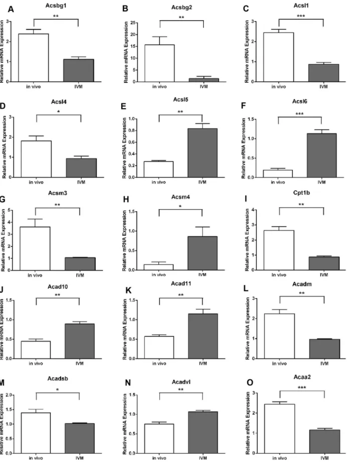
Fatty Acid Oxidation Pathway Genes are Dysregulated in COCs Matured In Vitro
We next assessed whether COCs matured in vitro had altered expression of these dynamically regulated fatty acid oxidation genes. The time point of 10 h maturation was chosen for comparison since the majority of genes were significantly different at this time point in vivo compared to levels at 0 h (Fig. 2). There was no difference in cell number between COCs from the two maturation conditions (in vivo: 30406205.5; IVM: 24576377.3 cells/COC). Analysis of COCs matured for 10 h in vitro showed that Acyl-CoA synthetase expression was significantly altered in IVM COCs compared to in vivo matured COCs with Acsbg1, Acsbg2,Acsl1,Acsl4andAcsm3expression significantly decreased in IVM COCs at 10 h whileAcsl5,Acsl6andAcsm4were significantly higher compared to in vivo matured COCs (Fig. 3 A–H).Acsl3, Acsm2, andAcsm3were not different in COCs matured in vivo for 10 h compared to non-matured COCs (Fig. 2 and data not shown) nor compared to COCs matured in vitro (data not shown). Expression of the isoformCpt1bwas significantly reduced in IVM COCs (Fig. 3I) whileCpt1a,Cpt1candCpt2were unaltered (data not shown). Genes encoding isoforms for acyl-CoA dehydroge-nases which perform the first step in the FAO spiral showed disparate expression patterns with Acad10, Acad11 and Acadvl significantly higher in IVM COCs whileAcadmand Acadsb were significantly reduced compared to in vivo matured COCs (Fig. 3 J–N).Acadl,Acad9 and Acads expression levels were not different between maturation treatment groups (data not shown). The expression of the acetyl-CoA transferaseAcaa2, which catalyzes the final step in the FAO spiral, showed a significant decrease following IVM (Fig. 3O).
We next compared functional rates of FAO in COCs that were matured in vitro for 10 h versus COCs isolated from follicles at 10 h post-hCG (i.e. matured in vivo). There was a significant 2.8-fold reduction in the level of FAO occurring in COCs matured in vitro (IVM) compared to those matured in vivo (Fig. 4A). Thus, IVM conditions result in COCs performing significantly less FAO than in vivo matured COCs at this timepoint.
Effect of PPAR Agonists on COCs during In Vitro Maturation
Because of the dramatically reduced level of FAO in IVM COCs, we next determined whether agonists of PPARs, specifi-cally bezafibrate and rosiglitazone, would increase FAO in COCs during in vitro maturation. Real-time RT-PCR was used to confirm the presence ofPpara,PpargandPpardmRNA transcripts in COCs prior to experiments examining the effects of the PPAR agonists (data not shown). Treatment of COCs with bezafibrate using doses ranging from 50–800mM had no significant effect on FAO rate during the maturation period (Fig. 4B). At 100mM bezafibrate did result in a 1.4-fold increase in FAO compared to control, however this dose also failed to have any significant effect over control conditions in subsequent assays investigating oocyte quality (data not shown). Conversely, treatment of COCs with rosiglitazone during IVM significantly reduced FAO in COCs in a dose dependant manner (Fig. 4C).
Interestingly, despite rosiglitazone treatment (20mM) signifi-cantly inhibiting FAO in COCs during IVM this dose caused a significant increase in the expression of genes involved in fatty acid Figure 5. Rosiglitazone treatment of COCs during in vitro
maturation significantly modulates expression of genes in-volved in fatty acid oxidation.Analysis ofAcsl1(A),Cpt1b(B),Cpt1c
(C),Cpt2(D) andAcaa2(E) expression in cumulus-oocyte complexes
following 10 h of in vitro maturation (IVM) in the absence (0) or presence of rosiglitazone (20mM). mRNA expression was normalized to
the geometric mean ofGapdhandHsp90ab1and presented as mean6
SEM (n = 3 experimental replicates, *P,0.05, **P,0.01 by unpaired t test).
activation (Acsl1), carnitine mediated transport (Cpt1b and Cpt2) and the FAO spiral (Acaa2) compared to untreated controls (Fig. 5). In contrast, expression ofCpt1cwas significantly decreased in the presence of rosiglitazone (Fig. 5C). The remaining 19 genes measured during in vivo maturation described above (or see Fig. 1) were also assessed but did not show significant modulation by rosiglitazone. Rosiglitazone treatment (20mM) also significantly increased the degree of cumulus expansion compared to control (cumulus expansion index: control: 3.4260.05 vs. rosiglitazone: 3.5860.05, P,0.05, Mann Whitney t test due to non-normal distribution of data, n = 7 experimental replicates, representative of$155 COCs per treatment).
These results show that rosiglitazone significantly inhibits FAO in the COC during IVM despite upregulating a subset of genes involved in the FAO metabolic pathway.
Effect of Rosiglitazone on Oocyte Quality
Oocyte mitochondrial membrane potential analyzed by JC-1 staining was altered by rosiglitazone (20mM) during in vitro maturation of COCs (Fig. 6 A–F). Quantification of fluorescence confirmed that rosiglitazone treatment of COCs resulted in oocytes with a higher red:green fluorescence ratio indicating increased mitochondrial activity and that this was due to significantly higher levels of both red and green
fluorescence(Fig. 6 G–I). Further, the distribution of green fluorescence appeared altered in rosiglitazone treated COCs with more of these mitochondria localised in the center of the oocyte (Fig. 6E) compared to a more homogeneous distribution observed in the oocytes matured in control conditions (Fig. 6B).
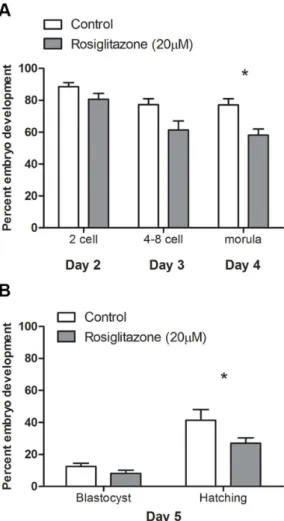
COCs matured in vitro in the presence of rosiglitazone (20mM) did not differ significantly in their ability to be fertilized and develop into a 2-cell embryo compared to COCs matured under control conditions (Fig. 7A). However, rosiglitazone treated COCs resulted in significantly fewer morula embryos on day 4 of embryo culture (Fig. 7A) and significantly fewer hatching blastocyst embryos on day 5 compared to control (Fig. 7B).
Together these data show that despite rosiglitazone treatment of COCs increasing oocyte mitochondrial membrane potential, indicative of mitochondrial hyperpolarization, this was associated with decreased oocyte developmental competence with fewer fertilized oocytes capable of developing to the blastocyst stage of preimplantation development compared to oocytes from COCs matured in control conditions.
Discussion
Little is known about the regulation of FAO in the COC during maturation and how it impacts subsequent embryo development. In the current study we have shown that genes involved in FAO Figure 6. Mitochondrial membrane potential is increased in oocytes following maturation of cumulus oocyte complexes in vitro in the presence of rosiglitazone.Mitochondrial membrane potential was assessed by JC-1 staining of oocytes following in vitro maturation of cumulus oocyte complexes in the absence (A–C) or presence of 20mM rosiglitazone (D–F). Red (G), green (H) and the ratio of red:green (I) fluorescence within oocytes was quantified and presented as mean 6 SEM, representative of 30–35 COCs per group, from 3 independent experiments. Data analyzed by unpairedttest on untransformed (H) or Log10 transformed (GandI) data, *P,0.05; ***P,0.001.
are hormonally regulated in the COC during maturation in vivo and are dysregulated during in vitro maturation. Since mature oocytes are transcriptionally inactive and oocyte mRNA would represent a small fraction of the mRNA in the COC, these dynamic changes in expression levels are attributed to changes in cumulus cells. This supports previous reports demonstrating that
b-oxidation by the oocyte represents a small fraction of that occurring in the whole COC [16,17]. Further, using a functional assay we show that FAO is decreased in COCs matured in vitro compared to those that mature in vivo within the follicular environment. Whether decreased FAO persists throughout mat-uration in vitro remains to be determined, as does whether this reduction is directly due to the identified alterations in gene expression or deficiencies in other factors, such as growth factors and specific metabolites that are lacking in in vitro systems. Lastly, we show that treatment with the PPARc agonist, rosiglitazone,
significantly inhibits FAO and results in poor developmental competence.
The significant upregulation of acyl-CoA synthetases Acsbg2, Acsl1, Acsl4, and Acsl5 in the COC during maturation in vivo indicate that the COC is preferentially utilizing fatty acids with chain lengths of C10-C20 [42–45]. This correlates well with the predominant forms of fatty acids present in human and bovine follicular fluid; palmitic, oleic, linoleic and arachidonic acid [46– 49] for which Acsl1, Acsl5 and Acsl4 have high affinity [43–45]. Significant 5-fold induction of Cpt1a during maturation in vivo indicates that there is increased capacity for carnitine-mediated transport of fatty acids into mitochondria which is required for the oxidation of long chain fatty acids. We have previously shown significant upregulation of the isoform Cpt1b during COC maturation in vivo [17] here we again saw a 2-fold increase in Cpt1bat 10 h post-hCG though this was not statistically significant (data not shown). However, the Cpt isoforms, Cpt1a, b and c do not differ in their specificity for different activated fatty acids only in their tissue localization. Thus both our previous and current study illustrate increased Cpt1 activity indicating increased capacity for carnitine-mediated transport of activated long chain fatty acids into mitochondria during maturation in vivo. Acyl-CoA dehydrogenases catalyze the first step in FAO and those encoded byAcadl,AcadmandAcadsbhave substrate optima of 16, 8, and 4 carbon chains, respectively [50,51]. Expression of these isoforms indicate that the COC is capable of metabolizing fatty acids of 4– 16 carbons long and increases its capacity to perform FAO during in vivo maturation.Acaa2codes for acetyl-CoA transferase, which catalyzes the fourth and final step in FAO, the formation of acetyl-CoA which is able to enter the TCA cycle for ATP production. Acaa2 was significantly induced further indicating an increased capacity for the COC to perform FAO for energy production during maturation in vivo.
The significant dysregulation of 15 genes in the FAO pathway in COCs matured in vitro compared to their in vivo matured counterparts (Fig. 3) may indicate a switch in preference of fatty acids during the two different maturation conditions. This may be due to the presence of different fatty acids during IVM which are supplied via bovine serum compared to those present in the follicular fluid in which the COC matures in vivo. It would be interesting to examine whether supplementation of IVM media with fatty acids found in follicular fluid normalizes the expression level of some of the genes examined here resulting in improved oocyte developmental competence.
Acsl4 has a particular preference for arachidonic acid, a precursor for prostaglandin synthesis, and is involved in prosta-glandin synthesis and release [52] with strong expression noted in steroidogenic tissues including the ovary [44]. Thus the significant down regulation ofAcsl4in IVM COCs observed in the current study may be partly responsible for the poor PGE2 secretion observed in these COCs [53] and the poorer developmental competence of these oocytes. In support of this female Acsl4 heterozygous null mice have reduced fertility [54] as do Ptgs2 deficient mice [55].
ThatAcaa2, which is responsible for the final step in FAO in the mouse, was significantly reduced in COCs matured in vitro indicates that FAO was deficient compared to COCs matured in vivo and this was confirmed using a functional assay. Previous studies have shown that IVM COCs have altered mitochondrial and glycolytic activity and poor oocyte developmental competence [3,4]. The current study demonstrates that the down regulation of FAO could be responsible for the subsequent developmental deficiencies that occur in response to IVM.
Figure 7. Rosiglitazone treatment during in vitro maturation of cumulus-oocyte complexes negatively affects oocyte develop-mental competence. Oocyte developmental competence was assessed following in vitro fertilization of cumulus-oocyte complexes matured in vitro in control conditions or in the presence of rosiglitazone (20mM). Embryo development was assessed on days, 2, 3, 4 and 5, with
The up-regulation of FAO by PPAR agonists has been reported in many different tissues and cells, however very little attention has been focused on their action within the ovary, especially the COC. Both PPARaand PPARcmRNA were confirmed to be expressed in the COCs used in these experiments (data not shown) however it is not possible to conclude whether this includes expression specifically in the oocyte. Bezafibrate is a potent PPARa agonist shown to increase FAO in other cell lines and tissues [35,36]. However, in the current study bezafibrate had no effect on FAO in the COC during in vitro maturation or on subsequent embryo development following IVF. Rosiglitazone has been shown to differentially modulate FAO in different cell types [56–58]. In the current study we show that rosiglitazone treatment of COCs during maturation in vitro dose dependently inhibited FAO despite upregulating genes involved in fatty acid activation, carnitine-mediated transport and metabolim. As a ligand for PPARc it is possible that rosiglitazone treatment also blocks distinct effects of its endogenous fatty acid ligands. Alternatively, since rosiglitazone is known to promote fat storage in some cell types while down regulating FAO [56], rosiglitazone may activate lipid storage rather than metabolism in the COC. The observation of increased cumulus expansion in the presence of rosiglitazone suggests that cumulus matrix production is increased, perhaps due to increased carbohydrate metabolism in the face of impairedb -oxidation. Indeed, elevated flux of glucose through the hexosa-mine biosynthesis pathway has been shown to increase extracel-lular matrix substrate supply and hence COC expansion [59]. Overall, these results indicate that compared to PPARa, PPARcis a more active regulator of COC functions during oocyte maturation.
We have previously demonstrated that in vivo administration of rosiglitazone in obese mice significantly improved oocyte devel-opmental competence [33]. What was unclear from this study however is whether the effects of rosiglitazone on oocyte developmental competence were due to direct effects on the ovarian cells or via systemic effects such as reduced circulating insulin and lipid. The results from the current study demonstrate rosiglitazone action directly on the COC inhibited FAO and impaired subsequent embryo development supporting the conclu-sion that in vivo administration of rosiglitazone has indirect beneficial effects on the oocyte via its actions on the maternal systemic environment. These contradictory effects of rosiglitazone
on oocyte developmental competence might also be due to differences in the availability of lipid substrate in the treated COCs. We have previously shown that mice fed high fat diet exhibit a marked increase in both oocyte and cumulus cell lipid content compared to mice fed a low fat diet [60] and perhaps in this cellular context rosiglitazone improves energy production and embryo development; but has a negative effect when lipid levels are limited.
Despite the negative effect of rosiglitazone on oocyte develop-mental competence we found an increase in the inner mitochon-drial membrane potential (MMP) of oocytes when COCs were matured in the presence of rosiglitazone. An increase in MMP in oocytes is usually associated with improved oocyte developmental competence [61–63], although more recent reports have associ-ated increased hyper-polarized mitochondria with decreased oocyte developmental competence and increased embryo frag-mentation [64,65]. Perhaps there is a threshold for a positive association between MMP and oocyte developmental competence and raising the MMP too high is detrimental due to excessive levels of reactive oxygen species (ROS). How rosiglitazone raises oocyte MMP despite significantly down-regulating FAO in the whole COC is unclear but it is likely that rosiglitazone exerts cell specific effects on cumulus cells and the oocyte, perhaps due to differential receptor expression levels between these two cell types and/or the presence of distinct co-activators.
From this and previous studies [17–20] it is emerging that FAO is an important metabolic pathway in determining oocyte developmental potential in vitro. We show here that FAO is induced during oocyte maturation in vivo and is deficient during IVM. The PPAR agonists bezafibrate and rosiglitazone were not effective in restoring FAO in vitro. In conclusion, further optimization of in vitro maturation conditions to normalize levels of FAO to that observed in vivo may be necessary to improve the success of IVM.
Author Contributions
Conceived and designed the experiments: KRD RLR. Performed the experiments: KRD MRA VJZ. Analyzed the data: KRD MRA VJZ DLR RLR. Contributed reagents/materials/analysis tools: KRD DLR RLR. Wrote the paper: KRD MRA VJZ DLR RLR.
References
1. Edwards RG (1965) Maturation in vitro of human ovarian oocytes. Lancet 2: 926–929.
2. Gilchrist RB (2011) Recent insights into oocyte-follicle cell interactions provide opportunities for the development of new approaches to in vitro maturation. Reproduction, fertility, and development 23: 23–31.
3. Krisher RL (2004) The effect of oocyte quality on development. Journal of animal science 82 Suppl: E14–23.
4. Martino NA, Lacalandra GM, Filioli Uranio M, Ambruosi B, Caira M, et al. (2012) Oocyte mitochondrial bioenergy potential and oxidative stress: within-/ between-subject, in vivo versus in vitro maturation, and age-related variations in a sheep model. Fertility and sterility 97: 720–728 e721.
5. Dunning KR, Lane M, Brown HM, Yeo C, Robker RL, et al. (2007) Altered composition of the cumulus-oocyte complex matrix during in vitro maturation of oocytes. Hum Reprod 22: 2842–2850.
6. Buckett WM, Chian RC, Dean NL, Sylvestre C, Holzer HE, et al. (2008) Pregnancy loss in pregnancies conceived after in vitro oocyte maturation, conventional in vitro fertilization, and intracytoplasmic sperm injection. Fertil Steril 90: 546–550.
7. Child TJ, Phillips SJ, Abdul-Jalil AK, Gulekli B, Tan SL (2002) A comparison of in vitro maturation and in vitro fertilization for women with polycystic ovaries. Obstet Gynecol 100: 665–670.
8. Gilchrist RB, Thompson JG (2007) Oocyte maturation: emerging concepts and technologies to improve developmental potential in vitro. Theriogenology 67: 6– 15.
9. Biggers JD, Whittingham DG, Donahue RP (1967) The pattern of energy metabolism in the mouse oocyte and zygote. Proc Natl Acad Sci U S A 58: 560– 567.
10. Thompson JG, Lane M, Gilchrist RB (2007) Metabolism of the bovine cumulus-oocyte complex and influence on subsequent developmental competence. Soc Reprod Fertil Suppl 64: 179–190.
11. Downs SM (1995) The influence of glucose, cumulus cells, and metabolic coupling on ATP levels and meiotic control in the isolated mouse oocyte. Dev Biol 167: 502–512.
12. Preis KA, Seidel GEJ, Gardner DK (2007) Reduced oxygen concentration improves the developmental competence of mouse oocytes following in vitro maturation. Mol Reprod Dev 74: 893–903.
13. Sugiura K, Eppig JJ (2005) Society for Reproductive Biology Founders’ Lecture 2005. Control of metabolic cooperativity between oocytes and their companion granulosa cells by mouse oocytes. Reprod Fertil Dev 17: 667–674.
14. Van Blerkom J, Davis PW, Lee J (1995) ATP content of human oocytes and developmental potential and outcome after in-vitro fertilization and embryo transfer. Hum Reprod 10: 415–424.
15. Downs SM, Mosey JL, Klinger J (2009) Fatty acid oxidation and meiotic resumption in mouse oocytes. Mol Reprod Dev 76: 844–853.
16. Valsangkar D, Downs SM (2013) A requirement for fatty acid oxidation in the hormone-induced meiotic maturation of mouse oocytes. Biol Reprod 89: 43. 17. Dunning KR, Cashman K, Russell DL, Thompson JG, Norman RJ, et al. (2010)
18. Dunning KR, Akison LK, Russell DL, Norman RJ, Robker RL (2011) Increased beta-oxidation and improved oocyte developmental competence in response to l-carnitine during ovarian in vitro follicle development in mice. Biol Reprod 85: 548–555.
19. McKeegan PJ, Sturmey RG (2011) The role of fatty acids in oocyte and early embryo development. Reprod Fertil Dev 24: 59–67.
20. Hashimoto S (2009) Application of in vitro maturation to assisted reproductive technology. J Reprod Dev 55: 1–10.
21. Somfai T, Kaneda M, Akagi S, Watanabe S, Haraguchi S, et al. (2011) Enhancement of lipid metabolism with L-carnitine during in vitro maturation improves nuclear maturation and cleavage ability of follicular porcine oocytes. Reprod Fertil Dev 23: 912–920.
22. Takahashi T, Inaba Y, Somfai T, Kaneda M, Geshi M, et al. (2013) Supplementation of culture medium with L-carnitine improves development and cryotolerance of bovine embryos produced in vitro. Reprod Fertil Dev 25: 589–599.
23. Wu GQ, Jia BY, Li JJ, Fu XW, Zhou GB, et al. (2011) L-carnitine enhances oocyte maturation and development of parthenogenetic embryos in pigs. Theriogenology 76: 785–793.
24. You J, Lee J, Hyun SH, Lee E (2012) L-carnitine treatment during oocyte maturation improves in vitro development of cloned pig embryos by influencing intracellular glutathione synthesis and embryonic gene expression. Therioge-nology 78: 235–243.
25. Ferguson EM, Leese HJ (2006) A potential role for triglyceride as an energy source during bovine oocyte maturation and early embryo development. Mol Reprod Dev 73: 1195–1201.
26. Cabrero A, Cubero M, Llaverias G, Jove M, Planavila A, et al. (2003) Differential effects of peroxisome proliferator-activated receptor activators on the mRNA levels of genes involved in lipid metabolism in primary human monocyte-derived macrophages. Metabolism: clinical and experimental 52: 652–657.
27. Sprecher DL, Massien C, Pearce G, Billin AN, Perlstein I, et al. (2007) Triglyceride: high-density lipoprotein cholesterol effects in healthy subjects administered a peroxisome proliferator activated receptor delta agonist. Arteriosclerosis, thrombosis, and vascular biology 27: 359–365.
28. Froment P, Gizard F, Defever D, Staels B, Dupont J, et al. (2006) Peroxisome proliferator-activated receptors in reproductive tissues: from gametogenesis to parturition. The Journal of endocrinology 189: 199–209.
29. Issemann I, Green S (1990) Activation of a member of the steroid hormone receptor superfamily by peroxisome proliferators. Nature 347: 645–650. 30. Dreyer C, Krey G, Keller H, Givel F, Helftenbein G, et al. (1992) Control of the
peroxisomal beta-oxidation pathway by a novel family of nuclear hormone receptors. Cell 68: 879–887.
31. Vamecq J, Latruffe N (1999) Medical significance of peroxisome proliferator-activated receptors. Lancet 354: 141–148.
32. Kliewer SA, Umesono K, Noonan DJ, Heyman RA, Evans RM (1992) Convergence of 9-cis retinoic acid and peroxisome proliferator signalling pathways through heterodimer formation of their receptors. Nature 358: 771– 774.
33. Minge CE, Bennett BD, Norman RJ, Robker RL (2008) Peroxisome proliferator-activated receptor-gamma agonist rosiglitazone reverses the adverse effects of diet-induced obesity on oocyte quality. Endocrinology 149: 2646–2656. 34. Pfaffl MW, Tichopad A, Prgomet C, Neuvians TP (2004) Determination of stable housekeeping genes, differentially regulated target genes and sample integrity: BestKeeper – Excel-based tool using pair-wise correlations. Biotechnol Lett 26: 509–515.
35. Djouadi F, Bonnefont JP, Thuillier L, Droin V, Khadom N, et al. (2003) Correction of fatty acid oxidation in carnitine palmitoyl transferase 2-deficient cultured skin fibroblasts by bezafibrate. Pediatric research 54: 446–451. 36. Cabrero A, Alegret M, Sanchez RM, Adzet T, Laguna JC, et al. (2001)
Bezafibrate reduces mRNA levels of adipocyte markers and increases fatty acid oxidation in primary culture of adipocytes. Diabetes 50: 1883–1890. 37. Djouadi F, Aubey F, Schlemmer D, Bastin J (2005) Peroxisome proliferator
activated receptor delta (PPARdelta) agonist but not PPARalpha corrects carnitine palmitoyl transferase 2 deficiency in human muscle cells. The Journal of clinical endocrinology and metabolism 90: 1791–1797.
38. Vandewalle B, Moerman E, Lefebvre B, Defrance F, Gmyr V, et al. (2008) PPARgamma-dependent and -independent effects of rosiglitazone on lipotoxic human pancreatic islets. Biochemical and biophysical research communications 366: 1096–1101.
39. Wang P, Renes J, Bouwman F, Bunschoten A, Mariman E, et al. (2007) Absence of an adipogenic effect of rosiglitazone on mature 3T3-L1 adipocytes: increase of lipid catabolism and reduction of adipokine expression. Diabetologia 50: 654– 665.
40. Djaouti L, Jourdan T, Demizieux L, Chevrot M, Gresti J, et al. (2010) Different effects of pioglitazone and rosiglitazone on lipid metabolism in mouse cultured liver explants. Diabetes/metabolism research and reviews 26: 297–305. 41. Vanderhyden BC, Caron PJ, Buccione R, Eppig JJ (1990) Developmental
pattern of the secretion of cumulus expansion-enabling factor by mouse oocytes
and the role of oocytes in promoting granulosa cell differentiation. Dev Biol 140: 307–317.
42. Pei Z, Jia Z, Watkins PA (2006) The second member of the human and murine bubblegum family is a testis- and brainstem-specific acyl-CoA synthetase. J Biol Chem 281: 6632–6641.
43. Iijima H, Fujino T, Minekura H, Suzuki H, Kang MJ, et al. (1996) Biochemical studies of two rat acyl-CoA synthetases, ACS1 and ACS2. Eur J Biochem 242: 186–190.
44. Kang MJ, Fujino T, Sasano H, Minekura H, Yabuki N, et al. (1997) A novel arachidonate-preferring acyl-CoA synthetase is present in steroidogenic cells of the rat adrenal, ovary, and testis. Proc Natl Acad Sci U S A 94: 2880–2884. 45. Oikawa E, Iijima H, Suzuki T, Sasano H, Sato H, et al. (1998) A novel acyl-CoA
synthetase, ACS5, expressed in intestinal epithelial cells and proliferating preadipocytes. J Biochem 124: 679–685.
46. Homa ST, Brown CA (1992) Changes in linoleic acid during follicular development and inhibition of spontaneous breakdown of germinal vesicles in cumulus-free bovine oocytes. J Reprod Fertil 94: 153–160.
47. Jungheim ES, Macones GA, Odem RR, Patterson BW, Lanzendorf SE, et al. (2011) Associations between free fatty acids, cumulus oocyte complex morphology and ovarian function during in vitro fertilization. Fertil Steril 95: 1970–1974.
48. Tsujii H, Khandoker MAMY, Hamano K (2001) Lipid in mammalian embryo development. J Mamm Ova Res 18: 73–80.
49. O’Gorman A, Wallace M, Cottell E, Gibney MJ, McAuliffe FM, et al. (2013) Metabolic profiling of human follicular fluid identifies potential biomarkers of oocyte developmental competence. Reproduction.
50. Finocchiaro G, Ito M, Ikeda Y, Tanaka K (1988) Molecular cloning and nucleotide sequence of cDNAs encoding the alpha-subunit of human electron transfer flavoprotein. J Biol Chem 263: 15773–15780.
51. He M, Burghardt TP, Vockley J (2003) A novel approach to the characterization of substrate specificity in short/branched chain Acyl-CoA dehydrogenase. J Biol Chem 278: 37974–37986.
52. Golej DL, Askari B, Kramer F, Barnhart S, Vivekanandan-Giri A, et al. (2011) Long-chain acyl-CoA synthetase 4 modulates prostaglandin E(2) release from human arterial smooth muscle cells. J Lipid Res 52: 782–793.
53. Dunning KR, Watson LN, Sharkey DJ, Brown HM, Norman RJ, et al. (2012) Molecular filtration properties of the mouse expanded cumulus matrix: controlled supply of metabolites and extracellular signals to cumulus cells and the oocyte. Biol Reprod 87: 89.
54. Cho YY, Kang MJ, Sone H, Suzuki T, Abe M, et al. (2001) Abnormal uterus with polycysts, accumulation of uterine prostaglandins, and reduced fertility in mice heterozygous for acyl-CoA synthetase 4 deficiency. Biochem Biophys Res Commun 284: 993–997.
55. Lim H, Paria BC, Das SK, Dinchuk JE, Langenbach R, et al. (1997) Multiple female reproductive failures in cyclooxygenase 2-deficient mice. Cell 91: 197– 208.
56. Roberts LD, Murray AJ, Menassa D, Ashmore T, Nicholls AW, et al. (2011) The contrasting roles of PPARdelta and PPARgamma in regulating the metabolic switch between oxidation and storage of fats in white adipose tissue. Genome Biol 12: R75.
57. Wilmsen HM, Ciaraldi TP, Carter L, Reehman N, Mudaliar SR, et al. (2003) Thiazolidinediones upregulate impaired fatty acid uptake in skeletal muscle of type 2 diabetic subjects. Am J Physiol Endocrinol Metab 285: E354–362. 58. Benton CR, Holloway GP, Campbell SE, Yoshida Y, Tandon NN, et al. (2008)
Rosiglitazone increases fatty acid oxidation and fatty acid translocase (FAT/ CD36) but not carnitine palmitoyltransferase I in rat muscle mitochondria. J Physiol 586: 1755–1766.
59. Sutton-McDowall ML, Gilchrist RB, Thompson JG (2004) Cumulus expansion and glucose utilisation by bovine cumulus-oocyte complexes during in vitro maturation: the influence of glucosamine and follicle-stimulating hormone. Reproduction 128: 313–319.
60. Wu LL, Dunning KR, Yang X, Russell DL, Lane M, et al. (2010) High-fat diet causes lipotoxicity responses in cumulus-oocyte complexes and decreased fertilization rates. Endocrinology 151: 5438–5445.
61. Van Blerkom J, Davis P (2006) High-polarized (Delta Psi m(HIGH)) mitochondria are spatially polarized in human oocytes and early embryos in stable subplasmalemmal domains: developmental significance and the concept of vanguard mitochondria. Reprod Biomed Online 13: 246–254.
62. Van Blerkom J, Davis P, Alexander S (2003) Inner mitochondrial membrane potential (DeltaPsim), cytoplasmic ATP content and free Ca2+ levels in metaphase II mouse oocytes. Hum Reprod 18: 2429–2440.
63. Van Blerkom J, Davis P, Mathwig V, Alexander S (2002) Domains of high-polarized and low-high-polarized mitochondria may occur in mouse and human oocytes and early embryos. Hum Reprod 17: 393–406.
64. Acton BM, Jurisicova A, Jurisica I, Casper RF (2004) Alterations in mitochondrial membrane potential during preimplantation stages of mouse and human embryo development. Mol Hum Reprod 10: 23–32.