Lack of Cul4b, an E3 Ubiquitin Ligase Component, Leads
to Embryonic Lethality and Abnormal Placental
Development
Baichun Jiang., Wei Zhao., Jupeng Yuan, Yanyan Qian, Wenjie Sun, Yongxin Zou, Chenhong Guo,
Bingxi Chen, Changshun Shao*, Yaoqin Gong*
Key Laboratory of Experimental Teratology, Ministry of Education and Institute of Molecular Medicine and Genetics, Shandong University School of Medicine, Jinan, Shandong, China
Abstract
Cullin-RING ligases (CRLs) complexes participate in the regulation of diverse cellular processes, including cell cycle progression, transcription, signal transduction and development. Serving as the scaffold protein, cullins are crucial for the assembly of ligase complexes, which recognize and target various substrates for proteosomal degradation. Mutations in humanCUL4B, one of the eight members in cullin family, are one of the major causes of X-linked mental retardation. We here report the generation and characterization ofCul4bknockout mice, in which exons 3 to 5 were deleted. In contrast to the survival to adulthood of human hemizygous males withCUL4Bnull mutation,Cul4bnull mouse embryos show severe developmental arrest and usually die before embryonic day 9.5 (E9.5). Accumulation of cyclin E, a CRL (CUL4B) substrate, was observed in Cul4b null embryos. Cul4b heterozygotes were recovered at a reduced ratio and exhibited a severe developmental delay. The placentas inCul4bheterozygotes were disorganized and were impaired in vascularization, which may contribute to the developmental delay. As in humanCUL4Bheterozygotes,Cul4bnull cells were selected against in Cul4bheterozygotes, leading to various degrees of skewed X-inactivation in different tissues. Together, our results showed that CUL4B is indispensable for embryonic development in the mouse.
Citation:Jiang B, Zhao W, Yuan J, Qian Y, Sun W, et al. (2012) Lack of Cul4b, an E3 Ubiquitin Ligase Component, Leads to Embryonic Lethality and Abnormal Placental Development. PLoS ONE 7(5): e37070. doi:10.1371/journal.pone.0037070
Editor:Anton Wutz, Wellcome Trust Centre for Stem Cell Research, United Kingdom
ReceivedAugust 19, 2011;AcceptedApril 16, 2012;PublishedMay 14, 2012
Copyright:ß2012 Jiang et al. This is an open-access article distributed under the terms of the Creative Commons Attribution License, which permits unrestricted use, distribution, and reproduction in any medium, provided the original author and source are credited.
Funding:This work was supported by National Basic Research Program of China [2011CB966200] and National Natural Science Foundation of China [30830065, 30900804 and 81101522]. The funders had no role in study design, data collection and analysis, decision to publish, or preparation of the manuscript.
Competing Interests:The authors have declared that no competing interests exist.
* E-mail: [email protected] (YG); [email protected] (CS)
.These authors contributed equally to this work.
Introduction
Cullin-RING ligases (CRLs) complexes comprise the largest known class of ubiquitin ligases [1]. CRLs regulate diverse cellular processes, including cell cycle progression, transcription, signal transduction and development [2]. CRLs are multisubunit complexes composed of a cullin, RING protein and substrate-recognition subunit, which was linked by an adaptor. Human cullin family consists of eight members, CUL1, CUL2, CUL3, CUL4A, CUL4B, CUL5, CUL7 and PARC [3], among them, CUL4A and CUL4B have the highest degree of homology, with 83% identity in protein sequences [4]. There is only one ortholog, Cul4, in lower organisms. CUL4A CRL complexes contained Rbx1 and the adaptor protein DDB1. DDB1 interact with substrate recognition subunits, which determine the substrate specificity of the CUL4A CRL complexes [5,6,7,8,9]. The substrates of CUL4A CRL complexes include CDT1, p21, p27, p53, c-Jun, HOXA9, H3 and CHK1 that play important roles in cell cycle regulation, chromosome remodeling, and differentiation [10,11].
Compared to CUL4A, CUL4B is less studied, and so far very few substrates of CUL4B CRL complexes have been identified [4,12,13,14,15]. However, mutations in humanCUL4Bappear to
be a common cause of X-linked mental retardation (XLMR). To date, at least 12 families of XLMR have been reported to be
attributable to base substitutions or deletions in CUL4B
[16,17,18,19,20]. In addition to mental retardation, those patients also manifest short stature, abnormal gait, impaired speech and other abnormalities. These findings suggest that CUL4B and CUL4A do not necessarily play redundant roles during neurogen-esis and other developmental processes.
defects in embryonic fibroblasts and hepatocytes, and an increase in genome instability [25].
In this study, we generatedCul4bfloxed mice and crossed it to
EIIa-Cretransgenic mice to produceCul4bnull mice. We observed that Cul4b null mice are embryonic lethal. Cul4b null embryos displayed decreased proliferation and increased apoptosis.Cul4b
heterozygotes were also affected, as reflected by their recovery at a reduced ratio at birth and by their developmental delay. Cells expressing Cul4b null allele in Cul4b heterozygous mice were selected against, to different degrees in different tissues, from early embryogenesis to early postnatal development. The embryonic lethality ofCul4bnull mice, when compared to the lack of gross abnormalities inCul4anull mice, indicated thatCul4bhas diverged fromCul4ato carry out some essential and unique functions during embryogenesis.
Results
Generation ofCul4bfloxed mice
Because CUL4B-deficient cells are strongly selected against [17], we envisaged that it might be difficult to generate Cul4b-deficient embryonic stem (ES) cells via conventional knockout technology. We therefore used the Cre/loxP strategy to generate
Cul4b floxed ES cells. First, a Cul4b floxed targeting vector was constructed (Fig. 1A). In this vector, exons 3–5 were floxed by two loxP sites. Aneogene flanked by two FRT sites and aTKgene were also insert into intron 5 and vector backbone, respectively, for ES cells selection.
The targeting vector was linearized and electroporated into 129 male ES cells (RW.4) for homologous recombination. Ninety six clones were screened by long-range PCR (data not shown) and Southern blot (Fig. 1B). Two correctly targeted clones, 2G and 4E, were identified. Targeted ES cells were injected into C57BL/6J blastocysts to produce chimaeric mice, which were then used for germline transmission to produceCul4bfloxed mice.
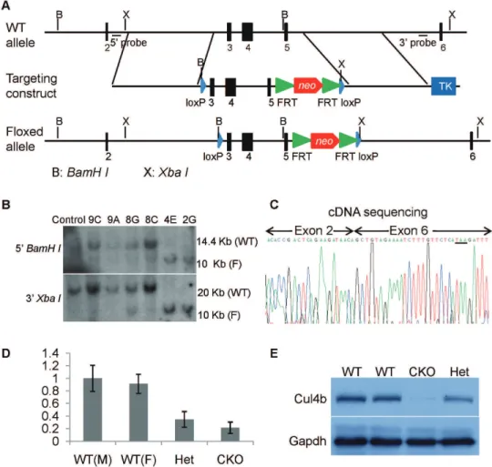
Figure 1. Generation ofCul4bflox mice.(A) Strategy for generation ofCul4bfloxed targeting vector. On the top was shown the wild-type allele ofCul4bgene. The targeting vector and targeted allele were shown in the middle and at the bottom, respectively.BamHI(labeled B) andXbaI (labeled X) sites are indicated. Black bars indicating the positions of the probes used in Southern blots are also indicated. (B) Southern blot analysis of genomic DNA isolated from wild-type ES cells and selected ES cell clones.BamHIdigested DNA was hybridized with 59probe andXbaIdigested DNA was hybridized with 39probe. WT, wild-type allele; F, flox allele. (C) cDNA sequencing ofCul4bgene from the brain tissue of brain-specific knockout mice (Cul4bflox/Y;Nesin-Cre). As predicted, exon 2 was spliced onto exon 6 after the excision of exons 3–5. (D) Real-time RT-PCR analysis ofCul4bmRNA isolated from brain tissues of wild-type males (Cul4b+/Y;Nestin-Cre+/2), wild-type females (Cul4b+/+;Nestin-Cre+/2), heterozygous females (Cul4b+/ flox
;Nestin-Cre+/2), and conditional knock-out males (Cul4bflox/Y
;Nestin-Cre+/2). (E) Western blot analysis of Cul4b protein isolated from brain tissues of wild-type, heterozygous and conditional knockout mice using an anti-Cul4b antibody. Gapdh was used as a loading control.
doi:10.1371/journal.pone.0037070.g001
Neither male hemizygous (Cul4bflox/Y) nor female heterozy-gous/homozygous (Cul4b+/flox
/Cul4bflox/flox) for the Cul4b floxed allele showed any apparent phenotype, suggesting that the flox allele did not disturb the normal function ofCul4bgene. To verify thatCul4bfloxcan be rendered nonfunctional by the expression of Cre, due to the removal of exons 3–5, we generated brain-specific knockout mice Cul4bflox/Y;Nestin-Cre mice and sequenced the cDNA ofCul4bprepared from the brain. We observed that exon 2 was indeed spliced onto exon 6, as a result of removal of exons 3–5 (Fig. 1C). The deletion would also result in a frameshift, generating 8 missense condons followed by a stop codon (Fig. 1C, underlined). The mutant allele is thus predicted to generate a peptide of merely 28 amino acids, if it can be expressed. Thus, deletion of exons 3–5 created a null mutation. Furthermore, the
Cul4b mRNA level in brains of the conditional knockout mice is much lower than that of littermate wild-type control (Fig. 1D), suggesting that the truncation mutation also resulted in
nonsense-mediated decay (NMD), as observed in patients with CUL4B
nonsense mutation [17]. Western blot analysis showed that Cul4b protein was nearly absent in brain tissues of conditional knockout mice (Fig. 1E). Together, these results indicate that deletion of exons 3–5 inCul4bresults in a null mutation.
Cul4bnull mice are not viable
To investigate the function ofCul4bduring mouse development,
Cul4b floxed mice were crossed to EIIa-Cre transgenic mice to generate constitutive Cul4b null mice. The Cre transgene in the
EIIa-Cremice is driven by the adenovirus EIIa promoter and is expressed in a wide range of tissues, including germ cells. Because
EIIa-Creis not expressed in all cells (mosaic expression),Cul4b+/flox
females were first crossed to EIIa-Cre+/+
males to obtain mosaic females that are Cul4b+/flox/Cul4b+/null;EIIa-Cre+/2. Mosaic fe-males were then crossed to wild-type fe-males to generate progeny of
Cul4b knockout male mice (Cul4bnull/Y), Cul4b heterozygous females (Cul4b+/null
) and wild-type mice (Cul4b+/+
andCul4b+ /Y). While Cul4b+/null
females were recovered, no Cul4bnull/Y mice were present in the weaned pups (Table 1), suggesting that
Cul4bnull/Y conceptuses can not develop to term or die before weaning.
Cul4bnull embryos showed early developmental arrest To determine the exact time point when Cul4bnull/Y con-ceptuses die, timed mating was performed and embryos at different developmental stages were dissected. DNA was extracted from yolk sac or total embryo and used for PCR genotyping.
Genotyping of E9.5, 10.5, 12.5 and 14.5 embryos showed that no
Cul4bnull/Y conceptuses were recovered on 9.5 day post coitum (dpc) and beyond. Instead, a proportion of embryos, ,25%,
appeared to have been absorbed and only placentas or empty deciduas remained (Fig. 2A), suggesting that these embryos implanted but died by E9.5.Cul4bnull embryos were recovered at a ratio expected of Mendelian inheritance at E7.5 and 8.5, and no empty deciduas were found at these developmental stages. However, theCul4b null embryos were much smaller (Fig. 2B). The recovery of Cul4b null embryos at the expected Mendelian ratio indicated that the mosaic females are functionallyCul4b+/null
in their capacity to transmitCul4bnullallele.
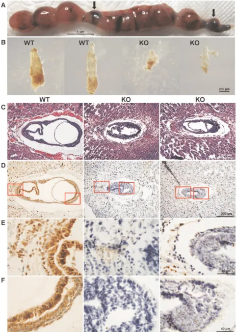
To gain further insight into the anomalies in the Cul4b null embryos, we performed H&E staining of paraffin-embedded sections of E7.5 embryos. While in wild-type embryos, ectoderm, mesoderm and endoderm appeared to have properly formed, the embryonic part of the cylinder was not expanded and boundaries between germ layers were indiscernible in theCul4bnull embryos (Fig. 2C), which were identified by their negative staining by anti-Cul4b antibody (see below). In addition, extraembryonic com-partment in Cul4b null embryos was also underdeveloped and poorly organized (Fig. 2C). Thus, in general, the Cul4b null embryos are developmentally retarded when compared to stage-matched wild-type embryos.
Immunohistochemistry, using an anti-Cul4b antibody, showed that Cul4b was ubiquitously expressed in wild-type E7.5 embryos (Fig. 2D–2F), including ectoplacental cone, trophoblast giant cells, chorion (Fig. 2E), and the embryonic region (Fig. 2F). Expression of Cul4b in extraembryonic regions suggests that Cul4b may be involved in placental development. As expected, the Cul4b null embryos were devoid of staining by anti-Cul4b.
Because a high degree of homology exists betweenCul4a and
Cul4b, we also examined the expression of Cul4a in 7.5 dpc embryos. Positive staining of Cul4a was detected throughout the whole embryos (Figure S1A). Furthermore, the expression of Cul4a was not found to differ betweenCul4b null and wild-type embryos, suggesting that deletion ofCul4bgene did not affect the expression ofCul4a gene. Apparently,Cul4afailed to compensate for the lack ofCul4bin early embryonic development.
Decreased proliferation and increased apoptosis inCul4b null embryos
To investigate what causes the developmental delay of theCul4b
null embryos, we analyzed the proliferative activity and apoptosis in E7.5 embryos. The proliferation was evaluated by
immunohis-Table 1.Distribution ofCul4bgenotypes in progeny ofCul4b+/flox/Cul4b+/null;EIIa-Cre+/2females.
Group Wild-Type Heterozygous Knockout Absorbed
Cul4bgenotype Cul4b+/+
orCul4b+/Y Cul4b+/null Cul4bnull/Y NDa
Expected Mendelian % 50% 25% 25%
-3 weeks (%) (n = 1-35) 107 (79%) 28 (21%) 0 (0%)
-14.5 dpc (%) (n = 61) 31 (51%) 11 (18%) 0 (0%) 19 (31%)
12.5 dpc (%) (n = 39) 17 (44%) 9 (23%) 0 (0%) 13 (33%)
10.5 dpc (%) (n = 38) 19 (50%) 12 (32%) 0 (0%) 7 (18%)
9.5 dpc (%) (n = 31) 15 (48%) 7 (23%) 0 (0%) 9 (29%)
8.5 dpc (%) (n = 28) 15 (54%) 6 (21%) 7 (25%) (0%)
Litters were dissected at the times shown and genotyped by PCR as described in Materials and Methods. a
tochemical staining of paraffin sections of E7.5 embryos for Ki67, a marker of proliferating cells. While there was a high level of proliferation in the wild-type embryos, proliferative cells were less abundant in Cul4b null embryos (Fig. 3A). BrdU incorporation assay confirmed the decrease in proliferation inCul4bnull embryos (Fig. 3B), suggesting that the proliferative activity was greatly compromised inCul4bnull embryos.
Apoptosis in E7.5 embryos was evaluated by TdT-mediated dUTP nick end labeling (TUNEL) assay. Apoptotic cells were rarely detected in wild-type E7.5 embryos; however, the number of apoptotic cells was remarkably increased in the Cul4b null
embryos (Fig. 3C). Apoptotic cells were also observed to line along the Reichert’s membrane.
It was previously reported that CUL4B can interact with cyclin E and the CUL4B immunocomplexes can polyubiquitinate cyclin E in vitro [26]. We previously showed that silencing of CUL4B
could lead to increased accumulation of cyclin E [4]. To determine whether deficiency of Cul4b would also cause increased accumu-lation of cyclin E in theCul4bnull embryos, the cyclin E level was examined using immunohistochemistry. Indeed, cyclin E was accumulated inCul4bnull embryos compared to littermate wild-type embryos (Fig. 3D).
Figure 2. Morphology and histology ofCul4bnull embryos.(A) Uterus excised from pregnant female at 12.5 dpc. Arrows indicate absorbed embryos. (B) Photomicrographs of E7.5 embryos dissected from surrounding deciduas tissue of the same uterus. The two embryos on the left appear normal in size and morphology and the two on the right were much smaller and were partially deteriorated. The bar represents 200mm. (C) H&E
staining of paraffin sections of wild-type andCul4bnull embryos at 7.5 dpc. Littermate embryos at 7.5 dpc were paraffin embedded and cross sectioned together with their surrounding deciduas. The genotype of each embryo was determined by immunohistochemistry using an anti-Cul4b antibody, as shown below. (D) Immunohistochemistry of paraffin sections of wild-type and Cul4bnull embryos at 7.5 dpc with an anti-Cul4b antibody. (E–F) Photomicrographs with higher magnification of the stained section shown in (D).
doi:10.1371/journal.pone.0037070.g002
The levels of other known substrates of CRL4 complex, p27 and p53, were also examined in wild-type andCul4bnull embryos at 7.5 dpc. They were not found to differ between Cul4b null embryos and wild-type embryos (Figure S1B–S1C), suggesting that Cul4b probably does not play a critical role in the proteosomal degradation of those proteins during embryonic development. Alternatively, Cul4a may have compensated for the lack Cul4b in lowering the levels of those substrates.
Taken together, these results suggested that the developmental delay in Cul4b null embryos is attributable to a reduction in proliferation and an increase in apoptosis.
Reduced recovery and growth retardation ofCul4b heterozygous mice
While noCul4bnull conceptuses could survive to E9.5, theCul4b
heterozygous females were also recovered at a lower than expected ratio at weaning (Table 1). This deficit was probably caused by an increased prenatal lethality inCul4b heterozygotes, because their ratio was reduced even at E14.5. For theCul4bheterozygotes that survive to term, they were significantly smaller than their wild-type littermates. The average body weight of heterozygous newborns (1.1360.17 g) was much smaller than that of wild-type controls (1.7960.12 g). However, Cul4b heterozygous mice were able to gradually catch up after birth, and the body weight difference became narrow in adults (Fig. 4A). Except for growth retardation,
Cul4b heterozygous mice showed no gross abnormalities for the first 18 months.
The fact that Cul4bheterozygous mice were smaller in size at birth but could catch up gradually suggests that developmental delay is primarily due to factor(s) that operate during prenatal development. Therefore, heterozygous embryos at different de-velopment stages were dissected and examined (Fig. 4B–4E).Cul4b
heterozygous embryos were smaller than wild-type controls at all development stages examined, suggesting that growth retardation began at early developmental stage. In addition, the Cul4b
heterozygous embryos appeared pale (Fig. 4D and 4E). Blood vessels on yolk sac were not well developed (Fig. 4D), and the blood flow appeared to be reduced (Fig. 4E). Poor vascularization and shortage in blood supply may have caused undernourishment in the heterozygous embryos.
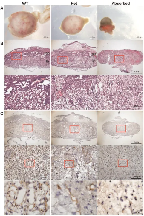
Placental defects ofCul4bheterozygous embryos To further delineate the mechanism underlying the growth retardation ofCul4b heterozygotes during prenatal development, we examined the morphology and histology of placentas at E14.5. Placentas of Cul4b heterozygous embryos appeared smaller and paler than those of wild-type (Fig. 5A). H&E staining of placental sections showed that the labyrinth layer of Cul4b heterozygous placentas was more loosely formed compared to that of wild-type controls, and was deeply invaded by the spongiotrophoblast layer, leaving the demarcation between labyrinth and spongiotropho-blast layers indiscernible (Fig. 5B, upper panels). While the network in the labyrinth layer of placentas in the wild-type was highly compacted and densely branched; the labyrinth layer in
Cul4b heterozygotes was laid out very loosely (Fig. 5B, lower
Figure 3. Decreased proliferation and increased apoptosis inCul4bnull embryos.(A) Paraffin sections of wild-type andCul4bnull embryos at 7.5 dpc were stained with an antibody against Ki67, a proliferation marker. Sections were counterstained with haematoxylin. Lower panels are the higher magnification of the upper panels. (B) Paraffin sections of wild-type andCul4bnull embryos at 7.5 dpc were analyzed by immunostaining against BrdU, and counterstained with DAPI. Lower panels are the higher magnification of the upper panels. (C) Paraffin sections of wild-type and Cul4bnull embryos at 7.5 dpc were analysed by TUNEL assay for labeling apoptotic cells, and counterstained with DAPI. Lower panels are the higher magnification of the upper panels. (D) Paraffin sections of wild-type andCul4bnull embryos at 7.5 dpc were stained with an antibody against cyclin E. Sections were counterstained with haematoxylin. Lower panels are the higher magnification of the upper panels.
panels). The labyrinth and spongiotrophoblast layers were in-distinguishable in the placentas of absorbed embryos.
The vascular network in placenta was further characterized by immunohistochemistry using an antibody to platelet/endothelial cell adhesion molecule-1 (PECAM), which marks endothelial cells in blood vessels. In contrast to normal placentas in which the blood vessels were well formed and uniformly distributed (Fig. 5C, left panels), PECAM staining signals in Cul4b heterozygous placentas only appeared in rather isolated regions (Fig. 5C, middle panels). Furthermore, vascular structures were not readily recognizable within the PECAM-positive islands. Only back-ground staining of PECAM was detected in placentas of absorbed embryos (Fig. 5C, right panels).
Taken together, these results showed that the placentas ofCul4b
heterozygous embryos were generally disorganized and were impaired in vascularization, which may contribute to the growth retardation during prenatal development of Cul4b heterozygous mice.
Pattern of X-chromosome inactivation (XCI) inCul4b heterozygous mice
Because Cul4b is X-linked, Cul4b heterozygous females are functional mosaics in terms of the expression ofCul4b. The somatic cells are eitherCul4bfunctional or null depending on the choice of the X-chromosome that becomes inactivated. In the XLMR family withCUL4Bmutation, X-chromosome inactivation (XCI) was extremely skewed in peripheral blood cells of carriers, due to the selection that favors the cells in which the mutantCUL4Ballele is inactivated [17,20]. We first determined the XCI pattern in 4-month-old adult Cul4b heterozygous females by immunohisto-chemical analysis for Cul4bexpression. If XCI were balanced in
Cul4b heterozygous females, the proportion of cells expressing
Cul4bshould be,50% of that in wild-type females. On the other
hand, if XCI is skewed towardCul4ballele, as in humanCUL4B
heterozygous carriers, the percentage of cells expressing Cul4b
should be closer to that in wild-type females. As shown in Fig. 6A, the percentages of cells expressingCul4bin kidney, liver and lung were identical betweenCul4bheterozygous females and wild-type females, suggesting that XCI is extremely skewed in those organs. XCI in hippocampus was also skewed, but to a lesser extent, since the percentage ofCul4b positive cells in heterozygotes remained lower than that in wild-type. Western blot assay showed a similar trend (Figure S2). While the expression levels of Cul4b in liver and lung inCul4bheterozygous mice were comparable to those in wild type mice, the expression levels in cortex and hippocampus were decreased inCul4bheterozygous mice compared to those in wild type mice.
The skewed XCI towardCul4bnull-bearing chromosome could be caused either by an initiation of XCI that favors theCul4bnull -bearing X or by a selection againstCul4bnull-expressing cells during development. To distinguish between those two possibilities, we next examined the XCI pattern in youngerCul4b heterozygous females. As shown in Fig. 6B–C, the percentages of Cul4b -expressing cells in lung were not significantly different between
Cul4b heterozygous and wild-type females even in the newborn. However, the percentages of Cul4b positive cells in kidney and liver were significantly lower in heterozygotes than in wild-type (Fig. 6B–C). These results suggest that the skewed XCI is caused by a gradual selection against the Cul4b negative cells and that different organs face differential selection pressure during prenatal and postnatal development. WhileCul4b null cells in lung were nearly all eliminated before birth, those cells remained in
Figure 4. Growth retardation ofCul4bheterozygous mice during embryonic development.(A) Bodyweights ofCul4bheterozygous mice and littermate wild-type females after birth. Data were presented as mean6SD. N = 8, *: p,0.05; **: p,0.01; ***: p,0.001. (B–E) Representative photographs ofCul4bheterozygous embryos and littermate wild-type controls at 9.5 (B), 10.5 (C), 12.5 (D) and 14.5 (E) dpc. The bar represents 1 mm in (B–C) and 2 mm in (D) and (E), respectively.
doi:10.1371/journal.pone.0037070.g004
hippocampus four months after birth (Fig. 6A–C, 6E). The less stringent selection against Cul4b null cells in hippocampus is consistent with the clear difference in the Cul4b expression level betweenCul4bheterozygotes and wild-type in the brain (Fig. 1D and 1E, Fig. S2).
To gain insight into the XCI pattern at embryonic stage, we examined the distribution of Cul4b-positive and –negative cells in E7.5 embryos. XCI is usually completed by E6.5 in epiblasts. As shown in Fig. 6F, Cul4b-positive and –negative cells were not evenly distributed in the embryonic regions of the E7.5 heterozygous embryos. While the two types of cells appeared in
an intermingled pattern and in roughly equal proportions in some areas (middle), indicative of the randomness of XCI before selection took place, Cul4b-positive cells were predominant in other areas (right), suggesting that selection for Cul4b-positive cells started before E7.5 wherein. The differential selection in different embryonic regions at early developmental stage may contribute to the distribution pattern of Cul4b-negative cells and the selection dynamics in various tissues at later developmental stages.
Figure 5. Morphology and histology of placentas of wild-type, Cul4b heterozygous and absorbed embryos at 14.5 dpc. (A) Representative photographs of placentas of wild-type,Cul4bheterozygous and absorbed embryos at 14.5 dpc. (B) H&E staining of radial sections of placentas. sp, spongiotrophoblast layer; la, labyrinthine layer. Lower panels are the higher magnification of the upper panels. (C) Immunohistochemisty of radial sections of placentas with an antibody to PECAM, an angiogenesis marker. Middle panels are the higher magnification of the upper panels, and lower panels are the higher magnification of the middle panels.
Discussion
The cullin members are critical for the assembly of CRL complexes that play important roles in multiple cellular processes. Lack of cullin function has indeed been shown to have severe consequences in cellular function and organism development. Disrupting either Cul1 orCul3genes in mice caused embryonic lethality and dysregulation of cyclin E [27,28,29]. Deletion ofCul7
caused neonatal lethality, growth retardation and vascular abnor-mality [30]. In this study, we showed that deletion ofCul4bresulted in embryonic lethality in null hemizygotes. This finding is consistent with the embryonic lethality of Cul4b mutant mice that were generated by gene trap technology [31]. In addition, Cul4b
heterozygotes were recovered in deficit and those that survive to term exhibited a developmental delay. Underscoring the impor-tance of Cul4b function is the observed skewedness of X-inactivation inCul4bheterozygotes as a result of selection againstCul4bnull cells. The severe phenotype ofCul4bnull mice is in a sharp contrast to the lack of obvious phenotype inCul4anull mice, although the two cullin members are closely related. These findings withCul4bmutant mice,
together with the development of XLMR syndrome in humans
carrying CUL4B mutation, suggest that CUL4A cannot fully
compensate for the absence of CUL4B activity in mammals. While mutations in CUL4B cause mental retardation, short stature, impaired speech, and other abnormalities, the patients usually survive to adulthood. Thus, the Cul4b mutant mice are more severely affected. The mechanism underlying this species-specific difference in phenotypic severity caused by CUL4B deficiency remains to be elucidated. It is possible that certain mutations in humans may retain residual function of CUL4B and thus have less deleterious effect than theCul4bnull mutation in the mouse. However, several mutations in human CUL4Bresemble null mutations. The p.R388X mutation, for example, would have truncated the whole CULLIN domain and the rest of the C-terminus [16,17]. Moreover, this mutation caused nonsense mediated decay ofCUL4BmRNA [17]. Two reported deletions inCUL4Bcausing XLMR would have been devoid of most of the CULLIN domain, which is essential for CRL activity [19,20]. It should be noted that the more severe phenotype in mouse mutants
Figure 6. Characterization of X chromosome inactivation by Cul4b expression in heterozygous mice.(A–C) Percentages of cells positive for Cul4b ofCul4bheterozygous mice and littermate wild-type female controls at 4 months (A), 3 weeks (B) and newborn (C). More than 2,000 cells of each tissue were scored. Hi, hippocampus; Ki, kidney; Li, liver; Lu, lung. Data were presented as mean6SD. *: p,0.05; **: p,0.01; ***: p,0.001. (D–E) Representative images of liver (D) and hippocampus (E) at 3 weeks stained with an antibody against Cul4b. Sections were counterstained with haematoxylin. Lower panels are the higher magnification of the upper panels. (F) Immunohistochemistry of paraffin sections ofCul4bheterozygous embryos at 7.5 dpc with an anti-Cul4b antibody. Embryos at 7.5 dpc were paraffin embedded and cross sectioned together with their surrounding deciduas. Middle and right panels are the higher magnification of the left panel.
doi:10.1371/journal.pone.0037070.g006
of genes responsible for XLMR is not restricted toCul4bnull mice. Loss ofAtrxfunction, which is the second most common cause of XLMR, next to fragile X gene, also resulted in early embryonic lethality in mice [32], and even deletion ofAtrx just in forebrain caused perinatal lethality [33]. Targeted inactivation ofHuwe1in the CNS also resulted in neonatal lethality [34]. It is also possible that certain functions of CUL4B are redundantly carried out by CUL4A in the humans, but not in the mouse. We are tempted to speculate that because extraembryonic development is particularly affected in Cul4b null and heterozygous embryos, CUL4B is probably more critical for placental development in the mouse than in the humans. Future studies employing ablation ofCul4bin embryo proper may help resolve this issue.
We previously showed that silencing of CUL4B led to an increased accumulation of cyclin E and a reduced cell proliferation that was accompanied by a prolonged S phase [4].
Correspond-ingly, we found that Cul4b null embryonic cells are also
accompanied by an increased accumulation of cyclin E. The inverse relationship between CUL4B and cyclin E was also observed in mouse liver in whichCul4bwas ablated (unpublished data). While several recent studies showed that excessive cyclin E may play negative roles in cell proliferation [35,36], it remains to be determined whether the developmental arrest in Cul4b null embryos is caused by excessive cyclin E.
Unexpectedly, we observed thatCul4bheterozygous mice were also affected, as reflected by their recovery at a lower than expected ratio and by their remarkable developmental delay at birth. They began to catch up with their wild-type littermates after birth. This delay was probably caused by the disorganization and impaired vascularization in placenta. These data suggested that
Cul4bplayed an important role in placental development, which is crucial for nutrition and oxygen supply to the developing embryos. Indeed, CUL7, another cullin family member, was found to be over-expressed in placentas associated with intra-uterine growth restriction [37]. Furthermore, targeted disruption of Cul7 gene resulted in abnormal vascular morphogenesis in placenta [30], a phenotype similar to what we observed inCul4b heterozygous embryos. This developmental delay inCul4bheterozygous mice is in contrast to human carriers, whose body sizes are in normal range in their childhood [17]. While it is possible that the proliferative disadvantage of the Cul4b null cells during late prenatal development may also contribute to the developmental delay ofCul4bheterozygous females, we think it less likely because selection against Cul4b null cells started rather early during embryogenesis, and the Cul4b functional cells would quickly compensate for the loss of theCul4bnull cells.
As reported forAtrxheterozygous mice in which selection against
Atrxnull cells followed different dynamics in different tissues [38], the selection pressure againstCul4bnull cells also varies among different organs. While the percentages ofCul4b-expressing cells in lung were not significantly different betweenCul4bheterozygous and wild-type females even in the newborn, a significant proportion ofCul4bnull cells still lingered in hippocampus four months after birth. The different selection pressure ofCul4bnull cells may reflect differential requirement of Cul4b function in proliferation and maintenance in various organs.
In summary, we showed thatCul4bnull mice were embryonic lethal at around 8.5 dpc.Cul4bnull embryos showed a reduction in proliferative capacity and an increase in apoptosis. Cul4b
heterozygous mice exhibited prenatal and early postnatal growth retardation that may be related to defective placental develop-ment. Our results illustrate the functional importance of Cul4b
gene in mouse embryonic development. The Cul4b floxed mice
should serve as a powerful tool for future study ofCul4bfunction in various cellular processes and in various tissues.
Materials and Methods
Generation of mice with the floxedCul4bgene
The animal work was approved by Animal Use Committee, Shandong University School of Medicine (Approval number:
ECAESDUSM2008005). Cul4b floxed mice were generated at
National Resource Center of Mutant Mice/Model Animal Research Center of Nanjing University. First, a Cul4b floxed targeting vector was constructed. Briefly, a 14.5 Kb DNA fragment of the mouse Cul4b gene, spanning the region from intron 2 to intron 5, was subcloned from a BAC clone (BMQ-455M17) into a vector which contained a TK cassette. A loxP site was inserted into intron 2 ofCul4bgene and a FRT-neo-FRT-loxP cassette was inserted into intron 5. (Fig. 1A)
The floxed targeting vector was linearized usingNotIrestriction enzyme and electroporated into 129 male ES cells (RW.4, obtained from ATCC), followed by selection in culture medium containing G418 and ganciclovir. To identify correctly targeted clones, 96 clones were selected, replicated and screened by long-range PCR using DNA isolated from each clone. Positive clones identified were further confirmed by Southern blot analysis. Southern blot of DNA digested with BamHIrestriction enzyme with the 59probe that hybridizes with the upstream of the targeted region yielded a 14.4 kb fragment for the wild-type allele and a 10 kb fragment for the targeted allele. Analysis using XbaI
restriction enzyme with the 39 probe that hybridizes with the downstream of the target region revealed a 20 kb fragment for the wild-type allele and a 10 kb fragment for the targeted allele.
Correctly targeted ES cells were injected into the blastocysts of E3.5 embryos from hyperovulated C57BL/6J mice. Surviving blastocysts were transferred into the oviducts of pseudopregnant recipient females to produce chimaeric mice, which were further crossed with wild-type mice for germline transmission to produce
Cul4bfloxed mice.
Generation of constitutive and brain-specificCul4b knock-out mice
To produce mice null forCul4bgene,Cul4b floxed mice were crossed withEIIa-Cretransgenic mice [39], in which Cre transgene was under the control of the adenovirusEIIapromoter that drives the expression of Cre recombinase in a wide range of tissues, including the germ cells that transmit the genetic alteration to progeny. Due to the mosaic pattern of Cre expression inEIIa-Cre
transgenic mice, deletion of the floxed fragment is usually not achieved in all the somatic cells, thus commonly resulting in genetically mosaic mice. To avoid mosaicism, the following strategy was used. Cul4b+/flox
females were crossed with EIIa-Cre+/+males to obtain mosaic females that areCul4b+/flox
/Cul4b+/ null
;EIIa-Cre+/2. Female mosaic mice were subsequently crossed with wild-type male mice. A germ cell produced by the mosaic may carry eitherCul4bnull or Cul4bflox allele, but not both. The Cul4bnull type of germ cells would give rise to progeny that were
Cul4bknockout males (Cul4bnull/Y) andCul4bheterozygous females (Cul4b+/null
). TheCul4bflox
type of germ cells, on the other hand, will give rise toCul4bflox/Y males orCul4b+/flox
females. As shown in the Results, even though the females were supposed to be
Cul4b+/flox
/Cul4b+/null
;EIIa-Cre+/2, noCul4bflox
allele was detected in the progeny, suggesting thatCul4bfloxallele is also converted into
Cul4bnullduring germline transmission.
To produce conditional knock-out mice in which Cul4b gene was specific deleted in brain tissue,Cul4b+/flox
cross with Nestin-Cre transgenic mice [40], in which Cre recombinase was under the control of the promoter and enhancer of rat nestinthat was primarily expressed in the nervous system. These crosses would be expected to yield wild-type females (Cul4b+/+
;Nestin-Cre+/2), heterozygous females (Cul4b+/flox
; Nestin-Cre+/2), wild-type males (Cul4b+/Y
;Nestin-Cre+/2), and conditional knock-out males (Cul4bflox/Y;Nestin-Cre+/2) in a ratio of 1:1:1:1.
The EIIa-Cre and Nestin-Cre transgenic mice were purchased form model animal research center of Nanjing University. All experiments involving animals were conducted in compliance with national regulations and by protocols approved by institutional animal care and use committee.
PCR genotyping
Genomic DNA was extracted from tails, whole embryos or yolk sac, and used for genotyping by PCR analysis. For the genotyping of Cul4b flox mice, primers p01 (59
-ACAGGTATTTGC-CAGTGCTGTC-39) and p02 (59
-TTCTGTTACCTTCC-TACCGAGAG-39), flanking the loxP site in intron 2, were used to amplifyCul4bflox allele (501 bp) and wild-type allele (383 bp) (Fig. S3A). For the genotyping ofCul4bnull mice, primers p03 (59
-GACTTTACAGAGTTTATCGTTGGT-39) and p04 (59
-ACAAGAGGGAGATGGTCAGC-39) were used for detection
of theCul4bnull allele (498 bp), and primers p05 (59
-AGCACG-CAGGCACATAAACG-39) and p06 (59
-CTGGAACCC-CAAGGCAGAAG-39) were used for detection of the Cul4b
wild-type allele (321 bp) (Fig. S3B).
Reverse transcription PCR and real-time RT-PCR
Total RNA from the brain tissues of 2-week mice of different genotypes was isolated using Trizol reagent (Invitrogen, Carlsbad, CA, USA), and treated with RQ1 RNase-Free DNase (Promega) to eliminate genomic DNA contamination. Freshly isolated RNA was reverse transcribed to generate cDNA using Super Script first-strand synthesis system (Invitrogen) following the manufacturer’s recommendations.Cul4bgene was amplified by PCR using cDNA as template, and PCR product were sequenced. Real-time PCR
was performed for quantitation of Cul4b mRNA using the
TagMan 7500 instrument (PE Applied Biosystems). The mRNA levels ofCul4bwas measured by SYBR Green I assay using SYBR Green Universal PCR Master Mix (Applied Bilsystems). Mice
Gapdh was used as endogenous control. The sequences of the
primers were for Cul4b, 59
-TATTAGTTGGCAAGAGTGCAT-39and 59-CCAGTAACCCATTGTCAGGAT-39, and forGapdh,
59-AGGTCGGTGTGAACGGATTTG-39 and 59
-TGTAGAC-CATGTAGTTGAGGTCA-39. Four independent measurements
per sample were performed. The quantified individual RNA expression levels were normalized toGapdh.
Western blot
Protein was extracted from brain tissues of 2-week mice of different genotypes. The concentration of tissue lysates was de-termined by using the BCA kit (Pierce, Rockford, IL, USA). Then equal amounts (50mg) of total protein was subjected to 12% SDS-polyacrylamide gel for electrophoresis, followed by blotting onto polyvinylidene difluoride (PVDF) membranes (Amersham Pharma-cia Biotech), and incubated with the anti-Cul4b primary antibody (Sigma; used at 1:1,000 dilution) overnight at 4uC. After washing, the membranes were incubated with a horseradish peroxidase (HRP) conjugated secondary antibody (Jackson ImmunoResearch; 1:10,000 dilution) for 1 hour at room temperature. Chemilumines-cence detection was performed by ECL PLUS kit (Amersham Pharmacia Biotech). The membranes then were exposed to X-Omat
Kodak film (Perkin Elmer) to visualize the bands. GAPDH was used as a loading control (Sigma, 1:5,000 dilution).
Timed pregnancies
To generate timed pregnancies, female mice were injected intraperitoneally with pregnant mare’s serum gonadotropin (PMSG, 5 IU per animal), followed by injection with human chorionic gonadotropin (HCG, 5 IU per animal) 48 hours later and mating overnight with males. The next morning, males were removed and females were examined for the presence of vaginal plugs. Females were sacrificed at 7.5, 8.5, 9.5, 10.5, 12.5 or 14.5 dpc and the embryos were dissected under a dissection microscope.
Histology
Specimens were dissected and fixed in 4% paraformaldehyde at 4uC overnight, followed by two different processes: (1) tissues of adult mice were cryo-sectioned for immunohistochemistry; (2) embryos were dehydrated, embedded in paraffin, and then sectioned for hematoxylin and eosin (H&E) staining, immunohis-tochemistry and TUNEL assay.
Immunohistochemistry
After deparaffinization and rehydration, the sections were boiled in citrate sodium buffer for 15 minutes for antigen recovery, and immersed in 3% H2O2for 10 minutes to quench endogenous
peroxidase. Sections were then blocked with 10% serum at 37uC for 1 hour. The primary antibodies were added to the sections and incubated overnight at 4uC. The primary antibodies used are anti-Cul4b (Sigma, 1:1,000 dilution), anti-ki67 (Abcam, 1:200 dilution), anti-cyclin E (Abcam, 1:200 dilution), anti-PECAM (Abcam, 1:50 dilution), anti-Cul4a (Abcam, 1:200 dilution), anti-p27 (Santa Cruz, 1:200 dilution), and anti-p53 (Santa Cruz, 1:200 dilution).
After washing, the sections were coated with a horseradish peroxidase (HRP) conjugated second antibody (Jackson Immu-noResearch; 1:200 dilution) and then incubated at 37uC for 1 hour. The DAB was used to visualize immunoreactions sites. Sections were counterstained with hematoxylin and mounted on glass slides. Negative controls were obtained by substituting the primary antibody with normal serum.
BrdU incorporation and immunofluorescence
For labeling of cells in S phase, BrdU (Sigma-Aldrich) was injected intraperitoneally into pregnant mice at 7.5 dpc, with 100 mg per Kg body weight. Animals were sacrificed after 2 hours by cervical dislocation and the embryos were recovered in ice cold PBS and were fixed in 4% paraformaldehyde. Incorporation of modified nucleotide was detected by staining with an anti-BrdU primary antibody (Abcam, 1:100 dilution) and Rhodamin-labeled secondary antibody (Jackson ImmunoResearch; 1:100 dilution). After staining, the slides were counterstained with DAPI and visualized under a fluorescence microscopy.
TUNEL assay
TUNEL assay was performed using the In Situ Cell Death
Detection Kit, TMR red (Roche) following the manufacturer’s recommendations. After labeling, the slides were counterstained with DAPI and visualized under a fluorescence microscopy.
Statistical Analysis
Data were expressed as the mean6SD. Data from the two groups were evaluated statistically by a two-tailed unpaired t test using SPSS13.0 for any significant differences. A p value of less than 0.05 was considered statistically significant.
Supporting Information
Figure S1 Expression of Cul4a, p27 and p53 in wild-type
andCul4bnull embryos at 7.5 dpc.Paraffin sections of
wild-type and Cul4b null embryos at 7.5 dpc were stained with an antibody against Cul4a (A), p27 (B) and p53 (C). Sections were counterstained with haematoxylin. Lower panels (1006) are the
higher magnification of the upper panels (206). (TIFF)
Figure S2 Western blot analysis of Cul4b levels.Proteins prepared from tissues of wild-type and heterozygous mice at 4 months were subjected to Western blot analysis using an anti-Cul4b antibody. Gapdh was used as a loading control.
(TIFF)
Figure S3 PCR genotyping ofCul4bflox mice andCul4b
null mice.(A) PCR genotyping analysis of tail DNA from
wild-type (WT), Cul4b flox and heterozygous (Het) mice. (B) PCR genotyping analysis of wild-type mice (Cul4b+/+ and Cul4b+/Y
),
Cul4b knockout male mice (Cul4bnull/Y) and Cul4b heterozygous female mice (Cul4b+/null
). The null allele can only be amplified by primers p03 and p04 when exons 3–5 are deleted.
(TIFF)
Acknowledgments
We thank Dr. Jing Hao for technical advice on sectioning of early mouse embryos.
Author Contributions
Conceived and designed the experiments: BJ CS YG. Performed the experiments: BJ WZ JY YQ WS YZ CG BC CS. Analyzed the data: BJ WZ CS YG. Wrote the paper: BJ CS YG.
References
1. Petroski MD, Deshaies RJ (2005) Function and regulation of cullin-RING ubiquitin ligases. Nat Rev Mol Cell Biol 6: 9–20.
2. Bosu DR, Kipreos ET (2008) Cullin-RING ubiquitin ligases: global regulation and activation cycles. Cell Div 3: 7.
3. Sarikas A, Hartmann T, Pan ZQ (2011) The cullin protein family. Genome Biol 12: 220.
4. Zou Y, Mi J, Cui J, Lu D, Zhang X, et al. (2009) Characterization of nuclear localization signal in the N terminus of CUL4B and its essential role in cyclin E degradation and cell cycle progression. J Biol Chem 284: 33320–33332. 5. Higa LA, Zhang H (2007) Stealing the spotlight: CUL4-DDB1 ubiquitin ligase
docks WD40-repeat proteins to destroy. Cell Div 2: 5.
6. Angers S, Li T, Yi X, MacCoss MJ, Moon RT, et al. (2006) Molecular architecture and assembly of the DDB1-CUL4A ubiquitin ligase machinery. Nature 443: 590–593.
7. Higa LA, Wu M, Ye T, Kobayashi R, Sun H, et al. (2006) CUL4-DDB1 ubiquitin ligase interacts with multiple WD40-repeat proteins and regulates histone methylation. Nat Cell Biol 8: 1277–1283.
8. He YJ, McCall CM, Hu J, Zeng Y, Xiong Y (2006) DDB1 functions as a linker to recruit receptor WD40 proteins to CUL4-ROC1 ubiquitin ligases. Genes Dev 20: 2949–2954.
9. Jin J, Arias EE, Chen J, Harper JW, Walter JC (2006) A family of diverse Cul4-Ddb1-interacting proteins includes Cdt2, which is required for S phase destruction of the replication factor Cdt1. Mol Cell 23: 709–721.
10. Leung-Pineda V, Huh J, Piwnica-Worms H (2009) DDB1 targets Chk1 to the Cul4 E3 ligase complex in normal cycling cells and in cells experiencing replication stress. Cancer Res 69: 2630–2637.
11. Waning DL, Li B, Jia N, Naaldijk Y, Goebel WS, et al. (2008) Cul4A is required for hematopoietic cell viability and its deficiency leads to apoptosis. Blood 112: 320–329.
12. Nakagawa T, Xiong Y (2011) X-Linked Mental Retardation Gene CUL4B Targets Ubiquitylation of H3K4 Methyltransferase Component WDR5 and Regulates Neuronal Gene Expression. Mol Cell 43: 381–391.
13. Li X, Lu D, He F, Zhou H, Liu Q, et al. (2011) Cullin 4B protein ubiquitin ligase targets peroxiredoxin III for degradation. J Biol Chem 286: 32344–32354. 14. Kerzendorfer C, Whibley A, Carpenter G, Outwin E, Chiang SC, et al. (2010)
Mutations in Cullin 4B result in a human syndrome associated with increased camptothecin-induced topoisomerase I-dependent DNA breaks. Hum Mol Genet 19: 1324–1334.
15. Ohtake F, Baba A, Takada I, Okada M, Iwasaki K, et al. (2007) Dioxin receptor is a ligand-dependent E3 ubiquitin ligase. Nature 446: 562–566.
16. Tarpey PS, Raymond FL, O’Meara S, Edkins S, Teague J, et al. (2007) Mutations in CUL4B, which encodes a ubiquitin E3 ligase subunit, cause an X-linked mental retardation syndrome associated with aggressive outbursts, seizures, relative macrocephaly, central obesity, hypogonadism, pes cavus, and tremor. Am J Hum Genet 80: 345–352.
17. Zou Y, Liu Q, Chen B, Zhang X, Guo C, et al. (2007) Mutation in CUL4B, which encodes a member of cullin-RING ubiquitin ligase complex, causes X-linked mental retardation. Am J Hum Genet 80: 561–566.
18. Badura-Stronka M, Jamsheer A, Materna-Kiryluk A, Sowinska A, Kiryluk K, et al. (2010) A novel nonsense mutation in CUL4B gene in three brothers with X-linked mental retardation syndrome. Clin Genet 77: 141–144.
19. Isidor B, Pichon O, Baron S, David A, Le Caignec C (2010) Deletion of the CUL4B gene in a boy with mental retardation, minor facial anomalies, short stature, hypogonadism, and ataxia. Am J Med Genet A 152A: 175–180. 20. Ravn K, Lindquist S, Nielsen K, Dahm T, Tumer Z (2012) Deletion of CUL4B
leads to concordant phenotype in a monozygotic twin pair. Clin Genet.
21. Li B, Ruiz JC, Chun KT (2002) CUL-4A is critical for early embryonic development. Mol Cell Biol 22: 4997–5005.
22. Liu L, Lee S, Zhang J, Peters SB, Hannah J, et al. (2009) CUL4A abrogation augments DNA damage response and protection against skin carcinogenesis. Mol Cell 34: 451–460.
23. Kopanja D, Roy N, Stoyanova T, Hess RA, Bagchi S, et al. (2011) Cul4A is essential for spermatogenesis and male fertility. Dev Biol 352: 278–287. 24. Yin Y, Lin C, Kim ST, Roig I, Chen H, et al. (2011) The E3 ubiquitin ligase
Cullin 4A regulates meiotic progression in mouse spermatogenesis. Dev Biol 356: 51–62.
25. Kopanja D, Stoyanova T, Okur MN, Huang E, Bagchi S, et al. (2009) Proliferation defects and genome instability in cells lacking Cul4A. Oncogene 28: 2456–2465.
26. Higa LA, Yang X, Zheng J, Banks D, Wu M, et al. (2006) Involvement of CUL4 ubiquitin E3 ligases in regulating CDK inhibitors Dacapo/p27Kip1 and cyclin E degradation. Cell Cycle 5: 71–77.
27. Dealy MJ, Nguyen KV, Lo J, Gstaiger M, Krek W, et al. (1999) Loss of Cul1 results in early embryonic lethality and dysregulation of cyclin E. Nat Genet 23: 245–248.
28. Singer JD, Gurian-West M, Clurman B, Roberts JM (1999) Cullin-3 targets cyclin E for ubiquitination and controls S phase in mammalian cells. Genes Dev 13: 2375–2387.
29. Wang Y, Penfold S, Tang X, Hattori N, Riley P, et al. (1999) Deletion of the Cul1 gene in mice causes arrest in early embryogenesis and accumulation of cyclin E. Curr Biol 9: 1191–1194.
30. Arai T, Kasper JS, Skaar JR, Ali SH, Takahashi C, et al. (2003) Targeted disruption of p185/Cul7 gene results in abnormal vascular morphogenesis. Proc Natl Acad Sci U S A 100: 9855–9860.
31. Cox BJ, Vollmer M, Tamplin O, Lu M, Biechele S, et al. (2010) Phenotypic annotation of the mouse X chromosome. Genome Res 20: 1154–1164. 32. Garrick D, Sharpe JA, Arkell R, Dobbie L, Smith AJ, et al. (2006) Loss of Atrx
affects trophoblast development and the pattern of X-inactivation in extraembryonic tissues. PLoS Genet 2: e58.
33. Berube NG, Mangelsdorf M, Jagla M, Vanderluit J, Garrick D, et al. (2005) The chromatin-remodeling protein ATRX is critical for neuronal survival during corticogenesis. J Clin Invest 115: 258–267.
34. Zhao X, D DA, Lim WK, Brahmachary M, Carro MS, et al. (2009) The N-Myc-DLL3 cascade is suppressed by the ubiquitin ligase Huwe1 to inhibit proliferation and promote neurogenesis in the developing brain. Dev Cell 17: 210–221.
35. Doronkin S, Djagaeva I, Beckendorf SK (2003) The COP9 signalosome promotes degradation of Cyclin E during early Drosophila oogenesis. Dev Cell 4: 699–710.
36. Spruck CH, Won KA, Reed SI (1999) Deregulated cyclin E induces chromosome instability. Nature 401: 297–300.
37. Gascoin-Lachambre G, Buffat C, Rebourcet R, Chelbi ST, Rigourd V, et al. (2010) Cullins in human intra-uterine growth restriction: expressional and epigenetic alterations. Placenta 31: 151–157.
38. Muers MR, Sharpe JA, Garrick D, Sloane-Stanley J, Nolan PM, et al. (2007) Defining the cause of skewed X-chromosome inactivation in X-linked mental retardation by use of a mouse model. Am J Hum Genet 80: 1138–1149. 39. Lakso M, Pichel JG, Gorman JR, Sauer B, Okamoto Y, et al. (1996) Efficient in
vivo manipulation of mouse genomic sequences at the zygote stage. Proc Natl Acad Sci U S A 93: 5860–5865.