Effect of Solder Fumes
on Liver Serum Markers and Hepatic
Vascular Elements in Rats
Mohammad Reza Arab, Ph.D.1*, Ramezan Mirzaei, Ph.D.2, Feridoon Sargolzaei Aval, Ph.D.1, Ali Reza Nakhaei, Ph.D.3, Mehrbod Karimi, M.D.4, Rezvaneh Mashhadi, M.Sc.5
1. Cellular and Molecular Research Center, Faculty of Medicine, Zahedan University of Medical Sciences, Zahedan, Iran
2. Occupational Health Department, Faculty of Health, Zahedan University of Medical Sciences, Zahedan, Iran 3. Biochemistry Department, Zahedan University of Medical Sciences, Zahedan, Iran
4. Pathology Department, Zahedan University of Medical Sciences, Zahedan, Iran 5. Zahedan University of Medical Sciences, Zahedan, Iran
* Corresponding Address: P.O.Box: 98165-493, Cell and Molecular Biology Research Center, Faculty of Medicine, Zahedan University of Medical Sciences, Zahedan, Iran
Email: [email protected]
Received: 12/Oct/2008, Accepted: 19/May/2009
Abstract
Objective: This study was performed to determine the effects of solder fume on liver serum markers and vascular elements in rats
Materials and Methods: A total number of 48 rats were randomly divided into experi-mental (n=30) and control (n=18) groups. Based on exposure time, each group was further divided into 2, 4 and 6 weeks of exposure subgroups. Rats in the experimental subgroups were placed in an exposure chamber and exposed to solder fume for one hour per day. The concentration of metal fumes and gases in the exposure chamber were measured daily by atomic absorption spectrophotometry. Blood and liver samples were collected from all rats in the experimental and control subgroups to determine the concentration of alkaline phosphatase, alanine aminotransferase, aspartate ami-notransferase, as well as total and conjugated bilirubin levels. Histological examinations of specimens were performed under a stage-micrometer calibrated microscope. Results: Despite some alterations in enzyme and bilirubin levels between control and experimental groups, the differences were not statistically significant. However, his-topathological examinations of liver tissues revealed significant differences in the di-ameters of cross sectioned sinusoids of rats in the 4-week experimental and control subgroups (p<0.001). Furthermore, there were significant differences in the diameters of cross-sectioned central venules of all experimental and control subgroup members (p<0.001).
Conclusion: The results of this study suggest that although solder fume in the acute phase of exposure of up to 4 weeks does not significantly change liver serum marker enzymes, it can greatly increase the diameters of cross-sectioned hepatic sinusoids and the central venule.
Keywords: Fumes, Central Vein, Liver, Rat
Introduction
Soft soldering is a widespread industrial activity, most typically performed manually using a flux cored solder wire commonly known as colophony (1). Welders and solderers use heat to permanently join pieces of metal (2). The soldering process, if not properly controlled (3), produces various con-taminants at concentrations sufficient to cause both short-term and long-term health effects. Activated rosin core soldering wires contain stannum (Sn),
lead (Pb) and formaldehyde. The metal compo-sition of the generated fumes during welding of metals is mostly derived from the welding elec-trode or wire, which is consumed during the proc-ess (4, 5).
Epidemiologic studies have shown higher rates of liver disease in certain occupations (6). Toxic gases, vapors and particles emitted during the sol-dering process are part of occupational and envi-ronmental substances associated with liver injury
(7). Soldering, welding, farming, chemistry and dry cleaning are examples of occupations with high risk of inhalation exposure to fumes (8). Ex-posure to welding fumes has been considered po-tentially carcinogenic by the International Agency for Research on Cancer (8). Epidemiological stud-ies have shown that exposure to welding fumes is associated with metal-fume fever, increased preva-lence of lung disease, and increased risk of mortal-ity from ischemic heart disease(2). Previous stud-ies demonstrated that 22% of solderers or persons working near solder were found to be suffering from occupational asthma and other lung diseases. While the adverse toxic effects of welding fumes on pulmonary function have been well document-ed, knowledge of toxic effects of these fumes on the liver is limited (1).
Liver is an important internal organ with many crucial functions on health maintenance; therefore, any level of liver dysfunction could be problematic (9). Extent of hepatotoxicity is usually determined by the measurement of certain enzymes in plasma. These include liver serum markers such as alanine aminotransferase (ALT), aspartate aminotrans-ferase (AST), alkaline phosphatase (ALP) and bi-lirubin and are used to diagnose and evaluate liver disease (5, 9). Any increase in liver enzyme levels reflects acute liver damage (10, 11). Furthermore, any increase in the concentration of conjugated bi-lirubin is a result of hepatic cell dysfunction and an increase in the concentration of nonconjugated bilirubin is due to billiary tree problems (12). Based on epidemiologic data, more than two mil-lion workers worldwide perform welding as part of their work duties (5). One of the main limi-tations in studying the adverse health effects of welding fumes is the variability of work-place environmental conditions. This includes differ-ent vdiffer-entilation qualities as well as additional ex-posure to other toxic materials such as asbestos, silica and solvents (3).
Despite the wide use of soldering in small indus-tries, the toxicity of inhalation of vapors produced during soft soldering is poorly understood. The aim of this study was to examine the potential toxic effects of manual soldering fume using flux cored (rosin based) solder wire on the rat liver.
Materials and Methods
A total number of 48 Sprague Dawley adult male rats weighing 125 ± 15 g were divided into ex-perimental (n=30) and control (n=18) groups. Af-ter adaptation to standard laboratory conditions, each group was equally subdivided into 2, 4 and 6-week subgroups. Experimental groups were
ex-posed to colophony solder flux fume for 1 hour/ day (13:00-14:00), directed into the plexyglass ex-posure chamber. The chamber had an internal vol-ume of 0.83 m3, was connected to a 200 cm long
hood inlet and outlet duct, and was ventilated 5-6 timers per hour. The feeding rate of manual solder wire was 5m/min. The chamber temperature was maintained at 22 ± 3 °C. This experiment design was approved by Zahedan University of Medical Sciences' Ethics Committee.
The exposure chamber’s fume concentrations of stannum (Sn), lead (Pb) and formaldehyde dur-ing the experiment were 0.35 mg/m3, 3 mg/m3
and 0.193 mg/m3 respectively. Air samples from
included instructions. Conjugated and non-conju-gated bilirubin was also measured (Pars Azmun Kinetic method). Normal distribution of the data was confirmed using the Kolmogorov-Smirnov test for all obtained data. Analysis of the collected data was performed using Statistical Package for Social Sciences (SPSS v.12), and differences be-tween the means were compared using ANOVA [with Least Significant Difference (LSD) as post-hoc test and 0.05 significance level].
Results
After measuring the serum levels of liver mark-ers [alkaline phosphatase (ALP), alanine ami-notransferase (ALT), aspartate amiami-notransferase (AST), as well as conjugated and non-conjugat-ed bilirubin], analysis of variance revealnon-conjugat-ed that despite the existence of some degree of marker concentration differences between the control and experimental groups, there was no statisti-cally significant difference among them (experi-mental and control groups of 2, 4 and 6 weeks of exposure time). There was also no significant liver weight difference between the control and experimental groups (Table 1).
The comparison of diameters of the cross sec-tioned sinusoids and cross secsec-tioned central
venules of the experimental and control group specimens (using ANOVA and LSD as post-hoc test) demonstrated a significant difference in cross sectioned sinusoidal diameters of only the 4-weeks of exposure-time group (p<0.001). Furthermore, similar data analysis for the cross-sectioned central venule diameters revealed a significant difference among all exposure-time categories in the control and experimental groups (p<0.001) (Table 2).
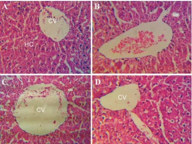
Histopathological examination of control speci-mens showed branched hepatic cell cords with intervening sinusoids in a radial arrangement directed from periphery of the lobule to the central venule at its center. In the experimental group, dilatation of the sinusoids (with an elon-gated endothelium nucleus) was prominent es-pecially in the 4-week exposure-time subgroup. There were also signs of inflammatory process and congestion of the central vein in its periph-ery (infiltration of lymphocyte) in the experi-mental group, especially in the 4- and 6-week exposure-time subgroups (Fig 1).
In contrast to the control group, in the experi-mental group, the diameters of sinusoids and central venules were increased in a time-de-pendent manner.
Table 1: Effect of solder fume on liver serum markers in rats
Six-week group Four-week group
Two-week group Groups
Mean ± SD Mean ± SD
Mean ± SD Variants Experimental Control Experimental Control Experimental Control
495.7 ± 71.2 366.6 ± 45.3
777.8 ± 189.7 739.8 ± 168.7
892.9 ± 232.22 768.6 ± 130.3
ALP IU/L
139.8 ± 38.2 119.6 ± 18.37
140.3 ± 23.41 144.5 ± 17.05
120.3 ± 31.54 129.3 ± 36.78
AST IU/L
45.2 ± 6.79 39.6 ± 6.08
47.3 ± 8.49 48.66 ± 5.18
70.3 ± 13.43 49.2 ± 16.9
ALT IU/L 3.09 3.02 2.96 2.96 1.71 2.62 AST/ALT
0.19 ± 0.08 0.2 ± 0.09
0.2 ± 0.07 0.166 ± 0.09
0.076 ± 0.026 0.086 ± 0.001
Direct Billirubin (mg/dl)
0.856 ± 0.1 0.862 ± 0.06
0.849 ± 0.067 0.866 ± 0.17
0.315 ± 0.05 0.382 ± 0.07
Total bilirubin (mg/dl)
10.55 ± 1.31 9.05 ± 0.48
10.2 ± 0.94 9.5 ± 1.65
9.58 ± 1.64 9.85 ± .17
Weight of Liver (g)
p>0.05: Not significant in all of experimental and control subgroups
Table 2: Effect of solder fume on sinusoid and central venule diameters of the rat liver
Six-week group Four-week group
Two-week group Groups
Mean ± SD Mean ± SD
Mean ± SD Variants Experimental Control Experimental Control Experimental Control
8.45 ± 1.87 8.4 ± 1.97
10.25 ± 2.16 8.66 ± 1.7
10.13 ± 2.26 9.8 ± 2.06
Diameter of sinusoid (μm)
60.44 ± 21.24 49.43 ± 17.05
65.88± 22.4 55.77 ± 21.59
65.12 ± 23.24 44.66 ± 14.99
Diameter of central venule (μm)
p<0.001 between control and experimental 4-week groups
Discussion
The obtained data from this study relating to defined fume-exposure times (1 hour/day for 2, 4 and 6 weeks) and concentration (0.193 mg/m3 for
formal-dehyde, 0.35 mg/m3 for Stannum and 3 mg/m3 for
lead) revealed that despite some differences in liver function test (LFT) results, there was no statistically significant difference between control and experi-mental groups (LSD test) in all studied subgroups. This finding is in disagreement with the Uboh et al. report showing that 4 hours/day of kerosene and pe-troleum fume inhalation in a period of 2 weeks sig-nificantly increased the concentration of ALP and aminotransferase in an experimental rat group in comparison to the control group (10). In their study, Uboh et al. have proposed that increased activity of aminotransferase, especially of ALT, in the ex-perimental group is caused by liver cell injury due to increased permeability of hepatocyte cell mem-branes or focal necrosis of hepatic cells. They docu-mented that the increase in concentration of AST in the experimental group may be due to abnormal dy-namic properties of cellular membranes following
exposure to inhaled fumes. In this regard, Uboh and colleagues introduced the ratio of AST/ALT as an important index of hepatic cell injury (10).
Our results demonstrated a moderate increase in the AST/ALT ratio in the experimental group in a time-dependent manner. They also showed a slight increase in the amount of bilirubin. Bilirubin, as a by-product of red blood cell (RBC) breakdown, could be increased following hepatic cell injury. The increased concentration of ALP in the experi-mental group reported in this study is in accord with the results of Jadhav et al. and Marques et al. (16, 17). It is probable that a lack of signifi-cant difference in the LFT marker in our study is attributable to the low duration of exposure time. Jadhav and colleagues’ study showed that intoxi-cation of rats by heavy metals induced an increase in the concentration of serum transferase, which is statistically significant only after 60 days of expo-sure (16).
Histopathological examination of specimens showed that a significant difference in sinusoi-dal diameters (p<0.001) was detected in the
perimental group compared to the control group of only the 4-week exposure-time subgroup. It seems that there was a tendency for time-dependent in-crease in the sinusoid diameters of the experimen-tal groups. In accordance with our results, Kutlu and colleagues reported a sinusoidal dilatation and liver congestion in rats exposed to polychlorinated biphenyl mixture (18). The appearance of inflam-matory cells in the liver as a sign of inflammation after exposure to solder fume is attributable to a general response of the organism to this toxic com-pound. The study of Kim and colleagues showed that acute exposure to welding fumes was associ-ated with increased levels of systemic inflamma-tory markers in welders (2). Furthermore, statisti-cal analysis demonstrated a significant difference in the central venule diameters among control and experimental groups of 2, 4 and 6-week exposure time. The study of Marroni and colleagues showed histopathological changes such as periportal fibro-sis in the human liver (19). The study of Uboh et al. showed changes in the rat liver cell integrity after inhalation exposure to toxic fumes (10).
According to a study reported by Marques and colleagues on Pb treated mice, despite a reduction in the RBCs, hematocrit and mean corpuscular hemoglobin, an increase in aminotransferase did not show a statistically significant difference. Ad-ditionally, in another study on intoxication of mice with Pb, they proved that mice possess a remark-able poisoning resistance (17). The latter finding might also be true for similar resistance in the rats exposed to soldering fume in our study. It seems that reaction of the rat liver vascular elements to solder fume followed an adaptive pattern. It also revealed that there were no significant changes in the liver after the first two-weeks of fume inhala-tion. However, in animals in the six-week exposure subgroup, there was a new fume resistance due to animal adaptation; significant changes were also seen in the liver vascular elements.
Based on previous studies, the toxicity of organic Sn compounds is much higher than that of inorgan-ic Sn compounds. Inhibition of adenosine triphate hydrolysis and uncoupling of oxidative phos-phorylation taking place in the mitochondria have been suggested as the cellular mechanisms of tin toxicity (20). Aminotransferases include aspartate aminotransferase (AST) and alanine aminotrans-ferase (ALT) which catalyze the transfer of amino groups from aspartic acid and pyrovic acid, respec-tively. ALT is a cytosolic enzyme, whereas AST is both a cytosolic and a mitochondrial enzyme (21). According to the obtained results, it seems that with this fume concentration and time exposure,
there was some potential for adaptation of the ani-mals before any dramatic changes in liver serum markers appeared. In all the studied parameters, a degree of increase was seen in the experimental group when compared to the control group. ALT is specific to the liver, whereas AST is also found in the cardiac muscle, the kidneys and the brain (22). Alkaline phosphatase (AP) comprises a group of enzymes present in a variety of tissues including those of the liver and kidney. An increase of up to three times in AP levels is relatively non-specific. Redlich and colleagues reported that occupational liver diseases may occur more frequently in work-ers exposed to occupational fumes than in the gen-eral population, and stated that improvement in conditions with liver enzyme abnormalities were seen after modification of the workplace (23). Studies of Dossing and colleagues also demon-strated focal necrosis, portal tract enlargement and fibrosis in livers of workers exposed to organic solvents (24).
Conclusion
We can conclude that vascular structures of the liver respond to toxic fumes before the beginning of hepatic cell injury and before the appearance of significant elevation of serum transferase.
Acknowledgments
The authors thanks deputy for research of Zahedan University of Medical Sciences for financial sup-port (grant number 748, awarded on 12/2/2005) of this study and Dr. S. J. Mowla for his encourage-ment and editing of this manuscript. There is no conflict of interest in this article.
References
1. Palmer K, Crane G. Respiratory disease in workers exposed to colophony solder flux fumes: Continuing health concerns. Occ Med. 1997; 47(8): 491-498. 2. Kim JY, Chen JC, Boyce PD, Christiani DC. Expo-sure to welding fumes is associated with acute systemic inflammatory responses. Occup Environ Med. 2005; 62(3): 157-163.
3. Antonini JM, Clarke RW, Krishna Murthy GG, Sreekan-than p, Jenkins N, Eagar TW, et al. Freshly generated stainless steel welding fumes induces greater lung in-flammation in rats as compared to aged fume. Tocicol Lett. 1998; 98(1-2): 77-86.
4. Antonini JM, Lewis AB, Roberts JR, Whaley DA. Pul-monary effects of welding fumes: Review of worker and experimental animal studies. Am J Indust Med. 2003; 350-360.
6. Leikin JB, Davis A, Klodd DA, Thunder T, Kelafant GA, Paquette DL, et al. Selected topics related to occupa-tional exposures. Part IV. Occupaoccupa-tional liver disease. Dis Mon. 2000; 46(4): 295-310.
7. Heederik D, Sigsgaard T, Thorne PS, Kline JN, Avery R, Bonlokke JH, et al. Health effects of airborne expo-sures from concentrated animal feeding operations. En-viron Health Perspect. 2007; 115(2): 298-302.
8. Gustavson P, Jakobson R, Nyberg F, Pershagen G, Jarup L, Scheele P. Occupational exposure and lung cancer risk: a population based case referent study in Sweden. Am J Epidemiol. 2000; 152(1): 32-40.
9. Kakar S, Batts KP, Poeterucha JJ, Burgart LJ. His-tologic changes mimicking billiary disease in liver biop-sies with venous outflow impairment. Mod Pathol. 2004; 17(7): 874-878.
10. Uboh FE, Akpanabiatu MI, Eyong EU, Ebong PE, Offiong O. Evaluation of toxicological implications of in-halation exposure to kerosene fumes and petrol fumes in rats. Acta Biol Szegediensis. 2005; 49(3-4): 19-22. 11. Sheweita SA, Abd El-Gabar M, Bastawy M. Carbon tetrachloride induced changes in activity of phase II drug metabolizing enzyme in the liver of male Rats: role of antioxidants. Toxicol. 2001; 165(2-3): 217-224.
12. Berk PD, Wolkff AW. Billirubin metabolism and the hyperbilirubinemias. In: Braunwald AS, Kasper DL, Hauser SL, Longo DL, Jamoson JL, editors. Harrison principle of internal medicine. 15th edition.; Mc Graw Hill Com; 2001; 3: 1715-1721.
13. American Society for Testing and Materials (ASTM), Annual book of ASTM standards: Atmospheric analysis and occupational health and safety; Standard practice for measurement of metals in work place atmosphere by atomic absorption spectrophotometer. Easton, USA: ASTM; 1996; Vol. 11.03, D 4185-90: 246-253.
14.http://www.cdc.gov/niosh/docs/2003%2D154/ pdf/3500.pdf.
15. Occupational Safety and Health Adminstration. Ana-lytical Methods Manual ICP Analysis of Metal/metalloid particulate from solder Operations. Method No ID 206. Osha, Salt Lake city, Utah 1991.
16. Jadhav SH, Sarkar SN, Patil RD, Tripathi HC. Effects of Subchronic exposure via drinking water to a mixture of eight water contaminating metals: a biochemical and histopathological study in male rats. Arch Environ Con-tam Toxicol. 2007; 53(4): 667-677.
17. Marques CC, Nunes AC, Pinheiro T, Lopes PA, San-tos MC, Viegas AM, et al. An assessment of time de-pendent effects of lead exposure in Algerian mice (Mus spretus) using different methodological approaches. Biol Trace Elem Res. 2006; 109(1): 75-89.
18. Kutlu S, Colakoglu N, Halifeoglu I, Sandal S, Seyran AD, Aydin M, et al. Comparative evaluation of hepato-toxic and nephrohepato-toxic effects of aroclors 1221 and 1254 in female rats. Cell Biochem Funct. 2007; 25(2): 167-172.
19. Maroni M, Mocci F, Visentin S, Preti G, Fanetti AC. Periportal fibrosis and other liver ultrasonography find-ing in vinyl chloride workers. Occup Environ Med. 2003; 60(1): 60-65.
20. Dossing M. Occupational toxic liver damage. J Hepatol. 1986; 3(1): 131-135.
21. Amdur OM. Toxicology the basic science of poisons. Singapore: McGraw-Hill; 1992; 670-671.
22. Massaro JE. Handbook of human toxicology. New York: CRC Press; 1997; 56-59.
23. Redlich CA, Beckett WS, Sparer J, Barwick KW, Ri-ely CA, Miller H, et al. Liver disease associated with oc-cupational exposure to the solvent dimethylformamide. Ann Intern Med. 1988; 108(5): 680-686.