ISSN 0102-695X
DOI: 10.1590/S0102-695X2013005000036 Received 4 Nov 2012
Accepted 23 Apr 2013 Available online 14 May 2013
coffees on the
in vivo
antioxidant activity and
prevention of liver injury in rats
Adriene R. Lima,
*,1Rosemary G. F. A. Pereira,
1Sheila A.
Abrahão,
2Márcio G. Zangeronimo,
3Fernanda B. A. Paula,
4Stella M. S. Duarte
41Departamento de Ciência dos Alimentos, Universidade Federal de Lavras, Brazil, 2Instituto Federal de Educação, Ciência e Tecnologia, Bom Jesus do Itabapoana-RJ, Brazil,
3Departamento de Ciências Veterinárias, Universidade Federal de Lavras, Brazil, 4Departamento de Análises Clínicas e Toxicológicas, Universidade Federal de Alfenas, Brazil.
Abstract: Decaffeination and roasting affects the composition of the chlorogenic acids in coffee, which have antioxidant potential. The aim of this study was to evaluate the effects of coffee decaffeination on the in vivo antioxidant activity and the prevention of liver damage. The Wistar rats received intraperitoneal doses of carbon tetrachloride and daily doses of Arabica coffee brews (whole and decaffeinated, both green and
roasted) by gavage for i fteen days. The activity of liver marker enzymes aspartate
aminotransferase, alanine aminotransferase and serum albumin were measured as
well as the quantii cation of the thiobarbituric acid reactive species and the content of
liver total lipids. Aspartate aminotransferase and alanine aminotransferase are good indicators of liver damage: the results showed that all studied coffee brews decreased the activity of aspartate aminotransferase and alanine aminotransferase, and liver levels of thiobarbituric acid reactive species and total lipids. The compounds presents in coffee brews are able to decrease the hepatic lipid peroxidation induced by carbon
tetrachloride, making a signii cant hepatoprotective effect, in accordance with the liver
function tests. The coffee brews are hepatoprotective regardless of the decaffeination process and our results suggest a better protection against liver damage for the roasted coffee brews compared with green coffee brews.
Keywords: antioxidant activity carbon tetrachloride decaffeination liver
chlorogenic acid caffeine
Introduction
Many studies have demonstrated that coffee, due to its antioxidant compounds, has protective effects in the
liver against diseases such as cirrhosis and reduces the risk
of developing hepatocellular carcinoma (Ozercan et al., 2006; Bravi et al., 2007; Muriel & Arauz, 2010; Masterton & Hayes, 2010; Carvalho et al., 2010; Shi et al., 2010), which is the most common form of cancer in the liver. Among the bioactive compounds investigated, we focused on the chlorogenic acids, melanoidins and caffeine.
Liver diseases are a public health problem worldwide. The evolution of liver disease begins with
steatosis, hepatitis, i brosis, cirrhosis following to
hepatocellular carcinoma. Research has shown that reactive oxygen species are related to the cascade of events that regulate the initiation and the progression of liver disease, regardless of the agent that originated (Loguercio & Frederico, 2003; Vitaglione et al., 2004).
Thus, the use of antioxidant therapies, drugs that interfere in the metabolism (CYP-450) and hepatotoxic agents that increase the activity of enzyme systems of defense are the options used in the study of hepatotoxicity (Weber et al., 2003; Porchezhian & Ansari, 2005; Lima et al., 2007).
The coni rmation of liver damage is carried out through specii c serological diagnostic tests, as
determination of the enzymatic activity of transaminases: alanine aminotransferase (ALT) and aspartate aminotransferase (AST), enzymes cellular function whose presence in the blood is a consequence of defective release into the circulation.
The use of carbon tetrachloride (CCl4) in experimental protocols helps to elucidate the mechanisms
of hepatotoxicity and its consequences: inl ammation, steatosis, hepatitis, i brosis, cirrhosis and carcinogenesis (Schatzki, 1963; Weber et al., 2003; Lima et al., 2007).
The quest for coffee without caffeine has mobilized researchers around the world to meet the
increasing demand of people wanting to eliminate the side effects, such as insomnia, caused by this stimulant substance (Lima et al., 2010). Many studies have been conducted on the physiological effects of this substance, but there is not yet a consensus on the positive and negative aspects of the results of these studies (Corrao et al., 2001; Ruhl & Everhart, 2005; Masterton & Hayes, 2010; Chu et al., 2012). Decaffeination and roasting processes caused changes in the levels of important bioactive compounds, such as chlorogenic acids (Toci et al., 2006; Lima et al., 2010).
This study may contribute to a deeper understanding of this issue, as research on the use of decaffeinated coffees as preventives of liver damage in
rats is scarce and is the main objective of this work.
Materials and Methods
Plant material
Samples of whole and decaffeinated coffee (blends of Coffea arabica L. grown in several areas of Brazil) were given by COCAM Industry, located in Catanduva-SP, Brazil. Decaffeination was performed using water and dichloromethane (Toci et al., 2006; Antonio et al., 2010). Samples of coffee (500 g) were roasted in a laboratory roaster (Probatino, Leogap
model, Brazil), with a capacity of 1 kg, at the average
degree of roasting. The tone of the final color of the grains was determined visually and with instruments
(Chomameter-2 Reflectance, Minolta, Osaka, Japan).
Next, the roasted beans were ground (Pinhalense electric grinder, ML-1, Brazil) into fine grains (20 mesh),
packed in polyethylene/aluminum, sealed and stored at
-20 °C until use. The green beans were finely ground in a mill cooled to 4 °C (Tecnal, model TE 631/2, Brazil) using liquid nitrogen (Lima et al., 2010).
Analysis of chlorogenic acid and caffeine by HPLC
For the determination of caffeine and chlorogenic acid levels in the coffee brews, a hot water extraction method was employed (Vitorino et al., 2001); the extraction was followed by dilution with 100 mL/0.5 g of distilled
water and, inally, HPLC analysis with a Shimadzu Brand
chromatograph (model M10AVP, Japan) equipped with a C-18 reverse-phase column (Shimadzu 100 mm long x 0.3 mm ID, 4,6 µm particle size, Japan). The HPLC was coupled to a UV/visible spectrophotometric detector (Shimadzu SPD-10A model) connected by an interface (CBM-101) to a microcomputer for data processing. The
conditions of analysis used were as follows: low (1 mL/
min); mobile phase (methanol, water and acetic acid in a ratio of 20:80:1); injection volume was 20 µL and wavelength detection at 272 nm. The concentrations of the
compounds were determined with standard concentration curves. Caffeine and 3-O-caffeoylquinic acid (chlorogenic acid, 3-CQA) were purchased from Sigma-Aldrich Chemical. Curves obtained had R2 greater than 0,999.
Preparation of brews
A ilter coffee brews was prepared according to
the methodology described by Lima et al. (2010), 10 g of
coffee powder were added in commercial ilter paper, and
then, 100 mL of deionized water at 90 °C were poured into
the coffee contained in the ilter. The coffee brews were
always prepared at the time of administration to animals.
Experimental animals
The study included 24 male adult Wistar rats (Rattus norvegicus), which were divided into six equal groups. The animals were maintained at 22±2 °C, 55±10%
humidity, with a 12-h light-dark cycle. The rats were
fed commercial Nuvilab CR-1® (Nuvital Nutriente S/A,
Colombo, PR, Brazil) 1®. To induce liver injury, CCl 4 was
injected through an intraperitoneal route at a dose of 1.5
mL.kg-1 (suspended with olive oil at a rate of 1:1) on days
11, 13 and 15 during the 15-day treatment.
The animals were divided into six groups of four animals each: Group 1, the negative control group (NC), was given water; Group 2, the positive control group (PC), was administered CCl4 to induce liver injury, along with water; Group 3 was administered whole green coffee and CCl4 (WGC); Group 4, decaffeinated green coffee and CCl4 (DGC); Group 5, whole roasted coffee and CCl4 (WRC) and Group 6, decaffeinated roasted coffee (DRC) and CCl4.
The coffee brews were administered to the
animals by gavage once daily for ifteen days and the dose used was 5.7 mL.kg-1 per day, which is equivalent to an
adult human drinking eight 50 mL cups of coffee brew
(10%) per day.
On the sixteenth day, the animals fasted for 12 h, were anesthetized for the removal of blood by cardiac puncture, and immediately after, the rats were euthanized in a CO2 chamber for liver removal. The blood was centrifuged to separate the serum. The livers were washed with 0.9% saline solution, stored at -20 °C and submerged in phosphate buffer (pH 7.4). The University Ethical Committee for Animal Research (Unifal, MG, Brazil) approved the protocols used in this study (protocol 155/2007).
Biochemical analysis: biochemical parameters in the serum and oxidative stress in the liver
The serum obtained by centrifuging the blood
aminotransferase (ALT), aspartate aminotransferase (AST) and albumin levels were determined by the colorimetric method.
The protein concentration of liver homogenate was determined by Bradford (1976), using bovine serum
albumin (BSA) as the standard. An aliquot of 10 μL
containing liver homogenate was added to 790 mL of distilled water and 200 mL of Coomassie Brilliant Blue G-250 in ethanol 95% w/v. Then, the optical density was measured at 595 nm in a spectrophotometer. Protein
concentration (μg/10 μL) was calculated by inserting a
calibration curve previously made with BSA (5-25 mg/ mL).
To determine whether drinking coffee was able to
reduce oxidative stress, we analyzed the lipid peroxidation of isolated rat livers. The lipid peroxidation was assessed by the formation of thiobarbituric acid reactive substances (TBARS) (Winterbourn et al., 1981). The lipid peroxidation products react with thiobarbituric acid to produce a compound that has an absorbance at 532 nm.
The livers were weighed and homogenized in a tissue homogenizer in an ice bath after the addition of 0.1 M PBS, pH 7.4 (with a volume equal to four times the weight of fresh tissue). The homogenate was centrifuged at 3000 x g for 10 min at 4 °C, and the supernatant that was
kept on ice was used in the tests. Aliquots of 500 mL of the
supernatant were mixed with 500 mL of hydrochloric acid 25% (v/v), 500 mL of thiobarbituric acid 1% (w/v in 0.05
M NaOH) and 45 μL of BHT 2% (w/v in ethanol).
The mixture was heated at 100 °C in a water bath for 10 min. After cooling in an ice bath for 10 min, 1.5 mL of
butanol as added, and the samples were shaken vigorously.
The samples were then centrifuged at 900 x g for 5 min, and the fraction containing butanol was collected and used to determine the absorbance at 532 nm. The TBARS concentration was calculated from the standard curve of malonic dialdehyde (MDA; tetraetoxipropano 1,1,3,3). The results were expressed as nmol MDA/g protein.
Determination of total liver lipids
Total liver lipids were measured according to the methodology described by AOAC (1995) with slight
modiications. The livers were lyophilized in a lyophilizer
Liobrás (L101) and crushed in a mortar; 1 g of the resulting powder was placed into cartridges cellulose, and lipids were extracted with diethyl ether for 6 h in a Soxhlet apparatus. The process is based on the weight loss of the material submitted to extraction with ether or the amount of material solubilized by the solvent. The results were expressed as a percentage of the lipids in relation to the total matter.
Statistical analysis
The values obtained for the chemical composition of the coffee samples were analyzed by one way ANOVA
followed by post hoc Tukey test. A global analysis of
variance was made with all the in vivo treatments, with the aim to obtain the residue average square to test the factorial and to apply Dunnett’s test at 5% to compare the control treatment (negative and positive) with the others. The F test was used to test treatments in a factorial way.
Results and Discussion
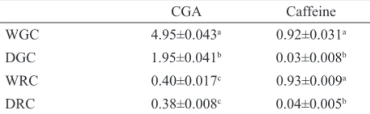
The results of the HPLC analysis are presented in Table 1. Bioactive compounds, such as chlorogenic acid (CGA) and caffeine were identified by comparison of the sample retention times with standards. The amount of each compound present was quantified using an external standard calibration. The retention times for the chlorogenic acid and caffeine standards, were 9 and 10 min, respectively.
Table 1. Concentrations of chlorogenic acid (CGA) and caffeine in the aqueous extracts of whole green coffee (WGC), decaffeinated green coffee (DGC), whole roasted coffee (WRC) and decaffeinated roasted coffee (DRC) (g/100 g of ground).
CGA Caffeine
WGC 4.95±0.043a 0.92±0.031a
DGC 1.95±0.041b 0.03±0.008b
WRC 0.40±0.017c 0.93±0.009a
DRC 0.38±0.008c 0.04±0.005b
*Values are expressed as mean±SD (n=3). Means within a column,
followed by different letters (a,b,c) are signiicantly different by Tukey’s
test, p<0.05.
The CGA content was reduced with the decaffeination process, with losses of 60% in the decaffeinated green sample. The decrease must have occurred mostly due to isomerization of the CGA when exposed to high temperatures for solvent evaporation and drying of the beans during decaffeination with dichloromethane (Toci et al. 2006).
The roasting led to losses of 92% in levels of CGA, which is consistent with other studies where decaffeinated and whole commercial coffees showed a
loss of up to 93% after roasting (Fujioka & Shibamoto,
2008). During the roasting of the beans, part of chlorogenic acids is transformed into other substances that are formed
during this process, and are related to color and lavor of
coffee (Farah et al., 2005).
AST and ALT when compared with the PC group (treated with carbon tetrachloride). However, these rates did not reach their baseline values (the negative control group). Among the groups treated with the different coffee brews,
there was no signiicant difference, which demonstrates
that regardless of the process of decaffeination and roasting, the coffee brews protected the liver from injury caused by carbon tetrachloride.
A study from Ozercan and coworkers (2006)
reports the effects of coffee on liver damage caused by carbon tetrachloride which are consistent with the results of this study. They found levels of AST and ALT in the
group treated with carbon tetrachloride to be signiicantly higher than in the control group. In this same work, the
observed levels of AST and ALT in the group treated
with carbon tetrachloride over coffee were signiicantly
lower than in the groups treated with carbon tetrachloride (p<0.05) (Ozercan et al., 2006).
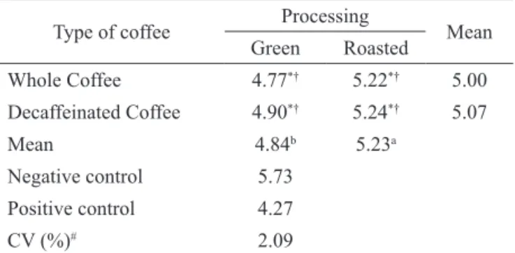
Albumin is the most abundant protein in the blood plasma, constituting approximately 50 to 65% of the total protein. Albumin is synthesized in the liver, and the most decisive factor for its concentration in the blood is the liver's ability to synthesize it. The accumulation of fat causes a reduction in the hepatic synthesis capacity of this body, reducing the concentration of albumin (Nicoluzzi et al., 2000). The concentrations of albumin are presented in Table 4.
Table 4. Serum albumin (g/dL) in the groups treated with the whole or decaffeinated samples of coffee (green and roasted), and the control groups.
Type of coffee Processing Mean
Green Roasted
Whole Coffee 4.77*† 5.22*† 5.00
Decaffeinated Coffee 4.90*† 5.24*† 5.07
Mean 4.84b 5.23a
Negative control 5.73
Positive control 4.27
CV (%)# 2.09
*differs from the positive control by Dunnett's test (p<0,05); †differs from
the negative control by Dunnett's test (p<0,05); #coeficient of variation.
Means within a line, followed by different letters (a, b) are signiicantly
different by Tukey’s test (p<0,05).
The positive control group showed signiicantly
lower albumin content than the negative control group, indicating that the carbon tetrachloride decreased the levels of serum albumin in rats. Tetrachloride-treated groups that
were given coffee drinks had signiicantly higher values
of albumin than the positive control group and had lower values than the negative group control.
The serum albumin levels in the groups treated
with roasted coffee (WRC and DRC) were signiicantly
values reported in the literature for green Arabica coffee
beans -0.9 to 1.3 g% (Ramalakshmi & Raghavan, 1999;
Toci et al., 2006; Lima et al. 2010). The caffeine content in the decaffeinated green sample (0.03%) is in accordance with Brazilian law, which sets a level of 0.1% caffeine in decaffeinated coffee (Anvisa, 1999).
Biochemical analyses
Acute administration of carbon tetrachloride causes centrilobular necrosis and steatosis, a fact
conirmed in this work, because their intra-peritoneal
injection caused liver injury in rats, which is evident
from the signiicant difference in serum aspartate
aminotransferase (AST) and alanine aminotransferase (ALT) between the negative and positive control groups. The results of AST and ALT are respectively shown in Tables 2 and 3.
Table 2. Concentration of the enzyme aspartate aminotransferase, AST (U/L) in the groups treated with the whole or decaffeinated samples of coffee (green and roasted), and the control groups.
Type of coffee Processing Mean
Green Roasted
Whole Coffee 248.05*† 259.22*† 253.63
Decaffeinated Coffee 243.47*† 244.88*† 244.18
Mean 245.76 252.05
Negative control 108.19
Positive control 327.49
CV (%)# 5.75
*differs from the positive control by Dunnett's test (p<0,05); †differs from
the negative control by Dunnett's test (p<0,05); #coeficient of variation.
Table 3. Concentration of the enzyme alanine aminotransferase, ALT (U/L) in the groups treated with the whole or decaffeinated samples of coffee (green and roasted), and the control groups.
Type of coffee Processing Mean
Green Roasted
Whole Coffee 141.24*† 141.91*† 141.57
Decaffeinated Coffee 144.57*† 145.39*† 144.98
Mean 142.91 143.65
Negative control 39.68
Positive control 233.0
CV (%)# 9.11
*differs from the positive control by Dunnett's test (p<0,05); †differs from the negative control by Dunnett's test (p<0,05); #coeficient of variation.
The group treated with carbon tetrachloride (PC)
showed AST and ALT levels signiicantly higher than the
group treated with water (NC). In all of the groups treated
higher than the groups treated with unroasted coffee (WGC and DGC). These results show that roasted coffee offered greater protection compared with the green coffee.
The process of decaffeination did not inluence the levels
of serum albumin.
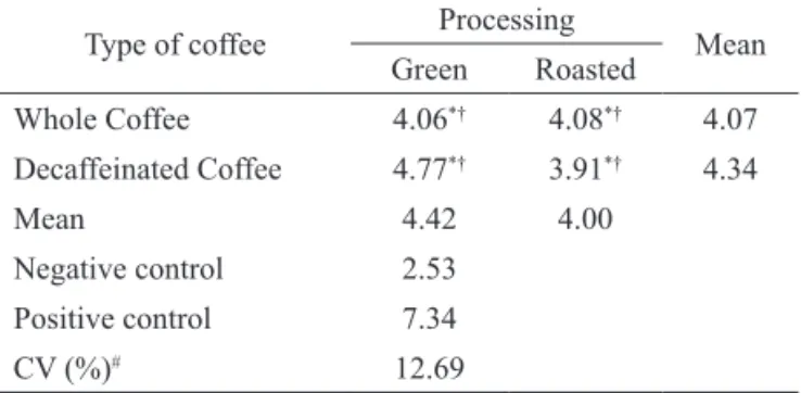
The presence of xenobiotics in the liver, such as carbon tetrachloride, caused injury and a consequent accumulation of lipids in the liver, which led to hepatic
steatosis (Ramakrishna et al., 2011). The results were
expressed as a percentage of fat in relation to the total matter and are presented in Table 5.
Table 5. Concentration of total lipids in the liver (%) in relation to the total matter in the groups treated with the different coffee samples and the control groups.
Type of coffee Processing Mean
Green Roasted
Whole Coffee 4.06*† 4.08*† 4.07
Decaffeinated Coffee 4.77*† 3.91*† 4.34
Mean 4.42 4.00
Negative control 2.53
Positive control 7.34
CV (%)# 12.69
*differs from the positive control by Dunnett's test (p<0,05); †differs from
the negative control by Dunnett's test (p<0,05); #coeficient of variation.
The total lipid content of the livers in the groups
treated with carbon tetrachloride was signiicantly higher
than that in the negative control group (treated with water), indicating that xenobiotics elevate liver lipid levels. The groups treated with carbon tetrachloride in conjunction with the coffee brews exhibited a decrease in lipid levels compared with the positive control group. There was no
signiicant difference among the coffee drinks. The coffee drinks, regardless of the type and the processing, prevented
the accumulation of fat in the liver but did not return to the baseline values found in the negative control group.
Substances capable of combating oxidative stress are able to protect the liver from improper fat accumulation
(Ramakrishna et al., 2011), thus the antioxidants present in
the coffee brews may have contributed to the decreased accumulation of hepatic fat.
Lipid peroxidation is a major consequence of liver injury caused by CCl4 and is mediated by the production of free radicals derived from CCl4. However, antioxidant activity and the inhibition of free radical generation are important in the protection against liver damage caused by
carbon tetrachloride (Teselkin et al., 2000; Campo et al., 2001; Ramakrishna et al., 2011).
The inhibition of lipid peroxidation was calculated as the concentration of thiobarbituric acid reactive substances (TBARS). The levels of MDA in the different groups are presented in Table 6.
All samples demonstrated inhibition of lipid
peroxidation, and roasting was a positive inluence in this inhibition. The decaffeination process did not inluence
lipid peroxidation.
Table 6. Content of thiobarbituric acid reactive substances (nmol MDA/mg protein) in the groups treated with the different samples of coffee and the control groups.
Type of coffee Processing Mean
Green Roasted
Whole Coffee 0.290*† 0.240*† 0.265
Decaffeinated Coffee 0.295*† 0.230*† 0.263
Mean 0.293a 0.235b
Negative control 0.148
Positive control 0.378
CV (%)# 12.22
*differs from the positive control by Dunnett's test (p<0,05); †differs from
the negative control by Dunnett's test (p<0,05); #coeficient of variation.
Means within a line, followed by different letters (a, b) are signiicantly different by Tukey’s test (p<0,05).
The coffee brews that were analyzed have phenolic compounds, mostly chlorogenic acids, which may contribute, at least in part, to the antioxidant activity demonstrated in this study. Moreover, the complexation of Fe+2 to phenolic compounds can reduce the availability of
the metal involved in the Fenton reaction in the initiation and propagation of lipid peroxidation (Lima et al., 2006).
Since the content of chlorogenic acid was reduced by decaffeination, other substances may have been formed during the roasting, that acted synergistically with the phenolic compounds for the inhibition of lipid peroxidation observed in the coffee brews (Lima et al., 2010). Several authors have suggested that the Maillard reaction products, which are formed during the roasting of the coffee, have antioxidant activity and may have contributed to the observed results (López-Galilea et al., 2006).
Furthermore, hydroxyl radical scavenging
characteristics of highmolecular weight melanoidin-like
compounds are stronger than those of low-molecular weight phenolic compounds (Delgado et al., 2005).
Conclusion and perspectives
Regardless of the decaffeination process, the decaffeinated coffee brews protected the rat livers, no
signiicant differences were observed between whole and
decaffeinated brews. Our results suggest a better protection against liver damage caused by carbon tetrachloride for the roasted coffee brews compared with green coffee brews.
compounds on liver metabolism. Because of the mixture of bioactive components present in the coffee, it is possible that more than one mechanism underlying this effect is involved.
Acknowledgements
This work was supported by CAPES, CNPq,
FAPEMIG and INCT/Café.
Authors’ contributions
ARL, SAA contributed to chromatographic
analysis running the laboratory work. ARL, SAA, FBAP
and SMSD contributed to biological studies analysis.
MGZ undertook the statistical analysis and interpretation
of data. ARL, SAA, RGFAP, FBAP and SMSD designed the study, analysis of the data and drafted the manuscript.
RGFAP, FBAP and SMSD supervised the laboratory work
and contributed to critical reading of the manuscript. All
the authors have read the inal manuscript and approved
the submission.
References
Antonio AG, Moraes RS, Perrone D, Maia LC, Santos KRN, Iorio NLP, Farah A 2010. Species, roasting degree and decaffeination influence the antibacterial activity of coffee against
Streptococcus mutans. Food Chem 118: 782-788.
Anvisa 1999. Agência Nacional de Vigilância Sanitária,
Legislação sobre descafeinação, Portaria nº 377, de 26 de abril de 1999. http://www.anvisa.gov.br/ anvisalegis/portarias/377_99.htm.
AOAC 1995. In: Cunniff P (Ed.) Oficial Methods of
Analysis of AOAC International, 16th Ed. AOAC
International, Arlington.
Bradford MM 1976. A rapid and sensitive method for the
quantiication of microgram quantities of protein
utilizing the principle of protein-dye binding. Anal Biochem 72: 248-254.
Bravi F, Bosetti C, Tavani A, Bagnardi V, Gallus S,
Negri E 2007. Coffee drinking and hepatocellular carcinoma risk: a meta-analysis. Hepatology 46: 430-435.
Campo GM, Squadrito F, Ceccarelli S, Calo M, Avenoso A, Campo S, Squadrito G, Altavilla D 2001. Reduction of carbon tetrachloride-induced rat liver injury by
IRFI 042, a novel dual vitamin E-like antioxidant.
Free Radic Research 34: 379-393.
Carvalho DC, Brigagão MRPL, Santos MH, Paula FBA, Paiva AG, Azevedo L 2011. Organic and conventional Coffea arabica L.: A comparative study of the chemical composition and
physiological, biochemical and toxicological effects in Wistar rats. Plant Foods Hum Nutr 66: 114-121.
Chu YF, Chen YM, Brown PH, Lyle BJ, Black RM, Cheng
IH, Ou BX, Prior RL 2012. Bioactivities of crude caffeine: Antioxidant activity, cyclooxygenase-2
inhibition, and enhanced glucose uptake. Food Chem 131: 564-568.
Corrao G, Zambon A, Bagnardi V, D'amicis A, Klatsky
A 2001 Collaborative SIDECIR Group. Coffee,
caffeine, and the risk of liver cirrhosis. Ann Epidemiol 11: 458-465.
Delgado-Andrade C, Ruian-Henares JA, Morales FJ 2005.
Assessing the antioxidant activity of melanoidins from coffee brews by different antioxidant methods.
J Agric Food Chem 3: 7832-7836.
Farah A, De Paulis T, Trugo LC, Martin PR 2005. Formation of chlorogenic acids lactones in roasted coffee. J Agric Food Chem 53: 1105-1113.
Fujioka K, Shibamoto T 2008. Chlorogenic acid and
caffeine contents in various commercial brewed coffees. Food Chem 106: 217-221.
Lima AR, Barbosa VC. Santos Filho P R, Gouvêa CMCP 2006. Avaliação in vitro da atividade antioxidante do extrato hidroalcoólico de folhas de bardana. Rev Bras Farmacogn 16: 531-536.
Lima AR, Pereira RGFA, Abrahão SA, Duarte SMS, Paula FBA 2010. Compostos bioativos do café: atividade antioxidante in vitro do café verde e torrado antes e após a descafeinação. Quim Nova 33: 20-24.
Lima CF, Fernandes-Ferreira M, Wilson CP 2007. Drinking
of Salvia oficinalis tea increases CCl4-induced hepatotoxicity in mice. Food Chem Toxicol. 45: 456-464.
Loguercio C, Federico A 2003. Oxidative stress in viral and alcoholic hepatitis. Free Radic Biol Med 34: 1-10.
López-Galilea I, Andueza S, Di Leonardo I, Peña MP, Cid C 2006. Influence of torrefacto roast on antioxidant and pro-oxidant activity of coffee.
Food Chem 94: 75-80.
Masterton GS, Hayes PC 2010. Coffee and the liver: a potential treatment for liver disease? Eur J Gastroenterol Hepatol 22: 1277-1283.
Muriel P, Arauz J 2010. Coffee and liver diseases.
Fitoterapia 81: 297-305.
Nicoluzzi JE, Barbu V, Baudrimont M 2000. Viabilité et état de différenciation des hépatocytes humains immunoprotégés par macroencapsulation et transplantés chez le rat. Gastroenterol Clin Biology 24: 342-348.
Ozercan IH, Dagli A, Ustundag B, Ozercan M, Bahcecioglu
I, Celik H, Yalniz M, Poyrazoglu O, Ataseven H
Res 35: 163-168.
Porchezhian E, Ansari Sh 2005. Hepatoprotective activity of Abutilon indicum on experimental liver damage in rats. Phytomedicine 12: 62-64.
Ramakrishna S, Geetha KM, Bhaskar Gopal PVVS, Ranjit
Kumar P, Charan Madav P, Umachandar L 2011. Effect of Mallotus philippensis Muell.-Arg leaves against hepatotoxicity of carbon tetrachloride in rats. Int J Pharm Sci Res 2: 74-83.
Ramalakshmi K, Raghavan B 1999. Caffeine in coffee: its
removal: why and how? Crc Cr Rev Food Sci 39: 441-456.
Ruhl CE, Everhart JE 2005. Coffee and caffeine
consumption reduce the risk of elevated serum
alanine aminotransferase activity in the United States. Gastroenterology 128: 24-32.
Schatzki PF 1963. Rat liver adenosine triphosphatase
changes following experimental carbon tetra chloride administration. Arch Pathol 75: 85-90. Shi H, Dong L, Zhang Y, Bai Y, Zhao J, Zhang L 2010.
Protective effect of a coffee preparation (Nescafe pure®) against carbon tetrachloride-induced liver
ibrosis in rats. Clin Nutr 29: 399-455.
Teselkin YO, Babenkova IV, Kolhir VK, Baginskaya AI, Tjukavkina NA, Kolesnik YA, Selivanova
IA, Eichholz AA 2000. Dihydroquercetin as a means of antioxidative defense in rats with tertrachloromethane hepatitis. Phytother Res 14: 160-162.
Toci A, Farah A, Trugo LC 2006. Effect of decaffeination using dichloromethane on the chemical composition of arabica and robusta raw and roasted coffees. Quim Nova 29: 965-971.
Vitaglione P, Morisco F, Caporaso V 2004. Dietary antioxidant compounds and liver health. Crit Rev Food Sci Nutr 44: 575-586.
Vitorino MD, França, AS, Oliveira, LS, Borges, MLA 2001. Metodologias de obtenção de extrato de café visando à dosagem de compostos não voláteis. Rev Bras Armazenamento 26: 17-24.
Weber, LWD, Boll M, Stampl A 2003. Hepatotoxicity and mechanism of action of haloalkanes: carbon
tetrachloride as a toxicological model. Crit Rev Toxicol 33: 105-136.
Winterbourn CC, Gutteridge JM, Halliwell B 1981. Doxorubicin-dependent lipid peroxidation at low partial pressures of O2. Free Rad Bio Med 2: 1119-1122.
*Correspondence
Adriene Ribeiro Lima
Departament of Food Science, Federal University of Lavras, Lavras, MG
Campus Universitário, Caixa Postal 3037, CEP 37200-000 Lavras MG, Brazil