PURIFICATION AND PROPERTIES OF A FUNGAL L-ASPARAGINASE FROM
TRICHODERMA VIRIDE
PERS: SF
GREY
Lynette Lincoln
1, Francois N. Niyonzima
2, Sunil S. More
1*
Address(es): 1
Department of Biochemistry, CPGS, Jain University, 18/3, Jayanagar 3rd block, Bangalore-560011.
2 Department of Biology-Chemistry, CE, University of Rwanda, Kigali-5039.
*Corresponding author: [email protected]
ABSTRACT
Keywords:Trichoderma viride Pers: SF Grey, L-asparaginase, antioxidant, acrylamide
INTRODUCTION
L-asparaginase (E.C.3.5.1.1) is an enzyme that catalyzes the hydrolysis of the amide bond in L-asparagine to form L-aspartic acid and ammonia. L-asparagine serves as an essential amino acid necessary for the growth of tumor cells; however, in normal cells L-asparagine is synthesized within a cell with the help of asparagine synthetase. The cancer cells therefore distinguish themselves from
normal cells in diminished L-asparagine expression (Manna et al., 1995). As the
lymphatic tumor cells require higher amounts of L-asparagine for survival, they deplete all the asparagine from the circulatory blood leading to death of leukemic
cells (Siddalingeshwara and Lingappa, 2010; Patro and Gupta, 2012). In the
treatment of acute lymphoblastic leukemia (ALL), L-asparaginase drug is used to reduce the circulatory L-asparagine level, thereby stopping tumor cell growth
(Gallagher et al., 1989). Bacterial L-asparaginase was essentially used as the potent antileukemic agent for over 3 decades; however, being a large protein, it was associated with minor side effects such as nausea, vomiting, fever, and skin
rash (Goodsell, 2005). The other side effects resulted from the contamination of
bacterial L-asparaginase by glutaminase (Gallagher et al., 1989). There is
therefore an extensive search for eukaryotic fungal L-asparaginases which have
less adverse effects compared to the bacterial enzymes (Sarquis et al., 2004).
Acrylamide is associated with potential human health risks. It is produced from asparagine and reducing sugars in baked or fried starchy foods rich in carbohydrates at temperatures higher than 120 °C. L-asparaginase serves as a processing agent during the manufacture of starchy food products as it converts L-asparagine to aspartic acid and thus decreases the level of acrylamide
formation (Pedreschi et al., 2008). Granda et al. (2004) reported the reduction
of acrylamide in foods by monitoring the cooking process. The two commercial
asparaginases used in the food industry are Acrylaway produced from Aspergillus
oryzae by Novozymes Company (Bagsvaerd, Denmark) and Preventase
manufactured from Aspergillus niger by DSM Company (DSM Food specialities,
Seclin Cédex, France). A 60% reduction in acrylamide formation in French fries
was recorded using 10000 U.mL-1 Acrylaway when applied before frying
(Pedreschi et al., 2008).
The production of an extracellular L-asparaginase under submerged fermentation at the commercial level requires optimization of the process parameters so that an appreciable yield can be achieved with a reduction of enzyme production cost. Reports on the L-asparaginase production by fungal species are scarce. There are
few reports on the L-asparaginase production by Trichoderma species but no
studies on complete purification and physicochemical properties. L-asparaginase is also in great demand in clinical applications and in food processing industries. In this paper, we therefore report the optimization of fermentation conditions for
L-asparaginase secretion by the marine soil Trichoderma viride Pers: SF Grey
using shake flask fermentation. The paper also reports the L-asparaginase purification to homogeneity, the biochemical and antioxidant studies of the enzyme. The application of the isolated L-asparaginase in acrylamide mitigation in fried potato was also highlighted.
MATERIALS AND METHODS
Screening and identification of L-asparaginase-producing fungi
Marine soil was collected from Mangalore beach and screened for L-asparaginase-producing fungi by serial dilution and spread plating on the modified Czapek Dox agar plate containing (w/v) glucose (0.2%), L-asparagine
(1%), agar (2%), MgSO4∙7H2O (0.052%), KCl (0.052%), KH2PO4 (0.152%),
CuNO3∙3H2O (trace), FeSO4 (trace) and ZnSO4∙7H2O (trace). Phenol red
(0.009%, v/v) was added to the medium of pH 6.2 before being poured into petri
plates. The plates were incubated for 3 days at room temperature (28 ± 2 °C).
The development of L-asparaginase producing fungi was indicated by a pink
zone around the fungal colonies (Gulati et al., 1997). The screened fungus was
identified on the basis of morphological and microscopic features (Koneman et
al., 1997). The fungal isolate was also sent to the National Fungal Culture Collection of India (Agharkar Research Institute, Pune) for identification.
Crude L-asparaginase production
The inoculum was prepared by growing the isolate on the modified Czapek-Dox
agar plates for 72 h at 28 °C. 2 inoculum discs taken from the culture plate edge
with a sterile 5 mm diameter cork-borer (Patro and Gupta, 2012) was
suspended in 100 ml of the modified Czapek-Dox medium. The incubation was carried out at 28 ± 2 °C. On the third day, the culture broth was filtered and the filtrate served as the crude enzyme solution.
A potent L-asparaginase-producing Trichoderma viride Pers: SF Grey was screened from the marine soil with the objective of studying
the enzyme properties. The maximum enzyme production occurred on the third day at pH 6.5 and 37 °C when 0.5% L-asparagine
supplemented with 0.5% peptone and 0.6% maltose. The enzyme was purified to homogeneity with a specific activity of 78.2 U.mg-1
and a molecular weight of 99 ± 1 kDa. It exhibited maximum activity at pH 7.0 and 37 °C. It was inhibited by Fe2+, Fe3+, Co2+ and Mn2+
but induced by Mg2+ and Na+. N-ethylemaleimide and phenylmethylsulphonylfluoride did not alter the enzyme activity, but strongly
inhibited by ethylenediaminetetraacetate. L-asparaginase showed high affinity for L-asparagine with a Km of 2.56 µM. Thin layer
chromatography confirmed the hydrolysis of L-asparagine. As the purified and characterized L-asparaginase of Trichoderma viride
showed a good scavenging activity and reduced acrylamide level in potato products, it can further serve as an antileukemic protein and an acrylamide mitigation agent in heat-treated food stuffs rich in carbohydrates, respectively.
ARTICLE INFO
Received 5. 11. 2014 Revised 13. 11. 2014 Accepted 4. 12. 2014 Published 1. 2. 2015
Regular article
L-asparaginase enzyme assay and protein estimation
L-asparaginase activity was determined according to the method of Imada et al.
(1973) with slight modifications. To 0.9 ml of 0.5 M phosphate buffer (pH 7.8), 0.1 ml of enzyme and 0.5 ml of 0.04 M L-asparagine were added and the volume was made up to 2.0 ml with distilled water. The reaction mixture was incubated
for 30 min at 37°C. After the incubation period, the reaction was stopped by
adding 0.5 ml of 1.5 M trichloroacetic acid (TCA). The reaction mixture (0.2 ml) was added to distilled water (7.5 ml) and to that Nessler’s reagent (0.4 ml) was
added and incubated for 10 min at room temperature (28 ± 2 °C). The absorbance
was recorded at a wavelength of 450 nm with a UV-spectrophotometer (SL 159 Elico, India). The enzyme was added after the TCA addition for the blank preparation. The ammonium sulfate standard curve was used to estimate the amount of the ammonia released. One unit of L-asparaginase is the amount of enzyme required to release 1 µmol of ammonia per min per ml under the assay
conditions. The amount of protein was evaluated by the method of Lowry et al.
(1951).
Optimization of process parameters for L-asparaginase production
Various process parameters were optimized one factor at a time, keeping all other variables constant except one. Once the optimization has been done for a factor, it was incorporated into the experiment for the optimization of the next parameter. The optimal incubation time for L-asparaginase production at pH 6.2 and room temperature (28 ± 2 °C) was evaluated by collecting the culture supernatant every day for 6 days. The effect of initial pH of the medium on enzyme production was investigated by adjusting the pH of the production medium in the range of 5.5 to 7.5. The effect of fermentation temperature on
L-asparaginase production was studied by incubating at 30°C, 37 °C and 45°C.
The influence of L-asparagine concentration on enzyme production was analyzed using different concentrations of L-asparagine (0.25-1%, w/v). The effect of nitrogen source (as supplement on 1 % asparagine) on enzyme production was investigated with 0.5% (w/v) peptone, tryptone, beef extract, yeast extract, urea and sodium nitrate. The effect of different carbon sources on enzyme production was evaluated by substituting 0.2% (w/v) glucose in the production medium with 0.2 % (w/v) D-galactose, fructose, maltose, lactose, sucrose and starch. The effect of concentration of the best carbon source (maltose) on L-asparaginase production by the isolate was analysed by varying concentration from 0.2 to 1% (w/v). The L-asparaginase activity was determined as mentioned previously.
L-asparaginase purification
The optimized fermentation medium was inoculated and incubated at 37 °C for 3 days. The broth was centrifuged at 12,000 rpm for 10 min and the supernatant served as the cell free extract. The acetone precipitation was used to partially purify the L-asparaginase. The resulted precipitate was dissolved in 0.05 M phosphate buffer (pH 7.0) and lyophilized in freeze dryer (Model LY3TTE, Snijders Scientific, Tilburg Holland). The lyophilized sample was subjected to anion exchange chromatography. The partially purified L-asparaginase was
loaded to the DEAE-cellulose column (2.0 × 25 cm) pre-equilibrated with 0.05 M
phosphate buffer (pH 7.0). A linear gradient of NaCl (0.1 - 1 M) in phosphate
buffer was used to collect fractions at a flow rate of 0.2 mL.min-1. The fractions
were analyzed for L-asparaginase activity and the ones with higher activities were pooled together and concentrated by lyophilization. The protein content and L-asparaginase activity of the crude, partially and purified enzyme were evaluated. The purified enzyme was used for L-asparaginase property studies.
SDS-PAGE and native PAGE
SDS-PAGE was carried out as per Laemmli (1970) with a 12.5% separating gel
and 6% stacking gel. The gel was silver stained (Blum et al., 1987).
L-asparaginase molecular weight was quantified in comparison with its mobility with that of the proteins in the marker. For native PAGE, the reagent preparation and procedure was similar to that of SDS-PAGE but SDS was not added in the buffers and the samples were not heated.
Activity staining of L-asparaginase
Activity staining was performed by adding 1% (w/v) L-asparagine to 1% (w/v) agarose dissolved in 25 ml 0.05 M phosphate buffer (pH 7.0) and pouring over
glass plates (5 × 5 cm) after heating. The wells were punched. 0.02 ml enzyme
solution was added in the well and incubated overnight in a moist chamber at 37
°C for 24 h. The gel was stained with Nessler’s reagent and the formation of a
brown zone was an indication of L-asparagine degradation.
Biochemical characterization of L-asparaginase
Effect of pH on L-asparaginase activity and stability
The activity of L-asparaginase was evaluated at different pH ranging from 2.0 to 13.0. The pre-incubation was done for 30 min at room temperature after mixing 0.15 ml of L-asparaginase and 0.85 ml of each of the buffers. The pH stability of L-asparaginase was evaluated by mixing 0.4 ml of enzyme and 7.6 ml of 0.05 M phosphate buffer, withdrawing 1 ml of the mixture at regular intervals and by determining the activity as before.
Effect of temperature on the L-asparaginase activity and stability
The temperature tolerance of L-asparaginase was investigated at different
temperatures ranging from 0 to 90 °C. The stability study of L-asparaginase was
performed at 37 °C. The enzyme - phosphate buffer mixture was incubated at 37
°C from 0 min up to 24 h and the mixture (1 ml) was withdrawn at desired time to determine the enzyme activity.
Effect of metal ions, specific inhibitors, and activators on L-asparaginase activity
The effect of various metal ions on L-asparaginase activity was analysed. 0.85 ml of the chloride of calcium, cobalt, copper, ferric and ferrous, magnesium, manganese, mercury, potassium, sodium or zinc ion (5 mM) was mixed with 0.15
ml of enzyme. The activity of L-asparaginase in the presence of N
-bromosuccinimide (NBS), urea, sodium azide (NaN3), EDTA, NEM, PMSF, sodium dodecyl sulfate (SDS), Triton X-100 and Tween 20 was also studied. The
activity was recorded as before after a pre-incubation of 30 min at 37 °C.
Substrate specificity of L-asparaginase and evaluation of kinetic constants
L-asparagine was substituted with L-aspartic acid, L-arginine, L-phenylalanine and L-histidine (Patro and Gupta, 2012) and the enzyme assay was carried out
as reported earlier. The Km and Vmax of the enzyme was determined by
Lineweaver and Burk (1934).
TLC
The standards L-aspartic acid and L-asparagine were prepared by adding 0.1 g in 1 ml distilled water. The hydrolysate mixture was prepared by thoroughly mixing 0.5 ml enzyme, 1 ml 0.05 M phosphate buffer (pH 7.0), 0.5 ml substrate and by incubating at 37 °C for 20 h. The test sample and the standards were applied onto the TLC plate. The plate was placed in a saturated solvent chamber consisting of butanol: acetic acid: water (5:1:4). After the solvent movement, it was liberally sprayed with 0.25% ninhydrin in acetone and air dried.
Antioxidant activity of L-asparaginase
The antioxidant activity of L-asparaginase was studied by 2,
2'-azino-bis(3-ethylbenzothiazoline-6-sulphonic acid (ABTS) assay as described by Re et al.
(1999).
Acrylamide reduction potential of L-asparaginase
The acrylamide reduction potential of L-asparaginase was carried out according to Kumar and Manonmani (2013) with slight modifications. It was conducted
at two different temperatures viz. room temperature (28 ± 2 °C) and at 37 °C with
raw and fried potatoes. The fried potato was prepared by finely slicing fresh potatoes into thin slices of 6 mm thickness with a slicer and pre-treated with the L-asparaginase for 45 min at room temperature. These slices were then deep fried
in oil at 150 °C for 6 min. 0.5 g of fried potato was then thoroughly crushed with
1 ml of 0.05 M phosphate buffer (pH 7.0) and incubated at room temperature and
at 37 °C for 1 h. After the incubation period, the enzyme reaction was stopped
with TCA and the liberation of ammonia was estimated before. The experiment was also performed with raw potato in the same condition without enzyme pre-treatment.
Statistical analysis
RESULTS AND DISCUSSION
Screening and identification of L-asparaginase producing fungi
Fungal screening from marine soil was done by serial dilution and plating on the modified Czapek-Dox agar medium supplemented with phenol red. The L-asparaginase production was indicated by the occurrence of an intense pink color around the fungal organism. The change of the indicator from yellow to intense pink was due to the ammonia released when the inducer L-asparagine was
converted to L-aspartic acid by L-asparaginase (Imada et al., 1973; Gulati et al.,
1997). The positive isolate was identified as Trichoderma sp. based on morphological and microscopic characteristics (Fig 1) and further confirmed as Trichoderma viride Pers: SF Grey at the National Fungal Culture collection of India (Pune). Few fungal species possessing L-asparaginase activity have been
previously screened from the marine soil. Balasubramanian et al. (2012)
reported Aspergillus terreus isolated from marine soil collected from various
locations around the Thanjavur district (Tamilnadu, India) that secreted significant amounts of the enzyme. There is therefore a big chance to screen L-asparaginase-producing fungi from marine soil. This may be attributable to the availability of extracellular metabolite essentially free amino acids including
L-asparagine. L-asparaginase secretion by Fusarium equiseti screened from
rhizosphere soil of various plants around Dharwad campus (Karnataka University, India) was due to the presence of amino acids present in root exudates
(Hosamani and Kaliwal, 2011).
Figure 1 L-asparaginase positive colony showing pink colour around the organism
Optimization of culture conditions for the production of L-asparaginase
The enzyme activity was checked for 6 days to determine the maximum enzyme
production and optimum activity (375 U.mL-l) was observed on the third day at
28 ± 2 °C (Tab 1). 3 days was also the maximum incubation period for various
fungal species producing L-asparaginase (Lapmak et al., 2010;
Siddalingeshwara and Lingappa, 2010; Baskar and Renganathan, 2011; Mushtaq et al., 2012). The short incubation period makes the present submerged fermentation inexpensive. The extended incubation time resulted in the reduction of enzyme activity (Tab 1), which may be ascribed to the decomposition of L-asparaginase by a protease, production of toxic substrates and/or due to the exhaustion of nutrients.
The effect of initial pH on the enzyme production was investigated to identify the most relevant pH to enhance the enzyme production. The optimum pH was recorded as 6.5 (Tab 1). Similarly, an initial pH of 6.2 was optimum for Fusarium sp.(Thiruvanakarasu et al., 2011). The initial pH 6.0 was optimum
for L-asparaginase production by Penicillum sp.(Mushtaq et al., 2012) and
Aspergillus terreus MTCC 1782 (Baskar and Renganathan, 2011). Fusarium equiseti exhibited an optimum of pH 7.0 (Hosamani and Kaliwal, 2011). Low value was noted at pH 5.5 and this may be ascribed to the acidity of the fermentation medium.
The effect of incubation temperature on enzyme production revealed an optimum temperature of 37 °C (Tab 1). Various fungal species have been found to
maximally produce L-asparaginase at 30 – 37 °C range (Gulati et al., 1997;
Sarquis et al., 2004; Lapmak et al., 2010; Baskar and Renganathan, 2011; Hosamani and Kaliwal, 2011; Rani et al., 2011). 45 °C was not suitable for the enzyme production since low activity value was observed due to partial enzyme
denaturation. Hosamani and Kaliwal (2011) reported the decline in enzyme
yield after optimum temperature due to the slowdown of the microbial metabolic activities.
L-asparagine plays a crucial role in the production of L-asparaginase because it is
an enzyme inducer (Imada et al., 1973; Gulati et al., 1997). The impact of
varying concentrations of the substrate L-asparagine was analysed. The highest yield was observed between 0.5 - 0.75% (w/v). Higher concentrations lowered the enzyme activity (Tab 1). The 0.5% L-asparagine also led to significant yield in Aspergillus oryzae(Gulati et al., 1997), Streptomyces gulbargenesis (Amena
et al., 2010). At 1% L-asparagine concentration, a decline in enzyme was observed which may be due to the nitrogen catabolite repression. Contrastingly, a considerable L-asparaginase production was noted with 1% L-asparagine
concentration in Aspergillus terreus MTCC 1782 (Baskar and Renganathan,
2011; Balasubramanian et al., 2012) and in Bipolaris sp. BR438 (Lapmak et al., 2010). The L-asparaginase yield may therefore depend on the inducer concentration and the fungal strain used.
Table 1 Effect of incubation time, initial pH, incubation temperature, L-asparagine, peptone and maltose concentration on L-asparaginase production by Trichoderma viride. The values with different letters in each row are significantly different at P0.05.
Incubation time (days) 1 2 3 4 5 6
Enzyme activity (U.mL-1) 165.2±12.7e 220.7±13.4d 375.5±6.3a 325.6±10.6b 249.52±13.4c 208±8.49d
Initial pH 5.5 6 6.2 6.5 7 7.5
Enzyme activity (U.mL-1) 327.6±10.6c 375.6±10.6b 391.4±13.7b 426.6±9.1a 389.2±15.8b 319±11c
Incubation temperature
(°C) 30 37 45
Enzyme activity (U.mL-1) 427.5±7.9b 480.8±6.2a 395.7±22.2c
Substrate conc. (%, v/v) 0.25 0.5 0.75 1
Enzyme activity (U.mL-1) 512.6±18.6 b,c 576.6±17.5a 544.3±20.7a,b 487±8.4c
Nitrogen supplement
(0.5%, w/v) L-asn Peptone Tryptone Beef extract Yeast extract Urea
Sodium nitrate
Enzyme activity (U.mL-1) 577.1±10.1b 642.3±24.6a 475.4±13.7c 543.2±35.7b 223.1±13.7e 368.6±27.4d 329.8±27.4d
Carbon source (0.2%,
w/v) Glucose D-Galactose Fructose Maltose Lactose Sucrose Starch
Enzyme activity (U.mL-1) 579.1±8.2c 247.7±29.5f 660.2±26.7b 759.5±19.3a 458.1±38.1d 528.1±21.5c 365.7±27.4e
Maltose conc. (%, w/v) 0.2 0.4 0.6 0.8 1
Enzyme activity (U.mL-1) 761.8±14.8b 788.8±12.1a,b 825.4±15.2a 794.5±10.3a,b 713.6±15.8c
The effect of nitrogen source as an additive was tested. 0.5% peptone was the best nitrogen source supplement (Tab 1). Likewise, peptone (0.6%) proved to be
the suitable nitrogen additive for L-asparaginase secretion in Aspergillus terreus
MTCC 1782 (Baskar and Renganathan, 2011). The enhancement of
The effect of various carbon sources on L-asparaginase production was analysed.
The highest enzyme activity (759 U.mL-1) was observed with maltose (Tab 1).
L-asparaginase yield increased with increase in maltose concentration up to 0.6%
(w/v) and decreased thereafter (Tab 1). A study on Streptomyces gulbargenesis
has shown maltose as the appropriate carbon source at 0.5% concentration
(Amena et al., 2010). Glucose (0.4%, w/v) was optimum for the growth of Aspergillus terreus MTCC 1782 (Baskar and Renganathan, 2011). In the present study, glucose has not increased the enzyme activity probably due to the fact that glucose acts as a repressor when used in higher concentration. The same
glucose repression was seen with L-asparaginase of Streptomyces gulbargenesis
(Amena et al., 2010).
L-asparaginase purification
Acetone precipitation was preferred over precipitation by ammonium sulfate due
to an improved yield. The L-asparaginase of Candida utilis was also partially
purified by acetone precipitation (Sakamoto et al., 1977). The DEAE cellulose
column was employed to purify the L-asparaginase. The column also gave a
significant yield when used for the purification of L-asparaginase of Aspergillus
flavus (Rani et al., 2011) and Penicillium sp. (Patro and Gupta, 2012). A
recovery of 30.6% and a specific activity of 78.2 (U.mg-1) was achieved for
L-asparaginase of Trichoderma viride (Tab 2). A yield of 13.73% with 518.45
U.mg-1 specific activity was noted for L-asparaginase of Aspergillusflavus(Rani
et al., 2011). A specific activity of 0.985 U.mg-1 with 85% recovery and
11.19-purification fold was recorded for the enzyme of Aspergillus terreus
(Balasubramanian et al., 2012).
Table 2 Purification summary of L-asparaginase of Trichoderma viride
Purification step Total activity
(U)
Total protein
(mg)
Specific activity
(U.mg-1)
Purification fold
Recovery (%)
Crude extracellular extract
1200.0 200.0 6.0 1.0 100.0
Acetone
precipitation 908.7 40.0 22.7 3.8 75.7
Ion exchange
chromatography 367.6 4.7 78.2 13.0 30.6
SDS-PAGE and native PAGE
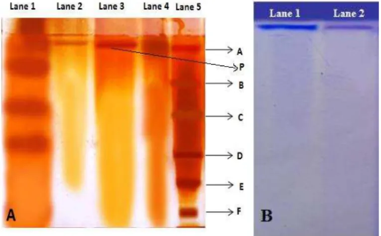
After silver staining, the molecular weight of L-asparaginase was found to be 99
± 1 kDa (Fig 2A). The molecular weight of 94 kDa was reported for
L-asparaginase of Penicillium brevicompactum NRC 829 (Elshafei et al., 2012)
and Aspergillus terreus (Balasubramanian et al., 2012). Another study showed
66 kDa for L-asparaginase from Penicillium sp (Patro and Gupta, 2012). The
L-asparaginase from Cladosporium sp.had a molecular weight of 121 kDa (Kumar
and Manonmani, 2013). Goodsell (2005) has reported the tetrameric nature of L-asparaginase and the present enzyme could be a homotetramer as a single band was observed not only in SDS-PAGE but also in native PAGE (Fig 2B). The L-asparaginase molecular weight variability may be attributable to the genetic differences.
Figure 2 (A) SDS-PAGE. with lane 1 – crude, lane 2 –purified (10 µg), lane 3 –
purified (20 µg), lane 4 –purified (40 µg) and lane 5 – molecular weight marker.
A: phosphorylase b (97.4 kDa), B: serum albumin (66.2 kDa), C: ovalbumin (45 kDa), D: carbonic anhydrase (31 kDa), E: trypsin inhibitor (21.5 kDa), F:
lysozyme (14.4 kDa), P: purified enzyme. (B) Native PAGE. Lane 1 and 2:
purified enzyme.
Activity staining of L-asparaginase
After staining with Nessler’s reagent, a brownish yellow colour was only developed in the purified and crude sample, indicating the L-asparagine degradation by L-asparaginase. The control did not show any variation due to the absence of the substrate (Fig 3).
Figure 3 Activity staining of L-asparaginase. Control (left), purified enzyme sample (center), crude sample (right).
Effect of pH on L-asparaginase activity and stability
The effect of pH on enzyme activity was analysed and the optimum was pH 7.0
and decreased thereafter (Fig 4). Comparable result was reported by Patro and
Gupta (2012) for L-asparaginase of Penicillum sp. The pH stability of the Trichoderma viride L-asparaginase at pH 7.0 was studied and retention of 82.49% asparaginase activity was observed after 24 h (data not shown).
Siddalingeshwara and Lingappa (2010) purified the L-asparaginase from Aspergillus terreus KLS2 that was only 100% stable at pH 8.0 for only 1 h.
Dange and Peshwe (2011) reported the L-asparaginase from Aspergillus aculeatus that was stable up to 8 h at pH 9.0. The pH stability of L-asparaginase of Trichoderma viride Pers: SF Grey is desirable in the pharmaceutical industries.
Figure 4 Effect of pH on L-asparaginase activity. The values with different letters on error bars are significantly different at P 0.05.
Effect of temperature on L-asparaginase activity and stability
The purified enzyme showed optimum activity at 37°C (Fig 5). The optimum
temperature in the 30 - 37 °C range was also reported for L-asparaginase of
various fungal species (Siddalingeshwara and Lingappa, 2010; Dange and
Peshwe, 2011; Patro and Gupta, 2012). The enzyme was unaffected up to 40
°C and a significant activity loss was evident at elevated temperatures (Fig 5).
Similar denaturation had been reported for L-asparaginase of fungal species
(Sakamoto et al., 1977; Dange and Peshwe, 2011). Stability studies at 37 °C revealed that there was no drastic change in the activity and the enzyme was 100% stable up to 4 h. However, a slight decrease of 8% was observed after 24 h
(data not shown). L-asparaginase of Penicillium brevicompactum NRC 829
maintained stability up to 1 h at 37 °C (Elshafei et al., 2012) while the enzyme of Aspergillus aculeatusremained stable for 2 h at 30 °C (Dange and Peshwe, 2011). The L-asparaginase of the present study can be applied as a therapeutic enzyme due to its thermostability and temperature-activity profiles.
j i
h d
c
a a
b c
e f
g
0 200 400 600 800 1000 1200
2 3 4 5 6 7 8 9 10 11 12 13
E
n
z
y
m
e
a
c
ti
v
it
y
(
U
/m
l)
Figure 5 Effect of temperature on L-asparaginase activity. The values with different letters on error bars are significantly different at P 0.05.
Effect of metal ions on L-asparaginase
Mg2+ and Na+ showed an enhancement in the L-asparaginase activity while K+,
Zn2+ did not have any effect on the enzyme. Ca2+, Hg2+ and Cu2+ moderately
inhibited the enzyme. However an important decline was seen with Fe3+, Co2+,
Mn2+ and Fe2+ (Fig 6). In contrast, Mg2+ acted as an inhibitor on L-asparaginase
of Cladosporium sp. (Kumar and Manonmani, 2013). Cu2+, Mg2+ also
inhibited L-asparaginase activity of Aspergillus aculeatus; however, Mn2+, Ca2+,
Fe2+ and Cu2+ did not alter the enzyme (Dange and Peshwe, 2011).
L-asparaginase of Cylindrocarpon obtusisporum was also inhibited by Fe2+, Ni2+,
Cu2+ and Hg2+ (Raha et al., 1990). The cations thus have different effects on
L-asparaginase activity.
Figure 6 Effect of metal ions on L-asparaginase activity. The values with different letters on error bars are significantly different at P 0.05.
Effect of specific inhibitors and activators
The inhibition studies provide an understanding of the active site amino acid residues responsible for L-asparaginase activity. PMSF and NEM did not have any effect on the enzyme, whereas NaN3 and NBS slightly inhibited the enzyme. A drastic inhibition was observed with EDTA (Fig 7) suggesting the purified
enzyme to be a metalloenzyme. The L-asparaginase purified from Penicillium
brevicompactum NRC 829 (Elshafei et al., 2012) was also metalloenzyme. Urea strongly inhibited the present enzyme. A similar inhibition was seen for
L-asparaginase of Pseudomonas stutzeri MB-405 (Manna et al., 1995). The
activity in the presence of non-ionic surfactants (Tween 20 and Triton X-100) was enhanced and this may be ascribed to their surfactant nature which is generally known to favor enzyme action. In contrast, the anionic surfactant SDS reduced the enzyme activity due to cleavage of disulphide bonds. A loss of the
L-asparaginase activity (81%) by SDS was also noted by Manna et al. (1995).
Figure 7 Effect of specific inhibitors and activators on L-asparaginase activity. The control was without any group reagent added. The values with different letters on error bars are significantly different at P 0.05.
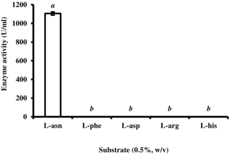
Substrate specificity and determination of kinetic constants of L-asparaginase
The affinity of L-asparaginase towards L-asparagine, L-aspartic acid, L-arginine, L-histidine and L-phenylalanine was studied. The enzyme only degraded its substrate (Fig 8). The result is in accordance with earlier reports where the fungal L-asparaginase exhibited higher affinity for L-asparagine than other amino acids
(Sakamoto et al., 1977; Siddalingeshwara and Lingappa, 2010; Dange and Peshwe, 2011; Patro and Gupta, 2012). The enzyme purified from this study did not act on L-glutamine. It may thus be used as an antileukemic and anti-lymphoma agent since it will not have the side effects related to L-glutamine specificity by L-asparaginase.
Figure 8 Substrate specificity of L-asparaginase. L-asparagine served as control. The values with different letters on error bars are significantly different at P0.05.
The Lineweaver-Burk plot was used to evaluate the affinity of L-asparaginase
towards L-asparagine. The Km and Vmaxvalues were 2.56 µM and 270.27 U.mL-1,
respectively (Fig 9). The Km value of 1.05 mM and 4 mM were reported by
Elshafei et al. (2012) and Patro and Gupta (2012), respectively. A few other
studies have shown 12.5 mM (and a Vmax of 104.16 I U.mL-1) (Dange and
Peshwe, 2011) and 1 mM (Raha et al., 1990). L-asparaginase with a low Km at
physiological pH is a candidate of choice to be used as antileukemic agent.
Figure 9 Lineweaver-Burk plot of L-asparaginase. c
b b a
a
d
e e
f g h 0 200 400 600 800 1000 1200
0 10 20 30 37 50 60 70 80 90 100
E n z y m e a c ti v it y ( U /m l)
Temperature (οC)
b a
a
b,c b,c c,d
e
d,e e,f
f,g g,h
h h h
0 20 40 60 80 100 120 140 160 180 Re la ti v e a c ti v ity (% ) Metal ions
a,b a a a,b b
c c
d
d d
0 20 40 60 80 100 120 R e la ti v e a c ti v it y ( % )
Specific activators and inhibitors (5 mM)
a
b b b b
0 200 400 600 800 1000 1200
L-asn L-phe L-asp L-arg L-his
E n z y m e a c ti v it y ( U /m l)
Substrate (0.5%, w/v)
y = 0,0096x + 0,0037 R² = 0,9978
-0,002 0,000 0,002 0,004 0,006 0,008 0,010 0,012
-0,6 -0,4 -0,2 0,0 0,2 0,4 0,6 0,8
TLC
TLC was performed to confirm hydrolysis of L-asparagine. The appearance of two separated spots indicated that L-asparaginase acted on its substrate and hydrolysed it (Fig 10). When the hydrolysate mixture was compared to the standards, it was confirmed that L-aspartic acid was formed due to the breakdown of the substrate. A comparable result was seen for L-asparaginase of Aspergillus terreus KLS2 (Siddalingeshwara and Lingappa, 2010).
Figure 10 Thin layer chromatography of L-asparaginase. Lane 1 – standard
L-aspartic acid; lane 2 – standard L-asparagine; lane 3 - enzyme hydrolysate
sample.
Antioxidant activity of L-asparaginase
ABTS assay was performed to check the antioxidant potential of L-asparaginase. The asparaginase was found to exhibit better antioxidant activity than
L-ascorbic acid which was used as a standard (Fig 11). ABTS•+ radical cation was
scavenged by the purified L-asparaginase. A linear correlation was observed between the scavenging activity of the ascorbic acid and that of
L-asparaginase. Similar correlation was observed for L-asparaginase of Aspergillus
flavus (KUFS20) (Rani et al., 2011). The enzyme of the present study can be used as an antileukemic agent as it can effectively remove free radicals.
Figure 11 L-asparaginase and L-ascorbic acid scavenging activities by ABTS assay
Acrylamide reduction potential of L-asparaginase
The effect of L-asparaginase on raw and fried potato was tested and higher activity was seen in raw potato (Fig 12). The L-asparagine present in potato gets converted into L-aspartic acid and ammonia. The difference in the enzyme activity can be attributed to higher content of free L-asparagine in raw potato, while in fried potato the free L-asparagine must have been converted into acrylamide when it was deep fried in oil. There was thus no residual L-asparagine
in the fried potato. It has been reported by Amrein et al. (2003) that the
L-asparagine concentration is normally higher than sugar content in potato. The
reduction of acrylamide formation by L-asparaginase was also reported (Kumar
and Manonmani, 2013). Reports have suggested that the formation of acrylamide can be prevented by lowering the frying temperature as this could
reduce the acrylamide content (Granda et al., 2004; Pedreschi et al., 2008). As
the formation of acrylamide is inevitable in fried potato chips when exposed to
high temperatures, the L-asparaginase of Trichoderma viride can be used as an
acrylamide reducing agent in the food industries.
Figure 12 Acrylamide reduction potential of L-asparaginase. The values with different letters or numbers on error bars are significantly different at P 0.05.
CONCLUSION
L-asparaginase was secreted by a marine soil screened Trichoderma viride Pers:
SF Grey. The properties of the enzyme such as high catalytic activity and stability at physiological pH and temperature, lack of glutaminase activity and high affinity towards L-asparagine, good free radical scavenging activity, and acrylamide mitigation makes it an ideal candidate to be exploited as a potent anticancer and acrylamide reduction agent.
REFERENCES
AMENA, S., VISHALAKSHI, N., PRABHAKAR, M., DAYANAND, A., LINGAPPA, K. 2010. Production, purification and characterization of
L-asparaginase from Streptomyces gulbargensis. Brazilian Journal of
Microbiology, 41, 173–178. http://dx.doi.org/10.1590/s1517-83822010000100025
AMREIN, T.M., SCHONEBACHLER, B., ROHNER, F., LUKAC, H., SCHNEIDER, H., KEISER, A., ESCHER, F., AMADO, R. 2003. Potential for
acrylamide formation in potatoes: data from the harvest. European Food
Research and Technology, 219, 572–578. http://dx.doi.org/10.1007/s00217-004-1025-z
BALASUBRAMANIAN, K., AMBIKAPATHY, V., PANNEERSELVAM, A.
2012. Production, isolation and purification of L-asparaginase from Aspergillus
terreus using submerged fermentation. International Journalof Advances inPharmaceutical Research, 3, 778–783.
BASKAR, G., RENGANATHAN, S. 2011. Optimization of media components and operating conditions for exogenous production of fungal L-asparaginase. Chiang Mai Journal of Science, 38, 270–279.
BLUM, H., BEIER, H., GROSS, H.J. 1987. Improved silver staining of
plant-proteins, RNA and DNA in polyacrylamide gels. Electrophoresis, 8, 93–99.
http://dx.doi.org/10.1002/elps.1150080203
DANGE, V.U., PESHWE, S.A. 2011. Production, purification and
characterization of fungal L-asparaginase. Bionano Frontier. 4, 162–167.
ELSHAFEI, A.M., HASSAN, M.M., ABOUZEID, M.A., MAHMOUD, D.A., ELGHONEMY, D.H. 2012. Purification, characterization and antitumor activity
of L-asparaginase from Penicillium brevicompactum NRC 82. British
Microbiology Research Journal, 2, 158–174.
GALLAGHER, M.P., MARSHALL, R.D., WILSON, R. 1989. Asparaginase as a
drug for treatment of acute lymphoblastic leukemia. Essays in Biochemistry, 24,
1–40.
GOODSELL, D.S. 2005. The molecular perspective: L-asparaginase. Oncolologist, 10, 238–239. http://dx.doi.org/10.1634/stemcells.FCM3
GRANDA, C., MOREIRA, R.G., TICHY, S.E. 2004. Reduction of acrylamide
formation in potato chips by low-temperature vacuum frying. Journal of Food
Science, 69, 405–411. http://dx.doi.org/10.1111/j.1365-2621.2004.tb09903.x
GULATI, R., SAXENA, R.K., GUPTA, R. 1997. A rapid plate assay for
screening L-asparaginase producing micro-organisms. LettersinApplied
Microbiology, 24, 23–26. http://dx.doi.org/10.1046/j.1472-765X.1997.00331.x
HOSAMANI, R., KALIWAL, B.B. 2011. Isolation, molecular identification and optimization of fermentation parameters for the production of L-asparaginase, an
anticancer agent by Fusarium equisetti. International Journal
ofMicrobiologyResearch, 3, 108–119.
IMADA, A., IGARASI, S., NAKAHAMA, K., ISONO, M. 1973. Asparaginase
and glutaminase activities of micro-organisms. Journalof General Microbiology,
76, 85–99. http://dx.doi.org/10.1099/00221287-76-1-85
KONEMAN, E.W., ALLEN, S.D., JANDA, W.M., SCHRECKENBERGER, P.C., WINN, W.C. 1977. Colour atlas and textbook of diagnostic microbiology.
5th J.B. Lippincott Company, p. 288–289, Philadelphia, USA.
KUMAR, N.S.M., MANONMANI, H.K. 2013. Purification, characterization and
kinetic properties of extracellular L-asparaginase produced by Cladosporium
sp. World Journal of Microbiology and Biotechnology, 29, 577–587.
http://dx.doi.org/10.1007/s11274-012-1213-0
a
b
1 1
0 100 200 300 400 500 600
Room temperature 37 °C
E
n
z
y
m
e
a
c
ti
v
it
y
(
U
.m
L
LAEMMLI, U.K. 1970. Cleavage of structural proteins during the assembly of
the heads of bacteriophage T4. Nature, 227, 680–685.
http://dx.doi.org/10.1038/227680a0
LAPMAK, K., LUMYONG, S., THONGKUNTHA, S., WONGPUTTISIN, P.,
SARDSUD, U. 2010. L-asparaginase production by Bipolaris sp. BR438 isolated
from brown rice in Thailand. Chiang Mai Journal of Science, 37, 160–164.
LINEWEAVER, H., BURK, D. 1934. The determination of enzyme dissociation
constants. Journal of the American Chemical Society, 56, 658–666.
http://dx.doi.org/abs/10.1021/ja01318a036
LOWRY, O.H., ROSENBROUGH, N.J., FARR, A.L., RANDALL, R.J. 1951.
Protein measurement with the folin phenol reagent. Journal of Biological
Chemistry, 193, 265–275.
MANNA, S., SINHA, A., SADHUKHAN, R., CHAKRABARTY, S.L. 1995. Purification, characterization and antitumor activity of L-asparaginase isolated
from Pseudomonas stutzeri MB-405. Current Microbiology, 30, 291–298.
http://dx.doi.org/10.1007/BF00295504
MUSHTAQ, M.S., SIDDALINGESHWARA, K.G., KARTHIC, J., SUNIL, D.P.L.N.S., NAVEEN, M., PRATIBHA, K.S. 2012. Rapid screening and
confirmation of L-asparaginase from Penicillium sp. International Journal of
Research in Pharmacology and Pharmacotherapeutics,1, 147–150.
PATRO, K.R., GUPTA, N. 2012. Extraction, purification and characterization of
L-asparaginase from Penicillium sp. by submerged fermentation. International
Journal of Biotechnology and Molecular Biology Research, 3, 30–34.
http://dx.doi.org/10.5897/IJBMBR11.066
PEDRESCHI, F., KAACK, K., GRANBY, K. 2008. The effect of asparaginase
on acrylamide formation in French fries. Food Chemistry, 109, 386–392.
http://dx.doi.org/10.1016/j.foodchem.2007.12.057
RAHA, S.K., ROY, S.K., DEY, S.K., CHAKRABARTY, S.L. 1990. Purification
and properties of an L-asparaginase from Cylindrocarpon obtusisporum MB-10.
Biochemistry International, 21, 987–1000.
RANI, S.A., SUNDARAMI, L., VASANTHA, P.B. 2011. In vitro antioxidant
and anticancer activity of L-asparaginase from Aspergillus flavus (KUFS20).
Asian Journal of Pharmaceutical and Clinical Research, 4, 174–177.
RE, R., PELLEGRINI, N., PROTEGGENTE, A., PANNALA, A., YANG, M., RICE-EVANS, C. 1999. Antioxidant activity applying an improved ABTS
radical cation decolorization assay. Free Radical BiologyandMedicine, 26,
1231–1237. http://dx.doi.org/10.1016/S0891-5849(98)00315-3
SAKAMOTO, T., ARAKI, C., BEPPU, T., ARIMA, K. 1977. Partial purification
and some properties of extracellular asparaginase from Candida utilis.
Agricultural and Biological Chemistry, 41, 1359–1364.
http://dx.doi.org/10.1080/00021369.1977.10862698
SARQUIS, M.I.M., OLIVIERA, E.M.M., SANTOS, A.S., DA-COSTA, G.L.
2004. Production of L-asparaginase by filamentous fungi. Memorias do Instituto
Oswaldo Cruz, 99, 489–492. http://dx.doi.org/10.1590/S0074-02762004000500005
SIDDALINGESHWARA, K.G., LINGAPPA, K. 2010. Key fermentation factors for the synthesis of L-asparaginase - An anti tumour agent through SSF
methodology. International Journal of Pharmaceutical Sciences, 1, 103–112.
THIRUNAVUKKARASU, N., SURYANARAYANAN, T.S., MURALI, T.S., RAVISHANKAR, J.P., GUMMADI, S.N. 2011. L-asparaginase from marine