Structural Model and Immunomodulatory Activities
Marle`ne Cot1,2, Aure´lie Ray1,2, Martine Gilleron1,2, Alain Vercellone1,2, Ge´rald Larrouy-Maumus1,2, Elise Armau3, Sophie Gauthier3, Ge´rard Tiraby3, Germain Puzo1,2, Je´roˆme Nigou1,2*
1CNRS, IPBS (Institut de Pharmacologie et de Biologie Structurale), Toulouse, France,2Universite´ de Toulouse, UPS, IPBS, Toulouse, France,3Cayla InvivoGen, Research Department, Toulouse, France
Abstract
Gram positive bacteria produce cell envelope macroamphiphile glycopolymers, i.e. lipoteichoic acids or lipoglycans, whose functions and biosynthesis are not yet fully understood. We report for the first time a detailed structure of lipoteichoic acid isolated from aStreptomycesspecies, i.e.Streptomyces hygroscopicus subsp. hygroscopicus NRRL 2387T. Chemical, MS and
NMR analyses revealed a polyglycerolphosphate backbone substituted witha-glucosaminyl anda-N-acetyl-glucosaminyl
residues but devoid of any amino-acid substituent. This structure is very close, if not identical, to that of the wall teichoic acid of this organism. These data not only contribute to the growing recognition that lipoteichoic acid is a cell envelope component of Gram positive Actinobacteria but also strongly support the recently proposed hypothesis of an overlap between the pathways of lipoteichoic acid and wall teichoic acid synthesis in these bacteria.S. hygroscopicuslipoteichoic acid induced signalling by human innate immune receptor TLR2, confirming its role as a microbe-associated molecular pattern. Its activity was partially dependant on TLR1, TLR6 and CD14. Moreover, it stimulated TNF-aand IL-6 production by a human macrophage cell line to an extent similar to that ofStaphylococcus aureuslipoteichoic acid. These results provide new clues on lipoteichoic acid structure/function relationships, most particularly on the role of the polyglycerolphosphate backbone substituents.
Citation:Cot M, Ray A, Gilleron M, Vercellone A, Larrouy-Maumus G, et al. (2011) Lipoteichoic Acid inStreptomyces hygroscopicus: Structural Model and Immunomodulatory Activities. PLoS ONE 6(10): e26316. doi:10.1371/journal.pone.0026316
Editor:Jean Louis Herrmann, Hopital Raymond Poincare - Universite Versailles St. Quentin, France
ReceivedAugust 3, 2011;AcceptedSeptember 23, 2011;PublishedOctober 18, 2011
Copyright:ß2011 Cot et al. This is an open-access article distributed under the terms of the Creative Commons Attribution License, which permits unrestricted use, distribution, and reproduction in any medium, provided the original author and source are credited.
Funding:The IPBS NMR equipment was financed by the French Research Ministry, CNRS, Universite´ Paul Sabatier, the Re´gion Midi-Pyre´ne´es, and European structural funds. The funders had no role in study design, data collection and analysis, decision to publish, or preparation of the manuscript.
Competing Interests:The authors have declared that no competing interests exist.
* E-mail: [email protected]
Introduction
Gram positive cell envelopes are characterized by the presence of cell-wall glycopolymers that are attached either to peptidogly-can or to membrane lipids. Lipid-linked glycopolymers are referred to as macroamphiphiles [1,2]. Many species contain both types of them [3–5]. The main macroamphiphiles are lipoteichoic acids (LTA), which are encountered in the majority of low G+C bacteria (Firmicutes) [6,7]. LTA is composed of a lipid anchor linked to a chain of poly-glycerol or poly-ribitol units separated by a phosphate group. Such repeated units are also characteristic of teichoic acids linked to peptidoglycan. In contrast, some of the high G+C Gram positive bacteria, i.e. Actinobacteria, do not produce LTA but rather lipoglycans [3,8–11]. Lipoglycans, of which mycobacterial lipoarabinomannan (LAM) is the archetype molecule [12], are macroamphiphiles made of a long chain of mannosyl units with possible arabinose and/or mannose ramifi-cations. They were proposed to discriminate bacteria of the phylum Actinobacteria [3,7]. However, LTA molecules have recently been characterized in a few number of Actinomycetes, such asAgromycesspecies [13] andThermobifida fusca[1] and several lines of evidence suggest that Streptomyces species would also produce LTA [14,15]. Identification of LTA in Actinobacteria raises the question of its biosynthesis in these bacteria. Indeed, the enzyme LtaS polymerase, which catalyses phospho-glycerol unit
polymerization and is essential for LTA synthesis inStaphylococcus aureus, lacks clear orthologues in the sequenced actinobacterial genomes [2]. Thus, LTA biosynthesis in Actinomycetes may proceed by an alternative pathway and Sutcliffe and colleagues [2] propose the attractive hypothesis that it could be achieved by a pathway that overlaps with that of teichoic acid biosynthesis, as suggested recently forStreptococcus pneumoniae[16].
activity of purified LTA has been a matter of controversy, contamination of LTA fractions by highly active lipopeptides being formally difficult to rule out [23]. However, immune activation is induced by synthetic LTA [24] and by LTA from a mutant S. aureusstrain lacking lipoproteins [25], confirming the role of LTA as a microbe-associated molecular patterns (MAMPs) of Gram-positive bacteria detected by the innate immune system. In the present study, we report for the first time a detailed structural model of LTA isolated from a Streptomyces species. Its capacity to induce TLR2 signaling and to stimulate cytokine production was investigated. Altogether, our results provide new clues on LTA biosynthesis in Actinobacteria and LTA structure/ function relationships.
Results
Extraction, purification and structural characterization of a LTA from S. hygroscopicus NRRL 2387 (ShLTA)
Macroamphiphile glycopolymers are classically extracted by a hot phenol-water procedure [26]. However, this might result in partially degraded LTA and most particularly in the loss of alanine substituents [27]. S. hygroscopicus NRRL 2387 cells were thus extracted by the more gentle butanol procedure introduced by Morathet al.[27]. After enzymatic degradation of the nucleic acid and protein contaminants, the fraction was purified by hydropho-bic interaction chromatography (HIC). This allowed removal of a glycopolymer, eluted with 10% isopropanol, composed of galactose and KDN (2-keto-3-deoxy-D-glycero-D-galacto-nononic acid) previously described inStreptomycesspecies [28,29] and that represented around 50% (w/w) of the fraction before HIC. SDS-PAGE analysis of the HIC fraction eluted with 35% of isopropanol revealed a compound with an apparent molecular weight of around 20 kDa and a migration pattern, as a broad band, similar to that of mycobacterial lipoglycans or LTA from S. aureus (SaLTA) (Figure 1). Its molecular mass distribution was estimated by MALDI-TOF mass spectrometry to be between 7 and 9 kDa for the major molecular species (not shown). Chemical analyses of the compound indicated a mean phosphorus content of 20 mol of
phosphorus per mol of molecule, suggesting a LTA rather than a lipoglycan structure. Accordingly, it was recognized in ELISA experiments by an antibody directed againstStaphylococcus epidermis LTA (Figure 2). This antibody also recognized SaLTA but neither mycobacterial lipoglycans nor the synthetic lipopeptide Pam3CSK4. In addition, glycerol (Gro) and glycerol-phosphate
(Gro-P) were detected by gas chromatography (GC)/MS suggest-ing a polyglycerolphosphate LTA (PGP-LTA). The compound was subsequently termed ShLTA. The predominant fatty acids detected by GC after alkaline hydrolysis were C16:0 (40%), isoC16 (11%), isoC17:0 (13%), anteisoC17:0 (28%), isoC15:0 (2%) and anteisoC15:0 (4%), a profile typical of Streptomyces species whole cells [30]. Glucosamine (GlcN) but neither mannosamine nor galactosamine were detected by GC and capillary electrophoresis monitored by laser-induced fluorescence (CE-LIF) after strong acid hydrolysis of ShLTA. Quantitative analyses by colorimetric assays indicated a ratio GlcN/phosphorus of 0.28/1 (mol:mol) (Table 1). No release of amino acids (,0.1% w/w), after acid hydrolysis, could be detected by a highly sensitive approach based on liquid chromatography (LC) monitored by LIF [31], indicating that ShLTA is neither substituted by amino acids nor contami-nated by traces amount of lipopeptides.
To get further insights into the distribution of the different structural motifs along the molecule backbone, ShLTA was submitted to 48% hydrofluoric acid (HF) hydrolysis for 48 h at 4uC, a reaction known to hydrolyze phosphodiester bonds. After a chloroform/methanol/water partition, the aqueous phase was analyzed by MALDI-TOF/MS and MS/MS. The data are summarized in Table 2. HF treatment did not completely hydrolyse all the phosphodiester bonds. Indeed, the positive MALDI mass spectrum showed peaks atm/z254.1, 336.1, 408.1, 569.2 and 611.2 attributed to (M+H)+
molecular ions that could be assigned to structures containing the motifs Gro-GlcN, P-Gro-GlcN, Gro-P-Gro-GlcN, Gro-GlcN-P-Gro-GlcN, and Gro-GlcNAc-P-Gro-GlcN respectively (Table 2). The assignments were confirmed by MS/MS analysis showing fragment ions characteristic of the loss of an amino sugar (i.e. GlcN) atm/z162.0 or a N-acetylated amino sugar (i.e. GlcNAc: N-acetyl-glucosamine) atm/z204.0 [32] (Table 2). Interestingly, the molecular ion atm/z 611.2 corresponded to a fragment containing both N-acetylated and non-N-acetylated amino sugars, suggesting that the PGP
Figure 1. SDS-PAGE analysis of a macroamphiphile glycopoly-mer fromS. hygroscopicus.Lane 1,Mycobactrium tuberculosisLAM and LM (top and bottom bands, respectively); lane 2,S. hygroscopicus macroamphiphile glycopolymer; lane 3, S. aureus LTA. The gel was revealed by periodic acid-silver nitrate staining. LAM, lipoarabinoman-nan; LM, lipomanlipoarabinoman-nan; LTA, lipoteichoic acid.
doi:10.1371/journal.pone.0026316.g001
Figure 2. An anti-LTA antibody recognizes S. hygroscopicus macroamphiphile glycopolymer.100 ng ofS. hygroscopicusorS. coelicolormacroamphiphile glycopolymers (ShLTA and ScLTA, respec-tively),S. aureus LTA (SaLTA), Pam3CSK4 synthetic lipopeptide or M.
tuberculosislipoglycans mixture were coated in microtiter plate wells and probed with an antibody directed againstS. epidermisLTA (anti-LTA). The results are mean 6 SD and are representative of three separate experiments.
backbone of ShLTA was substituted by both types of amino sugars. The control that a GlcNAc standard was not hydrolyzed into GlcN by HF treatment supported the above hypothesis.
Native ShLTA was subsequently analyzed by NMR. The1 H-NMR anomeric region exhibited two anomeric signals at d5.43
(GI1) andd5.10 (GII1) in a ratio 3.7/1 (Figure 3D), tentatively
attributed to both types of glucosamines (i.e. GlcN and GlcNAc). As revealed by 1H-13C HMQC spectrum, their corresponding anomeric carbons resonate at d 96.0 (GI1) and d 98.3 (GII1)
(Figure 3F). Proton and carbon resonances of both spin sys-tems were assigned from1H-13C HMQC and1H-1H HOHAHA experiments (partially shown in Figures 3F, 3G and 3E,
respectively). The assignments are summarized in Table 3. The chemical shift of C2 in both spin systems around 55 ppm was typical of amino sugars and the 1JC1,H1 coupling constant of
174 Hz indicated ana-anomeric configuration for both units. H2
resonances at d 3.36 (GI2) and d 3.94 (GII2) (Figure 3E) were
indicative of a non-N-acetylated and an N-acetylated glucosamine, respectively [33]. This was confirmed by a NOESY experiment recorded with ShLTA dissolved in H2O/D2O (9:1, v/v)
(Figures 3B and C). Indeed, an amide proton atd8.21 (GIINH)
(Figure 3A) showed intracyclic correlations with different protons that belonged to spin system GII (Figure 3C). In addition, another correlation was observed with methyl protons of an N-acetyl group at 2.09 ppm (Figure 3B). Altogether the data indicated that GI and GII spin systems correspond toa-GlcN and a-GlcNAc, respec-tively. Another amide proton resonance atd8.29 (GIIINH) with
very low intensity (,10%) was also observed and most probably corresponded to a minor form of N-acetylated amino sugar, whose characterization could not be carried out further.
Interestingly, nOe contacts were observed between anomeric protons of GI and GII and methine protons atd4.19 (gIII2) andd
4.05 (gIV2) respectively (Figure 3H), attributed to H2 protons of
1,3-diphospho-glycerol (P-1-Gro-3-P) units (Table 3) [33]. These data indicated that GlcN and GlcNAc units werea-glycosidically
linked to the C2 hydroxyl of the PGP repeating units. This was confirmed by an additional correlation on the NOESY spectrum between amide proton GIINH at d 8.21 and proton gIV2
(Figure 3D) and by HMBC experiment showing connectivities between anomeric protons of GI and GII and C2 of gIII and gIV units atd77.0 (gIII2) andd77.3 (gIV2) respectively (not shown)
(Table 3). The deshielding of the C2 resonances of these units (gIII2 and gIV2) as compared with C2 resonance of an
unsubstituted P-1-Gro-3-P unit at d 70.8 (gI2) (Dd 6.2 and
6.5 ppm, respectively) (Figure 3G; Table 3) was also in agreement with their substitution by glycosyl units. In contrast, C2 of Gro unit gII (gII2), whose resonance at d 76.6 was also deshielded as
compared to that of gI (Dd5.8 ppm) (Figure 3G; Table 3), did not
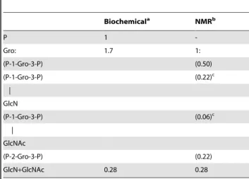
show in the HMBC experiment any correlation with protons, except with those of its own spin system (not shown). Similarly, H2 of gII unit did not correlate in NOESY or ROESY experiments with any protons other than those of its own spin system. Although clearly not glycosylated, the deshielding of C2 resonance indicated that this position was substituted and we hypothesized that gII spin system corresponded to 2,3-diphospho-glycerol (2-P-Gro-3-P) Table 1.Relative abundance of phosphorus, glycerol and
glucosamines inS. hygroscopicusLTA as determined by
biochemical and NMR analyses.
Biochemicala NMRb
P 1
-Gro: 1.7 1:
(P-1-Gro-3-P) (0.50)
(P-1-Gro-3-P) (0.22)c
|
GlcN
(P-1-Gro-3-P) (0.06)c
|
GlcNAc
(P-2-Gro-3-P) (0.22)
GlcN+GlcNAc 0.28 0.28
aRelative quantification of P and amino sugars (i.e. GlcN
+GlcNAc) was
performed by colorimetric assays, the amount of P being fixed to 1. The ratio Gro/amino sugars was determined by GC but is overestimated because of partial amino sugar degradation during strong acid hydrolysis.
bRelative quantification of the different Gro derivatives was performed by
integration of the corresponding H-2/C-2 cross peak on1H-13C HMQC spectrum (Figure 3G).
cThese values perfectly fit with the GlcN/GlcNAc ratio of 3.7/1 as determined by
integration of the corresponding anomeric signals (Figure 3D).
Gro, glycerol; GlcNAc, N-acetyl-glucosamine; GlcN, glucosamine; P, phosphate; P-1-Gro-3-P, P-2-Gro-3-P, 1,3- and 2,3-diphospho-glycerol units respectively. doi:10.1371/journal.pone.0026316.t001
Table 2.Positive ion MALDI-TOF/MS and MS/MS analyses of structural motifs generated by HF hydrolysis (48%, 48 h at 4uC) of ShLTA.
MSParent ionsm/z
(relative intensity) MS/MSFragments ionsm/z Composition M Molecular formula M
162.0 (M+
) (1) - Anhydro-GlcN C6H12O4N
204.0 (M+) (0.21) - Anhydro-GlcNAc C
7H14O5N 254.1 (M+H+
) (0.97) 162 Gro-GlcN C9H19O7N
336.1 (M+H+
) (0.11) 162 P-Gro-GlcN C9H22O10NP
408.1 (M+H+) (0.28) 162, 247a Gro-P-Gro-GlcN C12H26O12NP
569.2 (M+H+) (0.15) 162, 247a Gro-GlcN-P-Gro-GlcN C
18H37O16N2P
611.2 (M+H+
) (0.14) 162, 204, 408, 450a Gro-GlcNAc-P-Gro-GlcN C
20H39O17N2P
m/zof parent and fragment ions, possible composition and Molecular formula are shown. Gro, glycerol; GlcNAc, N-acetyl-glucosamine; GlcN, glucosamine; P, phosphate group.
aloss of GlcN.
units, as previously described [34]. This was confirmed by 31P NMR analysis of deacylated ShLTA. Indeed, on1H-31P HMQC spectrum, a phosphate resonance at 2.3 ppm correlated with H2 of gII unit at 4.33 ppm (not shown), indicating that the C2 hydroxyl of gII was directly substituted by a phosphate group.
Integration of the Gro unit H2-C2 cross-peaks on HMQC experiment (Figure 3G) allowed us to obtain the relative proportion of the different motifs on the PGP chain (Table 1) and propose the structural model depicted in Figure 4.
TLR2 activation and cytokine production
LTA has been reported to activate phagocytic cells via recognition by TLR2 and TLR6 [35–38]. The molecular bases of this recognition have been recently partially uncovered by the resolution of a crystal structure of a soluble form of TLR2 in complex withS. pneumoniaLTA [39]. We first tested the ability of ShLTA to stimulate HEK293 cells stably transfected with human TLR2 and CD14 genes and a NF-kB-inducible reporter system (HEK-TLR2 cells). As expected, ShLTA induced NF-kB
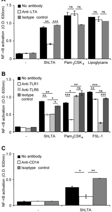
activa-tion in HEK-TLR2 cells (Figure 5) but not in the parent HEK cells (not shown). ShLTA stimulatory activity was inhibited by the anti-LTA antibody (Figure 5A). The latter did not show any effect on activation by the TLR2 ligands Pam3CSK4(Figure 5A) or FSL-1
(not shown), strongly suggesting that ShLTA activity was not due to contaminating lipopeptides. However, in agreement with a recent report by Seo et al. [40], ShLTA activity was altered by treatment by H2O2 (Figure 6A) or by a lipoprotein lipase
(Figure 6B). Indeed, although previously thought to be selective of lipopeptide/lipoproteins [22,23], these treatments have been
recently shown to also affect the chemical structure of pneumo-coccal and staphylopneumo-coccal LTA [40]. The requirement of TLR1 or TLR6 for recognition of ShLTA by TLR2 was tested using blocking antibodies. Whereas the activity of the triacylated Pam3CSK4 and diacylated FSL-1 lipopeptides was clearly
dependant on TLR1 and TLR6 respectively, activity of ShLTA was partially inhibited by both anti-TLR1 and anti-TLR6 antibodies (Figure 5B). ShLTA activity was also dependant on CD14 (Figure 5C). Finally, we investigated the capacity of ShLTA to activate the human THP-1 monocyte/macrophage cells, using a cell line derivative that stably expresses a NF-kB-inducible
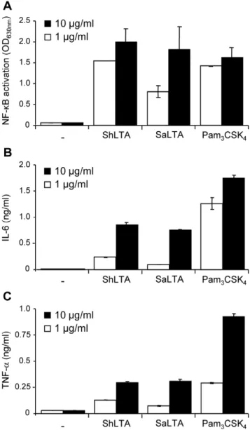
reporter system. ShLTA induced NF-kB activation (Figure 7A)
and IL-6 (Figure 7B) and TNF-a(Figure 7C) production by these
cells to an extent similar to that of SaLTA.
Discussion
In the present study, we report the first detailed structure of a LTA isolated from aStreptomycesspecies, i.e.S. hygroscopicus, and so confirms the presence of LTA in Streptomyces as previously suggested [14,15]. The assumption that LTA was absent from Actinomycetes most probably resulted from a sampling bias, since essentially families from the suborder Corynebacterineae, that all contain lipoglycans, have been analyzed so far [1]. In agreement with a preliminary report by Rahmanet al.[15], we also isolated LTA inStreptomyces coelicolor(ScLTA). It was recognized by the anti-LTA antibody (Figure 2) and preliminary structural analyses indicated that this LTA, as ShLTA, was of PGP type. However, surprisingly, we could not detect any macroamphiphile glycopo-Figure 3. 1D1H (A, D), 2D1H-13C HMQC (F, G),1H-1H HOHOHAtm110 ms (E) and1H-1H NOESYtm500 ms (B, C, H) spectra of ShLTA.
1
H (D),1H-13C HMQC,1H-1H HOHOHA and1H-1H NOESY (H) were recorded in D2O,1H (A) and1H-1H NOESY (B, C) in H2O/D2O (9:1) at 298K. Expanded
regions (d1H: 8.10–8.40) (A), (d1H: 8.10–8.40,d1H: 2.00–2.30) (B), (d1H: 8.10–8.40,d1H: 3.20–5.70) (C), (d1H: 5.05–5.50) (D), (d1H: 5.05–5.50,d1H: 3.20– 5.70) (E), (d1H: 5.05–5.50,d13C: 95–100) (F), (d1H: 3.90–4.40,d13C: 65–80) (G) and (d1H: 3.90–4.40,d1H: 3.20–5.70) (H) are shown. GI,a-GlcN, GII,a
lymer inStreptomyces verticilus. Similarly, Rahmanet al.reported the apparent absence of macroamphiphiles in the thermophilic Actinomycete Rubrobacter xylanophylus [1]. Our data thus extend their observation and demonstrate that within a given genus, some Table 3.Proton and carbon chemical shifts ofS.
hygroscopicusLTA.
Residue Name Protons d(ppm) Carbons d(ppm)
P-1)-Gro-(3-P- gI H-1, H-19 3.98/3.91 C-1 67.5
H-2 4.06 C-2 70.8
H-3, H-39 3.98/3.91 C-3 67.5
P-2)-Gro-(3-P- gII H-1, H-19 4.05 C-1 66.8
H-2 4.33 C-2 76.6
H-3, H-39 3.78/3.81 C-3 62.2
P-1)-Gro-(3-P-2) gIII H-1, H-19 4.04/4.13 C-1 66.0
H-2 4.19 C-2 77.0
| H-3, H-39 4.04/4.13 C-3 66.0
a-GlcN GI H-1 5.43 C-1 96.0a
H-2 3.36 C-2 55.3
H-3 3.97 C-3 71.1
H-4 3.50 C-4 70.9
H-5 3.96 C-5 73.7
H-6, H-69 3.91/3.81 C-6 61.7
P-1)-Gro-(3-P-2) gIV H-1, H-19 3.98 C-1 67.5
H-2 4.05 C-2 77.3
| H-3, H-39 3.90 C-3 67.5
a-GlcNAc GII H-1 5.10 C-1 98.3a
H-2 3.94 C-2 55.0b
H-3 3.82 C-3 72.4
H-4 3.50 C-4 70.9
H-5 3.97 C-5 73.7
H-6, H-69 3.91/3.81 C-6 61.7
Chemical shifts were measured at 298K in D2O and are referenced relative to internal acetone signals atdH2.225 anddC34.00. Gro, glycerol; GlcNAc, N-acetyl-glucosamine; GlcN, glucosamine; P, phosphate; P-1-Gro-3-P, P-2-Gro-3-P, 1,3- and 2,3-diphospho-glycerol units respectively.
a1
JH-1,C-1= 174 Hz. bCH
3CONH at 2.09 ppm and CH3CONH at 8.21 ppm. doi:10.1371/journal.pone.0026316.t003
Figure 4. Proposed structure of ShLTA. The PGP backbone is shown, with n, m, p and q estimated to be ca. 10, 4, 1 and 4 respectively. The relative position of the different motifs along the backbone is unknown. The LTA lipid anchor, presumptively a glycosyl-containing diacylglyceride glycolipid, is not shown. GI, GII and gI to gIV refer to spin systems as defined in Figure 3 and Table 3.
doi:10.1371/journal.pone.0026316.g004
Figure 5. ShLTA activity is inhibited by an anti-LTA antibody and is dependent on TLR1, TLR6 and CD14.A. Stimuli were pre-incubated for 30 min at 37uC with 5mg.ml21of anti-LTA or an IgG1
isotype control before HEK-TLR2 cells addition. ShLTA, Pam3CSK4andM.
tuberculosislipoglycans were tested at a concentration of 10 ng.ml21. B,
C. HEK-TLR2 cells were pre-incubated for 30 min at 37uC, before stimuli addition, with 5mg.ml21of various monoclonal antibodies: anti-TLR1
and anti-TLR6 (B), anti-CD14 (C) or IgG1 isotype control. ShLTA was tested at a concentration of 10 ng.ml21
. Pam3CSK4(5 ng.ml21) and
FSL-1 (0.5 ng.ml21) were used as positive controls of TLR2/TLR1 and TLR2/
TLR6 agonists, respectively. NF-kB activation was determined by
reading OD at 630 nm. The results are mean 6 SD and are representative of three separate experiments. *, P,0.05; **, P,0.01; ***, P,0.001; ns, not significant.
species can contain LTA and others not. This phenomenon has not been reported so far in lipoglycan-containing genera.
ShLTA was found to contain a PGP backbone, composed of 78% of 1-P-Gro-3-P and 22% of 2-P-Gro-3-P repeating units. A portion of the 1-P-Gro-3-P units werea-glycosylated at position 2
by GlcN or GlcNAc (Figure 4). Not unexpectedly, ShLTA was devoid of D-Alanine substituents found in many PGP-LTA. Indeed, this result is consistent with the fact that the genomes of Actinobacteria, includingStreptomyces spp., lack orthologues of the dlt system, which is dedicated to incorporation of D-Alanine into PGP-LTA and teichoic acids [1]. Interestingly, the PGP structure of shLTA is very similar, apart for lackingO-acetylation, to that of theS. hygroscopicusstrain ISP 5578 ( = NRRL 2387T) wall teichoic acid previously described by Tul’skaia et al. [34]. This contrasts with many Firmicutes, such asS. aureus orB. subtilis, for which LTA and teichoic acids have unrelated structures and are synthesized by two distinct pathways [3,41]. The genomes of Actinobacteria sequenced so far (including S. coelicolorand other Streptomyces spp) and of a few Firmicutes, such asS. pneumoniae, lack clear orthologues of the gene encoding LtaS polymerase [1]. This enzyme catalyses phospho-glycerol unit polymerization of LTA and is essential for its synthesis inS. aureus. Consequently, it has been suggested that in S. pneumoniae, LTA synthesis could be achieved by an alternative pathway that overlaps with that of teichoic acid biosynthesis [16,42] and Sutcliffe and colleagues [1] propose the attractive hypothesis that this pathway could also take place in Actinomycetes. Our finding that PGP moieties of S. hygroscopicusLTA and teichoic acid have a very similar structure further reinforces this hypothesis in which a PGP structure common to both molecules could be synthesized by the same
enzymatic machinery and then transferred to peptidoglycan as teichoic acid or to a lipid anchor to yield the PGP-LTA. Accordingly, orthologues of theB. subtilistagB/tagF teichoic acid biosynthesis genes are present in the genome ofS. himastatinicus(a genome sequenced member of the S. hygroscopicus phylogenetic subclade) and otherStreptomyces spp.(data not shown), supporting the occurrence of a pathway for teichoic acid biosynthesis in these species.
LTA is a powerful Gram-positive immunostimulatory compo-nent that induces cytokine production viabinding to TLR2 on phagocytic cells [43]. The structure/function relationships of this activity are not yet fully understood and possible contamination of LTA fractions by highly stimulatory lipoproteins have been advanced in the literature [44]. However, we found that TLR2 Figure 6. H2O2 (A) or lipoprotein lipase (B) treatments alter
ShLTA capacity to stimulate TLR2.HEK-TLR2 cells were stimulated with 10 ng.ml21ShLTA orM. smegmatislipoproteins (MsLP) previously
treated or not with 1% H2O2for 24 h at 37uC (A) or withPseudomonas
sp.lipoprotein lipase (B). NF-kB activation was determined by reading
OD at 630 nm. The results are mean6SD and are representative of three separate experiments. *, P,0.05; **, P,0.01; ***, P,0.001. doi:10.1371/journal.pone.0026316.g006
Figure 7. ShLTA stimulates NF-kB activation (A) and IL-6 (B) and TNF-a(C) production by human THP-1 monocyte/macro-phage cell line.A. THP-1 cells were stimulated with 1 or 10mg.ml21of
ShLTA, SaLTA or Pam3CSK4. NF-kB activation was determined by
reading OD at 630 nm. B, C. THP-1 cells were differentiated with 20 ng.ml21of PMA for 24 h and then stimulated with 1 or 10
mg.ml21
of ShLTA, SaLTA or Pam3CSK4. IL-6 and TNF-awere assayed in the
supernatant by sandwich ELISA. The results are mean6SD and are representative of three separate experiments.
activation by ShLTA could be inhibited by an anti-LTA antibody that had no effect on synthetic lipopeptide activity. Moreover, we failed to detect any amino acids (,0.1% w/w) in the ShLTA fraction used for bioassays, even by the use of a very sensitive technique based on LC-LIF [31]. Altogether, these data suggest that ShLTA is abona fideTLR2 ligand and they are in agreement with the demonstration by Morathet al.[24] that synthetic LTA is a potent stimulus of cytokine release. However, in contrast to the proposal by the same authors [24,27], our data demonstrate that D-alanination is not critical for LTA activity, or at least can be replaced. Indeed, ShLTA, although devoid of D-alanine substituents showed an activity similar to that of SaLTA. The role played by D-alanine in SaLTA activity is not clear. One hypothesis proposed [35] is that they could enable ionic interactions between LTA molecules forming multimers. If this is right, we can imagine that GlcN, rather uncommon in prokaryotes in its non-N-acetylated form, as a positively charged substituent, could functionally replace D-alanine in ShLTA. However, it is worth noting that only 22% of the PGP repeating units are substituted by GlcN in ShLTA as compared to 70% by D-alanine in SaLTA [27]. GlcNAc is supposed to have no impact on LTA activity [35]. ShLTA activity was altered by digestion with a lipoprotein lipase, which has been shown to deacylate not only lipoproteins but also LTA [40]. This observation is in agreement with the critical role played by fatty acids for LTA binding to TLR2 [39]. In agreement with a recent report by Seo et al. [40], ShLTA activity was also altered by treatment with H2O2. In contrast to what was previously thought, H2O2 and
lipoprotein lipase treatments are not selective at all of lipopep-tide/lipoproteins [22,23] and cannot be invoked to implicate the latter in TLR2 stimulating activities.
Using blocking antibodies experiments, we found ShLTA activity to be dependent on CD14 and partially on TLR6 and TLR1. Interestingly, these data are in agreement with the recent study by Bunket al.[25] demonstrating the required expression of TLR6 and CD14 for HEK-TLR2 cells activation by SaLTA devoid of lipopeptide contaminants. The choice of the TLR2 heterodimer, with either TLR1 or TLR6, can be dictated by subtle changes in the structure of the TLR2 ligand polar head [39] and is not yet completely understood.
Altogether, our findings contribute to the growing recognition that LTA is a cell envelope component of Gram-positive Actinobacteria [1]. Moreover, the detailed structure of ShLTA PGP moiety strongly supports the hypothesis of an overlap between the pathways of LTA and teichoic acid synthesis in Streptomyces. However, this has now to be definitely proved by genetic manipulation. Finally, our results provide new clues on LTA structure/function relationships, most particularly on the role of PGP substituents.
Materials and Methods
Srains and growth conditions
S. hygroscopicus subsp. hygroscopicusNRRL 2387 (ATCC27438),S. coelicolorM145 andS. verticillusATCC 21 890 II6-3 were grown at 27uC for 40 to 50 h under shaking in a culture medium which contained 5 g of yeast extract (BioSpringer), 5 g of bacto soytone peptone (Difco) and 20 g of glucose syrup (Roquette) per liter of deionized water.
Extraction and purification of LTA
Cells were delipidated at 50uC by mixing with chloroform/ methanol (1:2 and then 1:1) overnight. After lyophilisation, delipidated cells were resuspended in a 0.1 M citrate buffer
pH 4.7 and disrupted by sonication. The lysate was then extracted twice with 1 volume of butan-1-ol. Aqueous phase, containing LTA, was treated with DNAse, RNAse,a-amylase, pronase, a -chymotrypsin and trypsin. After ultrafiltration, samples were purified by hydrophobic interaction chromatography on an octylsepharose CL-4B column eluted with increasing concentra-tions of isopropanol (10, 15, 20, 25, 35, 40 and 80%) in 0.1 M ammonium acetate. Fractions were collected and analyzed by SDS-PAGE followed by periodic acid-silver nitrate staining. After dialysis and drying, they were submitted to a water/butan-1-ol (1:1 v/v) partition. The water phases were lyophilized and stored at
220uC. The fraction eluted with 35% isopropanol was used for further analysis.
H2O2and lipoprotein lipase treatments
20mg of LTA or lipoproteins (lipoproteins were extracted from a cleared lysate of Mycobacterium smegmatis by a phenol/water partition as previously described [45]) were resuspended in 100ml of 1% H2O2 and incubated at 37uC for 24 h. H2O2 was
evaporated by lyophilisation.
Lipoprotein lipase from Pseudomonas sp. (Fluka) was used at a ratio enzyme/LTA or lipoproteins 1:2 (w/w) in a 100 mM sodium phosphate buffer, 150 mM NaCl, 0.1% Triton X-100, pH 7.2 and incubated for 16 h at 37uC.
Elisa assay
ShLTA, ScLTA, commercial SaLTA (InvivoGen),M. tuberculosis lipoglycan mixture [12,46] or Pam3CSK4 (InvivoGen) (100 ng/
50ml of ethanol/water 1:1, v/v) were adsorbed in 96-wells microtiter plate by air drying and extensively rinsed with washing buffer (TBS, 0.5% BSA, 0.01% Tween 20). Wells were blocked for 2 h at room temperature with TBS, 5% BSA. After washing, 100ml of 5mg.ml21anti-LTA (Abcam, Ab12248) or IgG1 isotype control (eBioscience) antibodies in TBS, 1% BSA were added for 2 h at room temperature. After washing, 100ml of an HRP-conjugated goat anti-mouse antibody 1/3000 (Sigma) was added in TBS, 1% BSA for 1.5 h at room temperature. HRP activity was detected by addition of 100ml of Sure Blue TMB substrate (BD Biosciences).
Chemical analyses
Phosphorus content was determined after treatment of 50mg LTA by 260ml of perchloric acid at 170uC for 20 min. Then, 400ml of freshly prepared solutions of 1.25% molybdate and 5% ascorbate were added. After 5 min boiling in a water bath, samples were cooled in ice. Absorbance was read at 660 nm and phosphorus concentration was calculated relatively to a Na2HPO4
standard solution.
Amino sugars were analyzed after hydrolysis of 10mg LTA by 6N HCl at 110uC, overnight. They were detected either by CE-LIF using a 20 mM sodium borate buffer [47] after re-N-acetylation [48] and APTS derivatization [47] or by GC after trimethylsilylation by N,O-bis(trimethylsilyl)trifluoroacetamide/ trimethylchlorosilane (99:1, v/v) in pyridine (1:1, v/v) for 1 h at 110uC.
further added and after 30 min, OD was read at 520 nm. Amino sugar concentration was calculated relatively to a GlcN standard solution.
Amino acids were analyzed after hydrolysis of 100mg LTA by 6N HCl at 110uC, overnight by LC-LIF according to the method described by Siriet al.[31].
Fatty acids were analyzed by GC as their methyl esters after alkaline hydrolysis of 100mg LTA in 1N NaOH for 2 h at 37uC.
MALDI/MS
LTA molecular mass was determined on a Voyager DE-STR MALDI-TOF instrument (PerSeptive Biosystems) using linear mode detection. The matrix used was 2,5-dihydroxybenzoic acid (Sigma) at a concentration of 10mg.ml21in a mixture of ethanol/ water (1:1, v/v). 0.5ml of ShLTA, at a concentration of 10mg.ml21, were mixed with 0.5ml of the matrix solution. Mass spectra were recorded in the negative mode using a 350-ns time delay with a grid voltage of 90% of full accelerating voltage (25 kV) and a guide wire voltage of 0.15%.
ShLTA (1 mg) was depolymerized by 48% HF hydrolysis, 48 h at 4uC. After drying under a nitrogen stream, the sample was submitted to a methanol/chloroform/water (1:2:3, v/v/v) parti-tion. Aqueous phase, containing glycerophosphate motifs, was analyzed by MALDI/MS and MS/MS on a 4700 Proteomics Analyzer (with TOF/TOF optics, Applied Biosystems, Voyager DE-STR) using the reflectron mode. The matrix used was 2,5-dihydroxybenzoic acid at a concentration of 10mg.ml21 in a mixture of ethanol/water (1:1, v/v), 0.1% TFA. 0.3ml of the aqueous phase, at a concentration of 10mg.ml21, were mixed with 0.3ml of the matrix solution. Mass spectra were recorded in the positive mode. Collision-induced dissociation gas type was atmosphere, and the gas pressure was set to medium.
NMR
NMR spectra were recorded with an Avance DMX500 spectrometer (Bruker) equipped with an Origin 200 SGI using Xwinnmx 2.6. ShLTA was dissolved in D2O or H2O/D2O (9:1,
v/v) (D, 99.97%, Eurisotop) and analyzed in 20065 mm 535-PP NMR tubes at 298 K. Proton and carbon chemical shifts are expressed in parts per million and referenced relative to internal acetone signals at dH 2.225 and dC 34.00 ppm respectively. All
details concerning correlation spectroscopy and homonuclear Hartmann-Hahn spectroscopy sequences used and experimental procedures were as previously described [46].
HEK-TLR2 experiments
The HEK-Blue-2 cell line (InvivoGen), a derivative of HEK293 cells that stably expresses the human TLR2 and CD14 genes along
with a NF-kB-inducible reporter system (secreted alkaline phosphatase) was used according to the manufacturer’s instruc-tion. Cells were plated at 56104cells per well in 96-wells plates and the different stimuli were added at concentrations indicated in the figure legends in the HEK-Blue Detection medium (Invivo-Gen) that contains a substrate for alkaline phosphatase. Alkaline phosphatase activity was measured after 18 h by reading O.D. at 630 nm. To investigate the CD14 and TLR dependence of stimuli activity, HEK-TLR2 cells were pre-incubated for 30 min at 37uC, before stimuli addition, with various antibodies: 5mg.ml21 of monoclonal anti-CD14 (clone 134620, R&D Systems), monoclo-nal TLR1 (Clone H2G2, InvivoGen), monoclomonoclo-nal anti-TLR6 (Clone C5C8, InvivoGen) or an IgG1 isotype control (eBioscience). Anti-LTA (Abcam, Ab12248) antibody was used at a concentration of 5mg.ml21and pre-incubated with the different stimuli for 30 min at 37uC before HEK-TLR2 cells addition. Pam3CSK4and FSL-1 were purchased from InvivoGen.
THP1 experiments
THP-1-Blue cell line (InvivoGen), a derivative of THP-1 monocyte/macrophage human cells that stably expresses a
NF-kB-inducible reporter system (secreted alkaline phosphatase) was used according to the manufacturer’s instructions. Cells were added at 105 cells per well in 96-wells plates in the HEK-Blue Detection medium (InvivoGen) or differentiated with 20 ng.ml21 of PMA for 24 h in RPMI 1640 medium (Lonza) and the different stimuli were added at concentrations indicated in the figure legends. NF-kB activation was measured as described above. IL-6
and TNF-awere assayed in the supernatant by sandwich ELISA
using commercially available kits (Diaclone).
Statistical analysis
Results are expressed as a mean6SD and were analyzed using the Student’s t test to determine significant differences between samples.
Acknowledgments
We gratefully acknowledge Miss Audrey Noguera (IPBS) for helpful technical assistance and Dr Jean-Christophe Garrigue (IMRCP, CNRS, Toulouse) for amino acid analysis by LC-LIF. We thank Dr Iain Sutcliffe (Northumbria University, Newcastle upon Tyne, UK) for stimulating discussions.
Author Contributions
Conceived and designed the experiments: JN GP GT EA. Performed the experiments: MC AR MG AV GL-M EA SG. Analyzed the data: MC AR MG AV EA SG GT GP JN. Wrote the paper: MC JN.
References
1. Rahman O, Pfitzenmaier M, Pester O, Morath S, Cummings SP, et al. (2009) Macroamphiphilic components of thermophilic Actinomycetes: identification of lipoteichoic acid in Thermobifida fusca. J Bacteriol 191: 152–160.
2. Rahman O, Dover LG, Sutcliffe IC (2009) Lipoteichoic acid biosynthesis: two steps forwards, one step sideways? Trends Microbiol 17: 219–225.
3. Fischer W (1994) Lipoteichoic acids and lipoglycans. In: Ghuysen J-M, Hakenbeck R, eds. Bacterial cell wall. Amsterdam, The Netherlands.: Elsevier Science B.V. pp 199–215.
4. Brennan PJ (2003) Structure, function, and biogenesis of the cell wall of Mycobacterium tuberculosis. Tuberculosis 83: 91–97.
5. Weidenmaier C, Peschel A (2008) Teichoic acids and related cell-wall glycopolymers in Gram-positive physiology and host interactions. Nat Rev Microbiol 6: 276–287.
6. Sutcliffe IC, Shaw N (1991) Atypical lipoteichoic acids of gram-positive bacteria. J Bacteriol 173: 7065–7069.
7. Sutcliffe IC (1994) The lipoteichoic acids and lipoglycans of Gram-positive Bacteria: A chemotaxonomic perspective. Systematic and Applied Microbiology 17: 467–480.
8. Sutcliffe IC (1995) Identification of a lipoarabinomannan-like lipoglycan in Corynebacterium matruchotii. Arch Oral Biol 40: 1119–1124.
9. Gibson KJ, Gilleron M, Constant P, Sichi B, Puzo G, et al. (2005) A lipomannan variant with strong TLR-2-dependent pro-inflammatory activity inSaccharothrix aerocolonigenes. J Biol Chem 280: 28347–28356.
10. Whale GA, Sutcliffe IC, Morrisson AR, Pretswell EL, Emmison N (2004) Purification and characterisation of lipoglycan macroamphiphiles from Propioni-bacterium acnes. Antonie Van Leeuwenhoek 86: 77–85.
11. Garton NJ, Gilleron M, Brando T, Dan HH, Giguere S, et al. (2002) A novel lipoarabinomannan from the equine pathogenRhodococcus equi. Structure and effect on macrophage cytokine production. J Biol Chem 277: 31722–31733. 12. Nigou J, Gilleron M, Puzo G (2003) Lipoarabinomannans: from structure to
13. Gnilozub VA, Streshinskaia GM, Evtushenko LI, Shashkov AS, Naumova IB (1994) Lipoteichoic acids ofAgromycesspecies. Mikrobiologiya 63: 275–279. 14. Potekhina NV, Streshinskaia GM, Novitskaia GV, Naumova IB (1983)
[Isolation of lipoteichoic acid from Streptomyces levoris]. Mikrobiologiia 52: 434–437.
15. Rahman O, Cummings SP, Sutcliffe IC (2009) Phenotypic variation in Streptomyces sp. DSM 40537, a lipoteichoic acid producing Actinomycete. Lett Appl Microbiol 48: 226–229.
16. Seo HS, Cartee RT, Pritchard DG, Nahm MH (2008) A new model of pneumococcal lipoteichoic acid structure resolves biochemical, biosynthetic, and serologic inconsistencies of the current model. J Bacteriol 190: 2379–2387. 17. Fedtke I, Mader D, Kohler T, Moll H, Nicholson G, et al. (2007) AStaphylococcus
aureusypfP mutant with strongly reduced lipoteichoic acid (LTA) content: LTA governs bacterial surface properties and autolysin activity. Mol Microbiol 65: 1078–1091.
18. Grundling A, Schneewind O (2007) Synthesis of glycerol phosphate lipoteichoic acid inStaphylococcus aureus. Proc Natl Acad Sci U S A 104: 8478–8483. 19. Doran KS, Engelson EJ, Khosravi A, Maisey HC, Fedtke I, et al. (2005)
Blood-brain barrier invasion by group BStreptococcusdepends upon proper cell-surface anchoring of lipoteichoic acid. J Clin Invest 115: 2499–2507.
20. Ginsburg I (2002) Role of lipoteichoic acid in infection and inflammation. Lancet Infect Dis 2: 171–179.
21. Seo HS, Michalek SM, Nahm MH (2008) Lipoteichoic acid is important in innate immune responses to gram-positive bacteria. Infect Immun 76: 206–213. 22. Hashimoto M, Tawaratsumida K, Kariya H, Kiyohara A, Suda Y, et al. (2006) Not lipoteichoic acid but lipoproteins appear to be the dominant immunobio-logically active compounds inStaphylococcus aureus. J Immunol 177: 3162–3169. 23. Zahringer U, Lindner B, Inamura S, Heine H, Alexander C (2008) TLR2 -promiscuous or specific? A critical re-evaluation of a receptor expressing apparent broad specificity. Immunobiology 213: 205–224.
24. Morath S, Stadelmaier A, Geyer A, Schmidt RR, Hartung T (2002) Synthetic lipoteichoic acid fromStaphylococcus aureusis a potent stimulus of cytokine release. J Exp Med 195: 1635–1640.
25. Bunk S, Sigel S, Metzdorf D, Sharif O, Triantafilou K, et al. (2010) Internalization and coreceptor expression are critical for TLR2-mediated recognition of lipoteichoic acid in human peripheral blood. J Immunol 185: 3708–3717.
26. Fischer W, Koch HU, Haas R (1983) Improved preparation of lipoteichoic acids. Eur J Biochem 133: 523–530.
27. Morath S, Geyer A, Hartung T (2001) Structure-function relationship of cytokine induction by lipoteichoic acid fromStaphylococcus aureus. J Exp Med 193: 393–397.
28. Shashkov AS, Kosmachevskaya LN, Streshinskaya GM, Evtushenko LI, Bueva OV, et al. (2002) A polymer with a backbone of 3-deoxy-D-glycero-D-galacto-non-2-ulopyranosonic acid, a teichuronic acid, and a beta-glucosylated ribitol teichoic acid in the cell wall of plant pathogenicStreptomyces sp. VKM Ac-2124. Eur J Biochem 269: 6020–6025.
29. Shashkov AS, Tul’skaya EM, Evtushenko LI, Denisenko VA, Ivanyuk VG, et al. (2002) Cell wall anionic polymers ofStreptomyces sp.MB-8, the causative agent of potato scab. Carbohydr Res 337: 2255–2261.
30. Hoischen C, Gura K, Luge C, Gumpert J (1997) Lipid and fatty acid composition of cytoplasmic membranes fromStreptomyces hygroscopicusand its stable protoplast-type L form. J Bacteriol 179: 3430–3436.
31. Siri N, Lacroix M, Garrigues JC, Poinsot V, Couderc F (2006) HPLC-fluorescence detection and MEKC-LIF detection for the study of amino acids and catecholamines labelled with naphthalene-2,3-dicarboxyaldehyde. Electro-phoresis 27: 4446–4455.
32. Morelle W, Michalski JC (2007) Analysis of protein glycosylation by mass spectrometry. Nat Protoc 2: 1585–1602.
33. Shashkov AS, Streshinskaya GM, Senchenkova SN, Kozlova YI, Alferova IV, et al. (2006) Cell wall teichoic acids of streptomycetes of the phenetic cluster ‘Streptomyces fulvissimus’. Carbohydr Res 341: 796–802.
34. Tul’skaia EM, Shashkov AS, Evtushenko LI, Bueva OV, Naumova IB (1997) Strutural Identity of teichoic acids from Actinomycete species Strepotomyces hygroscopicus. Biochemistry (Mosc) 62: 289–293.
35. Deininger S, Stadelmaier A, von Aulock S, Morath S, Schmidt RR, et al. (2003) Definition of structural prerequisites for lipoteichoic acid-inducible cytokine induction by synthetic derivatives. J Immunol 170: 4134–4138.
36. Lehner MD, Morath S, Michelsen KS, Schumann RR, Hartung T (2001) Induction of cross-tolerance by lipopolysaccharide and highly purified lipoteichoic acid via different Toll-like receptors independent of paracrine mediators. J Immunol 166: 5161–5167.
37. Han SH, Kim JH, Martin M, Michalek SM, Nahm MH (2003) Pneumococcal lipoteichoic acid (LTA) is not as potent as staphylococcal LTA in stimulating Toll-like receptor 2. Infect Immun 71: 5541–5548.
38. Triantafilou M, Manukyan M, Mackie A, Morath S, Hartung T, et al. (2004) Lipoteichoic acid and toll-like receptor 2 internalization and targeting to the Golgi are lipid raft-dependent. J Biol Chem 279: 40882–40889.
39. Kang JY, Nan X, Jin MS, Youn SJ, Ryu YH, et al. (2009) Recognition of lipopeptide patterns by Toll-like receptor 2-Toll-like receptor 6 heterodimer. Immunity 31: 873–884.
40. Seo HS, Nahm MH (2009) Lipoprotein lipase and hydrofluoric acid deactivate both bacterial lipoproteins and lipoteichoic acids, but platelet-activating factor-acetylhydrolase degrades only lipoteichoic acids. Clin Vaccine Immunol 16: 1187–1195.
41. Fischer W (1988) Physiology of lipoteichoic acids in bacteria. Adv Microb Physiol 29: 233–302.
42. Fischer W (1993) Molecular analysis of lipid macroamphiphiles by hydrophobic interaction chromatography, exemplified with lipoteichoic acids. Anal Biochem 208: 49–56.
43. Draing C, Sigel S, Deininger S, Traub S, Munke R, et al. (2008) Cytokine induction by Gram-positive bacteria. Immunobiology 213: 285–296. 44. Hashimoto M, Furuyashiki M, Kaseya R, Fukada Y, Akimaru M, et al. (2007)
Evidence of immunostimulating lipoprotein existing in the natural lipoteichoic acid fraction. Infect Immun 75: 1926–1932.
45. Tapping RI, Akashi S, Miyake K, Godowski PJ, Tobias PS (2000) Toll-like receptor 4, but not toll-like receptor 2, is a signaling receptor forEscherichiaand Salmonellalipopolysaccharides. J Immunol 165: 5780–5787.
46. Gilleron M, Bala L, Brando T, Vercellone A, Puzo G (2000)Mycobacterium tuberculosis H37Rvparietal and cellular lipoarabinomannans. Characterization of the acyl- and glyco-forms. J Biol Chem 275: 677–684.
47. Gilleron M, Garton NJ, Nigou J, Brando T, Puzo G, et al. (2005) Characterization of a truncated lipoarabinomannan from the Actinomycete Turicella otitidis. J Bacteriol 187: 854–861.