UNIVERSIDADE NOVA DE LISBOA
REGULATION AND FUNCTION OF THE
CELL WALL POLYMER LIPOTEICHOIC
ACID IN
STAPHYLOCOCCUS AUREUS
CAROLINA PIÇARRA CASSONA
A THESIS SUBMITTED FOR THE DEGREE OF MASTER
IN MEDICAL MICROBIOLOGY
External supervisor:
Dr. Angelika Gründling
Internal supervisor:
Prof. Dr. Hermínia de Lencastre
Experimental work performed at the Centre for Molecular
Bacteriology and Infection, Imperial College London, Flowers
Building London SW7 2AZ, UK
Bibliographic elements resulting from this dissertation:
Published paper
Reichmann, N. T., Picarra Cassona, C. & Gründling, A. (2013). Revised mechanism of D-alanine incorporation into cell wall polymers in Gram-positive bacteria.
Acknowledgments
First, I would like to thank Angelika for the opportunity to work in her laboratory for her good supervision and guidance, and for all I have learned during this year.
I would like to thank everyone from CMMI3 who received me so well, and particularly to the members of Angelika’s lab: Rebecca, Matt, Ivan, Marta and Lauren and, although she is not in the lab anymore, to Nathalie, for all the help, good ideas and support she gave me during this year. Everyone made me fell part of the group and helped me when I needed advices.
I wish to thank my family and friends, especially Tiago, Joana, Verónica and Gabriela for all the support and friendship during this year.
I also thank Jan Marchant from the cross faculty NMR Centre at Imperial College London for running the NMR samples.
Abstract
Lipoteichoic acid (LTA) is an important polymer present in the envelope of various Gram-positive bacteria. In Staphylococcus aureus it is composed of a polyglycerolphosphate chain synthesised by the enzyme LtaS, and is anchored to the membrane via a glycolipid anchor. Depletion of LTA is known to cause growth arrest, aberrant positioning of septa and enlargement of cells leading to eventual cell lysis. This highlights the importance of LTA for bacterial growth and also suggests a possible link between LTA synthesis and cell division. Although key enzymes required for LTA synthesis have been identified, how this process is regulated has not been elucidated. This study investigates the mechanisms of regulation of LTA synthesis, as well as provides further insights into the roles of LTA in S. aureus.
In this study, nuclear magnetic resonance (NMR) was used to experimentally confirm a function for LTA in the incorporation of D-alanines into a second polymer, wall teichoic acid (WTA). LTA was also shown to be essential not only for bacterial growth, but also for bacterial survival, as cells become bactericidal in its absence. Furthermore, fluorescence microscopy using a GFP-tagged version of the synthase enzyme indicated that LtaS localises to the septum, suggesting a possible role for LTA in cell division.
Resumo
O ácido lipoteicóico (LTA) é um importante polímero presente no envelope de varias bactérias Gram-positivas. Em Staphylococcus aureus é composto por uma cadeia de poliglicerolfosfato sintetizada pela enzima LtaS e está ancorado na membrana através de uma âncora glicolipídica. Sabe-se que na ausência de LTA há interrupção do crescimento bacteriano, o septo deslocaliza e as células aumentam o seu tamanho e eventualmente lisam. Estas observações, mostram a importância do LTA para o crescimento bacteriano e sugerem uma ligação entre a sua síntese e a divisão celular. Apesar das principais enzimas necessárias para a síntese de LTA serem conhecidas, a regulação deste processo permanece por elucidar. Este estudo investiga a regulação da síntese do LTA e fornece novas perspectivas sobre as suas funções em S. aureus.
Neste estudo, ressonância magnética nuclear foi utilizada para confirmar experimentalmente a função do LTA na incorporação de D-alanina num segundo polímero, o ácido teicóico da parede (WTA). Mostrou-se também que o LTA não só é essencial para o crescimento, mas também para a sobrevivência bacteriana, tendo a sua ausência um efeito bactericida. Adicionalmente, microscopia de fluorescência utilizando fusões da sintase à proteína de fluorescência GFP, demonstrou que LtaS localiza no septo, sugerindo uma possível função do LTA na divisão celular.
Symbols and Abbreviations
% Percentage
°C Degrees Celsius
∆ Deletion
Ω Ohm
A Ampere
Amp Ampicillin
Atet Anhydrotetracycline
ATP Adenosine triphosphate
bp Base pairs
BSA Bovine serum albumin
c-di-AMP Cyclic diadenosine monophosphate CDP-Gro Cytidine diphosphoglycerol
D-Ala D-alanine
D-Gln D-glutamine
D-Lac D-lactate
D2O Deuterium oxide
DAG Diacylglycerol
ddH2O Double distilled water
DMSO Dimethyl sulfoxide
DNA Deoxyribonucleic acid
DNase Deoxyribonuclease
dNTP Deoxyribonucleotide triphosphate
ECL Enhanced chemi-luminescence
EDTA Ethylenediaminetetraacetic acid
Erm Erythromycin
F Farad
FucNAc N-acetyl-L-fucosamine
g Gram
GFP Green fluorescent protein
Glc-1-P Glucose-1-phosphate
Glc-6-P Glucose-6-phosphate
Glc2-DAG Diglucosyldiacylglycerol
GlcNAc N-acetylglucosamine
GlcNAc-1-P N-acetylglucosamine-1-phosphate
GroP Glycerol phosphate
h Hour(s)
IgG Immunoglobulin G
IPTG Isopropyl β-D-thiogalactosidase
Kan Kanamycin
kb Kilo base pair
kDa Kilodalton
L Liter
L-Lys l-lysine
LA Luria Bertani agar
LB Luria Bertani broth
LTA Lipoteichoic acid
Lys-PG Lysyl-phosphatidylglycerol
M Molar
ManNAc N-acetylmannosamine
ManNAcA N-acetylmannosaminuronic acid MBOAT Membrane-bound O-acetyltransferases
MIC Minimum inhibitory concentration
min Minute(s)
MOPS 3-(N-morpholino)propanesulfonic acid MRSA Methicillin resistant Staphylococcus aureus MSSA Methicillin sensitive Staphylococcus aureus
MU 4-methylumbilliferone
MUG β-D-galactopyranoside
MurNAc N-Acetylmuramic acid
NMR Nuclear magnetic resonance
nt Nucleotide(s)
OD Optical density
P-C55 Undecaprenyl phosphate
PBP Peptidoglycan binding protein
PBS Phosphate buffered saline
PCR Polymerase chain reaction
PG Phosphatidylglycerol
PGN Peptidoglycan
PGP Polyglycerolphosphate
PVDF Polyvinylidene difluoride
RboP Ribitol phosphate
rpm Rotations per minute
s Second(s)
SDS Sodium dodecyl sulfate
SOE PCR Splicing by overlap extension polymerase chain reaction
TA Teichoic acids
TCA Trichloroacetic acid
Tris Tris(hydroxymethyl)aminomethane
TSA Tryptic soy agar
TSB Tryptic soy broth
U Unit(s)
UDP Uridine diphosphate
UDP-Glc Uridine diphosphate glucose
UDP-GlcNAc Uridine diphosphate N-acetylglucosamine UDP-MurNAc Uridine diphosphate N-Acetylmuramic acid
UTR Untranslated region
V Volt
VISA Vancomycin-intermediate Staphylococcus aureus
VRSA Vancomycin-resistant Staphylococcus aureus
WT Wild-type
WTA Wall teichoic acid
X-Gal 5-bromo-4-chloro-3-indolyl-β-D-galactopyranoside
Table of Contents
Page
Acknowledgments ... v
Abstract ... vi
Resumo ... vii
Symbols and Abbreviations ... viii
Table of Contents ... xi
List of Figures ... xiii
List of Tables ... xiv
1. Introduction ... 1
1.1. Staphylococcus aureus ... 1
1.2. Components of the S. aureus cell wall ... 2
1.2.1. The polysaccharide capsule ... 4
1.2.2. Peptidoglycan structure and synthesis ... 5
1.2.3. Teichoic acids ... 7
1.2.3.1. WTA structure, synthesis and functions ... 8
1.2.3.2. LTA structure and synthesis ... 10
1.2.3.3. The function and localisation of lipoteichoic acid ... 13
1.2.3.4. D-Alanine incorporation into teichoic acids ... 14
1.2.3.5. Functions of D-alanine esters in teichoic acids ... 16
1.3. Objectives of this study... 17
2. Materials and Methods ... 19
2.1. Bacterial strains, growth conditions and storage ... 19
2.2. Recombinant DNA techniques ... 19
2.2.1. Purification of plasmid DNA from E. coli ... 19
2.2.2. Isolation of chromosomal DNA from S. aureus ... 20
2.2.3. Polymerase Chain Reaction (PCR) ... 20
2.2.5. Sequencing of clones ... 21
2.2.6. Preparation and transformation of chemically competent E.coli cells ... 22
2.2.7. Preparation and electroporation of electrocompetent S. aureus cells ... 22
2.2.8. Phage transduction in S. aureus cells ... 23
2.2.8.1. Preparation of phage lysates ... 23
2.2.8.2. Phage transduction of S. aureus ... 24
2.3. Strain and plasmid construction ... 24
2.3.1. ltaS promoter-lacZ fusions constructs ... 24
2.3.2. Construction of gfpP7-ltaSS218P fusions ... 26
2.4. S. aureus growth curves ... 27
2.5. Live/Dead bacterial viability assay ... 28
2.6. Membrane potential assay ... 28
2.7. β-Galactosidase activity assay in S. aureus ... 29
2.8. Sodium Dodecyl Sulphate Polyacrylamide Gel Electrophoresis (SDS-PAGE) and western immunoblot ... 30
2.8.1. Preparation of samples and detection of LTA by western immunoblot ... 31
2.8.2. Preparation of samples and detection of LtaS by western immunoblot ... 32
2.9. Preparation of cell wall and WTA purification for analysis by NMR ... 32
2.10. Fluorescence microscopy ... 33
3. Results ... 37
3.1. Cellular functions of LTA... 37
3.1.1. LTA is required for efficient incorporation of D-alanines into WTA ... 37
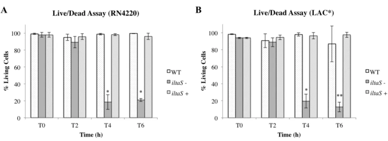
3.1.2. Depletion of LTA is bactericidal ... 38
3.1.3. Depletion of LTA causes membrane depolarisation ... 42
3.1.4. LtaS localisation studies ... 45
3.2. Regulation of ltaS expression and LTA production ... 51
3.2.1. Mapping and activity analysis of two ltaS promoters ... 51
3.2.2. LTA production is reduced under high salt conditions ... 55
4. Discussion and Conclusions ... 59
5. Bibliographic references ... 70
List of Figures
Page
Figure 1 –Schematic representation of the cell wall in Gram-positive bacteria...4
Figure 2 – WTA chemical structure in S. aureus...9
Figure 3 – LTA chemical structure in S. aureus...10
Figure 4 – Schematic representation of LTA synthesis in S. aureus...11
Figure 5 – NMR analysis of WTA isolated from WT and LTA negative S. aureus strains...38
Figure 6 – Determination of bacterial viability using a live/dead staining assay...41
Figure 7 – Quantification of the live/dead assay...42
Figure 8 – Membrane potential changes in RN4220-iltaS and LAC*-iltaS...44
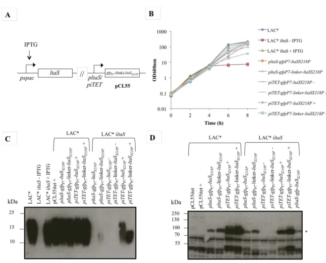
Figure 9 – Schematic representation of fluorescent protein fusions to the S. aureus LtaSS218P protein...47
Figure 10 – LtaS complementation analysis as assessed by LTA and LtaS western blot analysis and bacterial growth complementation analysis...48
Figure 11 – Localisation of LtaS in S. aureus as assessed by fluorescence microscopy...50
Figure 12 – Schematic representation oftwo ltaS promoter region...51
Figure 13 – Mapping and activity of two ltaS promoters as assessed by lacZ fusions and β-galactosidase activity...53
Figure 14 – Mapping and activity analysis of the ltaS promoters using mutagenesis approach as assessed by lacZ fusions and β-galactosidase activity...54
Figure 15 – Bacterial growth curves using wild-type and inducible ltaS strains in LB media containing a gradient of NaCl concentrations...56
Figure 16 – LTA and LtaS production under different NaCl concentration as assessed by western blot...58
List of Tables
Page
Table 1 –Strains used in this study...34
1.
Introduction
1.1.
Staphylococcus aureus
Staphylococcus aureus is a Gram-positive bacterium that is able to colonise and
infect human and animal hosts. It is non-motile, non-sporeforming, facultative anaerobe and a member of the Firmicutes that is distinguishable from other staphylococcal species by its ability to produce coagulase (coagulase-positive), a secreted protein that promotes clumping of the human blood cell (Gotz et al., 2006). S. aureus cells are 0.5-1.5 μm in diameter and can occur in various forms: singly, in pairs, tetrads or as grape-like clusters, which are formed by cells that fail to fully separate (Gotz et al., 2006; Tzagoloff & Novick, 1977). Cell division occurs in three orthogonal planes and the future cell division site is perpendicular to the previous septum (Tzagoloff & Novick, 1977).
On agar plates S. aureus forms golden-yellow round colonies, which is due to its ability to produce a carotenoid pigment, staphyloxanthin, that is important for its virulence and to evade the host innate immune system (Liu et al., 2005). Like other staphylococcal species, it has a low guanine and cytosine content in its genome (30-39 mol %) and the genome is approximately 3000 kb in size (Gotz et al., 2006).
S. aureus mainly colonises the nasal passages transiently, sometimes
permanently, and is a potential pathogen, which is able to cause both superficial skin infections as well as invasive infections such as toxic shock syndrome, endocarditis and osteomyelitis (Foster, 2005; Lowy, 2003).
The introduction of penicillin in the 1940s quickly promoted the proliferation of resistant S. aureusstrains expressing β-lactamase (Barber & Rozwadowska-Dowzenko, 1948), making the development of antibiotics against this Gram-positive pathogen a challenge. β-lactamase is encoded by blaZ that is part of a transposable element in a large plasmid. This enzyme acts by hydrolysing the β-lactam ring inactivating the antibiotic (Bondi & Dietz, 1945; Rowland & Dyke, 1989). With the introduction in the 1950s of the first semisynthetic penicillin, methicillin, methicillin-resistant S. aureus
(HA-MRSA), where the majority of infections still occur, but nowadays it is also community associated (CA-MRSA) (Fey et al., 2003; Okuma et al., 2002). β-lactam antibiotics act by mimicking the substrate of penicillin-binding proteins (PBPs) and, consequently, acylate and inactivate the PBPs, which are essential for the last steps of peptidoglycan synthesis (Chambers, 2003). The resistance mechanism in MRSA strains is due to the acquisition of an extra PBP, PBP2A encoded by the mecA gene (Beck et al., 1986). This PBP is able to perform the normal functions of the other PBPs in cell wall biosynthesis, but has low affinity for all β-lactam antibiotics (Beck et al., 1986; de Jonge et al., 1992). PBP2A acts by compensating for PBP2 transpeptidase (TPase) function, though the transglycosylase (TGase) function of PBP2 is still required (Pinho
et al., 2001) (see section 1.2.2. Peptidoglycan structure and synthesis).
The glycopeptide antibiotic vancomycin started to be used as an alternative treatment for MRSA infections. However, vancomycin-intermediate S. aureus (VISA) and fully vancomycin-resistant S. aureus (VRSA) have now been already detected. In VISA strains there is an increase in the quantity of peptidoglycan, which results in an increase of D-Ala-D-Ala residues exposed and, since these are the targets for vancomycin, the access to the bacterial target is prevented (Sieradzki & Tomasz, 1999). VRSA strains, on the other hand, emerged due to the acquisition of the vanA gene from vancomycin resistant enterococci. In these isolates, the terminal dipeptide in the peptidoglycan is D-Ala-D-Lac instead of D-Ala-D-Ala, the synthesis of which is catalysed by a ligase encoded by vanA (Showsh et al., 2001).
Given the emergence of resistance mechanisms to the available antibiotics, there has been a growing interest in studying the staphylococcal cell surface in order to find novel antibiotic targets.
1.2.
Components of the
S. aureus
cell wall
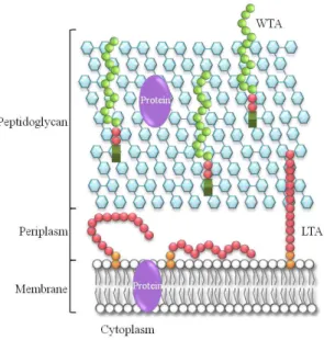
(TAs). TAs are composed of polyol-phosphate subunits and can be grouped into wall teichoic acids (WTAs), which are covalently attached to the peptidoglycan and lipoteichoic acids (LTAs), which are anchored to the cell membrane via a glycolipid anchor (Fischer, 1994; Neuhaus & Baddiley, 2003; Reichmann & Gründling, 2011; Xia
et al., 2010) (Figure 1).
The surface of Gram-positive bacteria also contains proteins that can be associated with the membrane through membrane spanning domains or attached to lipid anchors (Silhavy et al., 2010). Proteins can also be associated to the peptidoglycan layers or to other polymers such as TAs and polysaccharides (Navarre & Schneewind, 1999) (Scott & Barnett, 2006). It is still worth noting that the presence of a low-density region that can be considered to be the Gram-positive periplasm has been identified in these bacteria (Matias & Beveridge, 2005; Matias & Beveridge, 2006; Matias & Beveridge, 2008) (Figure 1).
The membrane is a phospholipidic bilayer with integrated membrane proteins and other components such as LTA and lipoproteins. In S. aureus, the four major lipids making up the membrane are diglucosyl diacylglycerol (Glc2-DAG), phosphatidyl
glycerol (PG), diacylglycerol (DAG) and a lysyl-phosphatidyl glycerol (Lys-PG) (Koch
et al., 1984; White & Frerman, 1967). LTA represents 6% of the membrane
composition and its synthesis is closely interconnected with the synthesis of the other membrane lipids (Koch et al., 1984) (see section 1.2.3.2 LTA structure and synthesis).
Figure 1 – Schematic representation of the cell wall in Gram-positive bacteria. The cell wall in Gram-positive bacteria is mainly composed of cross-linked peptidoglycan (blue hexagons), teichoic acids that can be divided into wall teichoic acids (WTAs) (green circles) and lipoteichoic acids (LTAs) (pink circles). Additionally, there are proteins associated to the membrane or to the peptidoglycan layer (purple). Adapted from Reichmann and Gründling, 2011.
1.2.1. The polysaccharide capsule
A capsule usually surrounds microorganisms that cause invasive infections. Encapsulation is an important characteristic as it enhances virulence and protects the bacteria from phagocytic uptake (O'Riordan & Lee, 2004). In S. aureus, polysaccharide capsules are grouped into serotypes and, although there are 11 serotypes, the serotype 5 and 8 are the most predominant ones in clinical isolates (Hochkeppel et al., 1987; Roghmann et al., 2005; Sompolinsky et al., 1985).
Of the S. aureus strains where the capsule biochemical structure has been ascertained all contain hexosaminuronic acids. Although types 5 and 8 belong to different serotypes, they have similar structures: both have a residue of N-acetylmannosaminuronic acid (ManNAcA) and two residues of N-acetyl-L-fucosamine (FucNAc) and they only differ in the way the sugars are linked and in the site of O-acetylation of the N-acetylamannosaminuronic acid residues (Fournier et al., 1984; Fournier et al., 1987).
also contain type-specific genes. The functions of these genes have not been experimentally demonstrated but the majority are likely to be involved in sugar synthesis, transfer, chain-length regulation and polymerisation (Sau et al., 1997).
It has been shown that the environment has an influence on polysaccharide capsule production. For example, growth in low concentrations of iron and on solid medium enhances the production of the serotype 8 capsule (Lee et al., 1993; Poutrel et al., 1995). Furthermore, serotype 5 capsule production is downregulated by CO2 levels
(serotype 8 capsule production is also regulated by CO2 levels but in a genetic
background dependent manner between different strains) and enhanced by NaCl concentration (Herbert et al., 2001; Sompolinsky et al., 1985).
1.2.2. Peptidoglycan structure and synthesis
The peptidoglycan structure is common to most bacteria, both Gram-positive and Gram-negative (Silhavy et al., 2010). It is composed of disaccharide-peptide repeats linked through glycolipid bonds [β-1,4 linkages between N-acetylmuramic acid (MurNAc) and N-acetylglucosamine (GlcNAc) subunits] forming glycan strands, which in turn are cross-linked through peptide stems attached to the MurNAc subunits (Ghuysen & Strominger, 1963; Navarre & Schneewind, 1999). This results in a structure that is flexible enough to ensure that the cell can resist changes in the internal osmotic pressure (Scheffers & Pinho, 2005). Although this structure is well dispersed amongst bacteria, its length is variable and in S. aureus most of the glycan strands have 3 to 10 subunits, but can reach up to 26 subunits (Navarre & Schneewind, 1999; Scheffers & Pinho, 2005).
Schneewind, 1999). S. aureus has a high degree of cross-linking when compared with other Gram-positive bacteria (up to 90% comparing with 56 to 63% in B. subtilis, for example), which is mainly attributed to PBP4 (Scheffers & Pinho, 2005).
As described below, the biosynthesis of peptidoglycan can be divided into three stages:
a) The first stage takes place in the cytoplasm and generates the precursors UDP-N-acetylmuramyl-pentapeptide (UDP-MurNAc-pentapeptide) and UDP-N-acetylglucosamine (UDP-GlcNAc). These steps involve the GlmM, GlmS and GlmU and the MurA-F proteins in a well known pathway (reviewed in Barreteau et al., 2008).
b) The second stage occurs in the cytoplasmic membrane, where precursor lipid intermediates are synthesised. Phospho-MurNAc-pentapeptide is transferred from UDP-MurNAc-pentapeptide to the membrane acceptor bactoprenol producing MurNAc-(pentapeptide)-pyrophosphoryl-undecaprenol, also called lipid I . The addition of GlcNAc to lipid I then yields GlcNAc-β -(1,4)-MurNAc-(pentapeptide)-pyrophosphoryl-undecaprenol, commonly known as lipid II (Scheffers & Pinho, 2005). The five glycine residues involved in cross-linking are added to the L-Lys residue by the nonribosomal peptidyl transferases Fem A, Fem B and Fem X (Berger-Bachi & Tschierske, 1998; Ton-That et al., 1998). These precursors are then translocated across the hydrophobic membrane to the outer leaflet of the membrane prior to being incorporated into the growing peptidoglycan chains (Scheffers & Pinho, 2005).
In methicillin susceptible S. aureus strains (MSSA) there are four native PBPs, which catalyse these reactions. PBP1 has an essential role in cell division and its transpeptidase domain has been shown to play a vital role during cell separation (Pereira et al., 2009). PBP2 has both TGase and TPase activity and localises at the cell division site (Scheffers & Pinho, 2005). However, in the presence of oxacillin, a β -lactam antibiotic which binds to the transpeptidase domain, PBP2 delocalises, indicating that binding to its substrate is necessary for correct localisation (Pinho & Errington, 2005). In methicillin resistant S. aureus strains there is an additional PBP, the PBP2A, which is encoded by the mecA gene (Beck et al., 1986; Hartman & Tomasz, 1984) and is able to restore PBP2 localisation even in the presence of oxacillin (Pinho & Errington, 2005). This extra PBP acts as a surrogate for the TPase activity of PBP2 in the presence of β-lactam antibiotics, but the TGase activity of the native PBP2 is still needed (Pinho et al., 2001). PBP3 is encoded by pbpC and its absence does not cause a change in peptidoglycan composition. However, the mutant of PBP3, once grown in the presence of sub-MIC levels of methicillin, displays increased cell size, aberrant cell shape and misplacement of the septa, indicating a role in cell division (Pinho et al., 2001). Finally, PBP4 is non-essential, is responsible for the high degree of peptidoglycan cross-linking observed in S. aureus and its mutant was found to be more susceptible to β-lactams (Memmi et al., 2008; Wyke et al., 1981).
Besides the enzymes necessary for PG synthesis it is also important not to forget that PG hydrolases have a vital role in cell growth, since it is important to cleave old PG in order to incorporate new material. Therefore, it has been proposed that PG synthases form a multienzyme complex together with hydrolases in order to maintain the equilibrium between these two activities (Scheffers & Pinho, 2005).
1.2.3. Teichoic acids
The cell surface of most Gram-positive bacteria including S. aureus and B.
subtilis also contain TAs. These components have a zwitterionic character and perform
least in S. aureus), and given its charge distribution, they are also possibly involved in regulating cation concentrations and enzymatic activities at the cell surface and in mediating interactions with receptors and biomaterials, which is relevant in biofilm formation (Schirner et al., 2009; Weidenmaier & Peschel, 2008; Xia et al., 2010).
TAs are composed of repeating polyol phosphate subunits such as ribitol phosphate (RboP) or glycerol phosphate (GroP) and can be grouped into WTA that is covalently linked to the peptidoglycan, and LTA that is anchored to the membrane through a glycolipid anchor and extends into the wall (Xia et al., 2010).
1.2.3.1.WTA structure, synthesis and functions
WTA is structural diverse in Gram-positive bacteria due to the nature of its monomeric forms and to modifications with glycosyl substituents and D-alanyl esters (Neuhaus & Baddiley, 2003). The monomers are polyol phosphate residues, which form linear chains covalently linked to the peptidoglycan through a phosphodiester bond. In the majority of S. aureus strains, the chains consist of RboP subunits, while in B.
subtilis WTA is made up of GroP residues (Bertram et al., 1981; Sanderson et al.,
Figure 2 – WTA chemical structure in S. aureus. The RboP chain can be modified with a D-alanyl ester (blue) or glycosyl group (green) and is linked to the peptidoglycan via a disaccharide composed of N-acetylglucosamine-1-phosphate (GlcNAc-1-P) and N-acetylmannosamine (ManNAc), and two GroP residues (pink).
The steps for S. aureus WTA synthesis are known and begin at the inner leaflet of the cytoplasmic membrane where the disaccharide linkage unit is produced. First, GlcNAc-1-P and ManNAc are transferred from UDP-activated precursor molecules to undecaprenylphosphate (C55-P), by TagO and TagA, respectively (Brown et al., 2008;
Xia et al., 2010). This first step is followed by the addition of the GroP residues,
involving TagB that adds the first GroP subunit to the disaccharidic linker, and TarF, which adds the second GroP subunit. The substrate used by these enzymes is the activated precursor cytidine diphosphoglycerol (CDP-Gro), which is generated by TagD (Badurina et al., 2003). TarL then polymerises the poly-RboP chain (Brown et al., 2008), using as its substrate the TarIJ generated precursor, CDP-Rbo (Meredith et al., 2008). Finally, this structure is transported to the outside of the cell through a two-component transporter (TagG and TagH), where it can be attached to the peptidoglycan (Brown et al., 2008). Besides D-alanylation, S. aureusWTA is also modified with α- or β-O-acetylgucosamine residues, which are added due the action of TarM and TarS as recently shown (Brown et al., 2012).
Although WTA is not essential for viability in both S. aureus and B. subtilis
1.2.3.2. LTA structure and synthesis
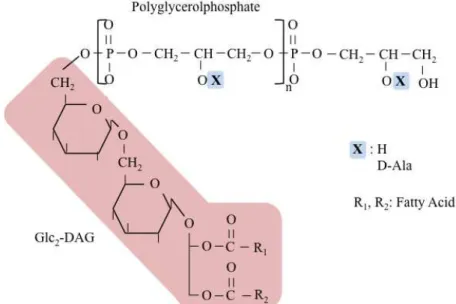
LTA consists of a polyglycerolphosphate (PGP) chain that is linked through a glycolipid anchor to the bacterial membrane and extends into the cell wall (Figure 3) (Archibald et al., 1968; Fischer, 1994). In staphylococci, bacilli and streptococci the glycolipid anchor is glucosyl (β1-6) glucosyl (β1-3) diacylglycerol (Glc2-DAG)
(Neuhaus & Baddiley, 2003; Reichmann & Gründling, 2011). Although LTA has a well-defined structure there is still variability due to glycosyl and D-alanine substitutions, fatty acid composition and length of the PGP chain (Neuhaus & Baddiley, 2003).
Figure 3 – LTA chemical structure in S. aureus. The polyglycerolphosphate (PGP) chain is linked to the membrane through a glycolipid anchor (Glc2-DAG) (pink). The PGP chain can be modified at the C2
position with D-alanine residues, as shown in blue. Adapted from Reichmann & Gründling, 2011.
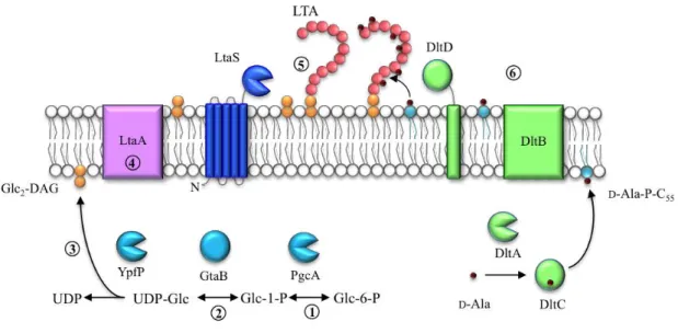
Figure 4 – Schematic representation of LTA synthesis in S. aureus. The glycolipid anchor (orange) is synthesised in the cytoplasm of the cell by PgcA, GtaB and YpfP (blue) (steps 1 to 3), and translocated across the membrane, which possibly involves LtaA (violet) (step 4). The polyglycerolphosphate chain (pink circles) is synthesised by the LTA synthase, LtaS (dark blue) (step 5). The DltA-D proteins (green) (step 6) are essential for the incorporation of D-alanine esters (dark red) into the LTA chain.
In S. aureus and B. subtilis, the Glc2-DAG anchor is synthesised in the
cytoplasm and this process involves the transference of two glucose moieties from uridine diphosphate glucose (UDP-Glc) to DAG (Fischer, 1994; Kiriukhin et al., 2001) (Figure 4). For this to occur, the action of three enzymes is required: PgcA (or Pgm), a α-phosphoglucomutase, which converts glucose-6-phosphate (Glc-6-P) to glucose-1-phosphate (Glc-1-P) (Gründling & Schneewind, 2007a; Lazarevic et al., 2005; Lu & Kleckner, 1994); GtaB (or GalU), which transfers UDP to glucose-1-phosphate, giving rise to UDP-Glc (Gründling & Schneewind, 2007a; Pooley et al., 1987); and YpfP (or UgtP), a processive glycosyltransferase which transfers two glucose moieties from UDP-Glc to DAG, generating Glc2-DAG and releasing UDP (Gründling & Schneewind,
2007a; Jorasch et al., 1998; Kiriukhin et al., 2001) (Figure 4, steps 1 to 4). Lastly, Glc2
-DAG needs to be transported across the membrane to the outer leaflet of the membrane, where the PGP chain will be polymerised on this glycolipid. It is thought that in S.
aureus LtaA a member of a major facilitator superfamily clan, and encoded in the same
All the enzymes involved in this process are not essential for cell viability since its mutants are still able to synthesise LTA but without a glycolipid anchor, resulting in LTA directly anchored to DAG (Gründling & Schneewind, 2007a; Lazarevic et al., 2005). Cells from S. aureus ypfP mutants were shown to be misshaped and enlarged (Kiriukhin et al., 2001) but still viable. However, these mutants demonstrate varying phenotypes depending on the strain background. For instance, RN4220 ypfP mutants produce a slightly increased amount of PGP-LTA that is released into the supernatant (Kiriukhin et al., 2001) while SA113 ΔypfP contains significantly reduced amounts of LTA (Fedtke et al., 2007).
As mentioned above, the PGP chain is polymerised on the outside of the cell and the LTA synthase (LtaS) is responsible for this reaction (Gründling & Schneewind, 2007b) (Figure 4). LtaS is a polytopic membrane protein with five N-terminal transmembrane helices (5TM domains) and a C-terminal extracellular enzymatic domain (eLtaS) (Gründling & Schneewind, 2007b; Lu et al., 2009) (Figure 4). This enzyme is cleaved in the linker region between these two domains by the protease SpsB, which is thought to inactivate the enzyme that is only active when in its full-length form (Wörmann et al., 2011b).
In S. aureus, LtaS depletion results in inability to synthesise LTA and growth arrest associated with severe envelope and division defects (Gründling & Schneewind, 2007b). In these conditions, cells present aberrant positioning of the septa, altered cell morphology and even lysed cells without cytoplasm or nucleic acids (Gründling & Schneewind, 2007b).
1.2.3.3.The function and localisation of lipoteichoic acid
In addition to WTA, LTA is also important to maintain the cell wall structure and has important functions in helping cells to adapt to different environmental factors.
In S. aureus it has been shown that LTA is important to protect the cell in low
osmolarity conditions, given the fact that an LtaS depleted strain is only able to survive at 37°C at high salt (7.5%) or sucrose (40%) (Oku et al., 2009). This may indicate that LTA act as an osmoprotectant under normal growth conditions.
As described above, S. aureus mutants with depleted LtaS have misplaced division septa, which seems to indicate a role in cell division (Gründling & Schneewind, 2007b; Oku et al., 2009). This may be due to interactions with the membrane-bound division machinery (Xia et al., 2010). Although in S. aureus it is still not proved that LtaS localises at the division septa, in B. subtilis both LtaS and YqgS localise to the site of cell division and the absence of LtaS results in FtsZ delocalisation and subsequent misplacement of septa (Schirner et al., 2009). Also, the YpfP homolog
in B. subtilis, UgtP, was shown to have a septal localisation, which is dependent on the
presence of UDP-Glc, the substrate for this enzyme (Weart et al., 2007). Together, this data seem to indicate a link between LtaS and cell division.
LTA, together with WTA, also has an important role in regulating autolytic activity in S. aureus, since in the absence of LtaS there is a decrease in autolytic activity
(Oku et al., 2009). Furthermore, an LTA suppressor strain with mutations affecting the
of LTA in autolysis regulation and may be related to the fact that LTA is essential for ion binding, particularly Mg2+ ions (Heptinstall et al., 1970).
Suggested function of LTA in the cell is closely related to its location and some suggested roles for this polymer are based on the assumption that this is a cell-surface exposed molecule. However, LTA is detected in the membrane and the detection of this polymer in whole cells of S. aureus is variable between strains (Aasjord & Grov, 1980). Furthermore, labelling experiments with positively charged gold nanoparticles suggested that the periplasmic space in B. subtilis is occupied by negatively charged components (Matias & Beveridge, 2008). These components could not be removed by protease treatment, indicating that these are not proteinaceous components, and could be detected by an LTA-specific monoclonal antibody (Matias & Beveridge, 2008). These data suggest that LTA might be the major component of the Gram-positive periplasm. Additionally, LTA PGP chain synthesis by LtaS that uses PG as a substrate, and D-alanylation by Dlt proteins suggests that LTA needs to remain close to the membrane during and after its synthesis (Reichmann & Gründling, 2011). However, it is important not to forget the possibility that LTA is also involved in WTA D-alanylation (Haas et al., 1984), which implies flexibility in the positioning of the LTA molecule.
It has also been suggested that LTA has roles in adherence, biofilm formation and interactions with the molecules from the host immune system (Xia et al., 2010) but these are not compatible with the idea that LTA is not exposed at the cell surface. These roles might be related with the fact that LTA is also released from the bacterial cell and found in the supernatant (Fischer, 1988).
1.2.3.4. D-Alanine incorporation into teichoic acids
Both LTA and WTA are modified with D-alanine esters and this process, adding positive charges to the otherwise negatively charged polymers, is important to modulate envelope properties (Neuhaus & Baddiley, 2003).
small protein with an expected size of 5.9 kDa and has been annotated to belong to the DUF3687 superfamily (Reichmann et al., 2013). In S. aureus and other gram-positive bacteria, this protein is encoded immediately upstream of dltA, but whether if functions in the D-alanylation process it is still unknown (Koprivnjak et al., 2006). The dltA gene encodes the D-alanine-D-alanyl carrier protein ligase (DltA) (Baddiley & Neuhaus, 1960), which belongs to a family of enzymes that activate and transfer amino or fatty acids to a thiol group of the 4-phosphopantetheine prosthetic group of a carrier protein
(Du et al., 2008; Osman et al., 2009). In the process of D-alanylation, its role is to ligate
D-alanine to the 4-phosphopantetheine of the carrier protein DltC, leading to the release
of adenosine monophosphate (AMP) (Heaton & Neuhaus, 1994; Neuhaus et al., 1996). DltB and DltD, encoded by dltB and dltD, respectively, have more questionable roles. DltB is predicted to be a polytopic membrane protein with 12 transmembrane domains and belongs to the membrane-bound O-acetyltransferases (MBOAT) family (Hofmann, 2000). DltD is also a membrane protein that is likely to be anchored to the membrane via its N-terminus (Perego et al., 1995). The roles these enzymes play in the process of D-alanylation are divergent between two proposed models, one forward by Fisher and
colleagues (1995) and the alternative proposed by Neuhaus & Baddiley (2003).
In the first model (Figure 4) it was proposed that DltB acts as a facilitator to transfer D-alanines from the carrier protein, DltC, to undecaprenyl phosphate, C55-P,
forming the lipid linked intermediate D-Ala-P-C55 (Perego et al., 1995). As a second
role for DltB this protein would aid in traversing the intermediate to the outer leaflet of the membrane. In a final step, DltD would act on the outside of the cell and, with its positive charge, would be able to detect the negatively charged PGP from LTA and bring it in close proximity to the intermediate D-Ala-P-C55 (Perego et al., 1995). The
Baddiley, 2003). Only recently it has been shown, using protein localisation and membrane topology analysis in S. aureus, that DltC remains within the cell, while DltD has an N-terminal embedded in the membrane and a C-terminus on the outer side of the membrane. These observations are in agreement with the model proposed by Fisher and colleagues (Reichmann et al., 2013).
For WTA D-alanylation it is known that the dlt operon is also necessary for this process, since it is abolished in B. subtilis dlt mutants (Perego et al., 1995). However, it has been proposed that the Dlt proteins may not be directly involved and instead D-Ala-LTA is the D-alanine donor for WTA (Haas et al., 1984). Pulse-chase experiments using [14C]-alanine showed that a decrease in radioactivity in LTA is coupled with an increase in radioactivity in WTA (Haas et al., 1984; Koch et al., 1985). However, there is still no direct evidence that D-Ala-LTA is the donor for WTA D-alanylation.
1.2.3.5.Functions of D-alanine esters in teichoic acids
Given the negative charges in TAs, their decoration with positively charged D-alanine residues plays an important role in the function of these cell wall components. In the absence of D-alanylation the cell wall net charge at the surface becomes more negative and this has the effect of making the cell more susceptible to positively charged molecules such as cationic antimicrobial peptides (CAMPs), defensins and nisin (Fabretti et al., 2006; Kovács et al., 2006; Kramer et al., 2008; Peschel et al., 1999), as well as to cells from the host immune system such as phagocytes and neutrophils, highlighting the importance of D-alanylation during infection (Collins et al., 2002; Kristian et al., 2005; Poyart et al., 2003; Walter et al., 2007). In S. aureus, it has also been observed that dlt mutants have an impaired ability to form biofilms and a reduced adherence to epithelial cells (Gross et al., 2001; Weidenmaier et al., 2004b).
The important role of TAs in scavenging ions, especially Mg2+ is related to D-alanylation (Fischer et al., 1981; Koprivnjak et al., 2006). In fact, the dlt operon in S.
aureus is downregulated in the presence of Mg2+ and also by an increase in NaCl
of the dlt gene is under the control of the two-component systems DltRS and LiaRS, respectively (McCormick et al., 2011; Poyart et al., 2001).
1.3.
Objectives of this study
Through the analysis of mutants defective in LTA synthesis, various functions have been attributed to this zwitterionic polymer. However, most of these roles are based on phenotypic analysis of mutants with altered LTA glycolipid anchor (Fedtke et al., 2007; Gründling & Schneewind, 2007a; Kiriukhin et al., 2001), since strains lacking the PGP chain are only viable under osmoprotective conditions or by the acquisition of suppressor mutations (Corrigan et al., 2011; Oku et al., 2009). Therefore little is known about the function of the PGP chain of LTA in S. aureus, including whether effects observed are direct or indirect, and so one key aim of this research was to provide further insights into roles of LTA. One such role is during D-alanine incorporation into WTA, whereby D-Ala-LTA has been proposed to act as the D-alanine donor (Haas et al., 1984). However, direct experimental evidence is lacking, and so this study investigated this hypothesis.
Under normal growth conditions, LTA is essential for S. aureus growth (Gründling & Schneewind, 2007b), though it is still not known if cells enter starvation and growth arrest under these circumstances or if the absence of this polymer causes actual cell death. In this study it is investigated whether the absence of LTA in S. aureus
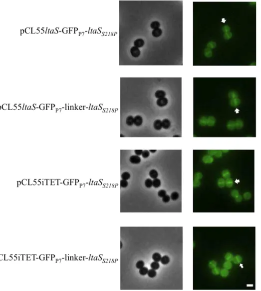
usually represented. At this location LTA is likely to have a physiological function during bacterial growth and cell division since the division machinery is mainly kept close to the membrane (Reichmann & Gründling, 2011). Therefore, in this study the localisation of LTA synthase, LtaS, was investigated using fusions to a green fluorescent protein (GFP).
2.
Materials and Methods
2.1.
Bacterial strains, growth conditions and storage
E. coli and S. aureus strains used in this study are shown in Table 1 with the respective references when applicable. The E. coli strain XL1 Blue was used for cloning and plasmid propagation. Strains were stored at -80°C by mixing 500 μl overnight culture with 500 μl freezer medium that consists of 10% bovine serum albumin and 10% monosodium glutamate. E. coli strains were grown at 37°C with shaking at 180 rpm in Luria Bertani (LB) broth or on LB agar. When necessary the medium was supplemented with the following antibiotics and inducers: Ampicillin (Amp), 100 μg/ml; Kanamycin (Kan), 30 μg/ml; 5-bromo-4-chloro-3-indolyl-β-D-galactopyranoside (X-gal), 40 µg/ml.
S. aureus strains were grown at 37°C with shaking at 180 rpm in Tryptic Soy
Broth (TSB) or on Tryptic Soy Agar (TSA). When necessary the medium was supplemented with the following antibiotics, inducers and indicators: Chloramphenicol (Cam), 5 to 10 μg/ml; Erythromycin (Erm), 10 μg/ml; Anhydrotetracycline (Atet), 100 to 200 ng/ml; Isopropyl β-D-thiogalactosidase (IPTG), 1 mM; 5-bromo-4-chloro-3-indolyl-β-D-galactopyranoside (X-gal), 100 µg/ml.
All optical density (OD) was measured using the Biochrom Libra S11 spectrophotometer.
2.2.
Recombinant DNA techniques
2.2.1. Purification of plasmid DNA from E. coli
Plasmid DNA was extracted from 5 ml E. coli overnight cultures using the Macherey-Nagel plasmid DNA purification kit, following the manufacturer’s instructions. The plasmid DNA was eluted in 40 μl ddH2O. When higher amounts were
plasmid midi kit, following the manufacturer’s instructions. The DNA was rehydrated in 150 μl ddH2O. Plasmid DNA was stored at -20°C.
2.2.2. Isolation of chromosomal DNA from S. aureus
In order to isolate S. aureus chromosomal DNA, a 4 ml overnight culture was centrifuged for 20 min at 1,303 xg. The pellet was suspended in 50 μl TSM buffer (50 mM Tris HCl pH 7.5, 0.5 M sucrose, 10 mM MgCl2 filter sterilised) and the cell wall
was digested for 30 min at 37°C using lysostaphin (AMBI Products) at a final concentration of 100 μg/ml. The subsequent steps of the chromosomal DNA isolation were performed using the Wizard Genomic DNA Purification kit (Promega, Madison USA) and following the manufacturer’s protocol. The DNA was rehydrated in 40 μl ddH2O for 1 h at room temperature and stored at -20°C.
2.2.3. Polymerase Chain Reaction (PCR)
All the primers used in this study were ordered from Sigma Aldrich and can be found in Table 2. In general all the reactions were performed in a total volume of 50 μl, adding 2 μl of 40 mM dNTPs, 1 μl of each primer at a concentration of 10 mM, 50 to 100 ng of template DNA, polymerase enzymes with their respective buffers, where for cloning 1 μl Herculase II (Agilent) was usedwith 10 μl 5X Herculase buffer and for all other PCR reactions, 0.5 μl Taq (New England Biolabs) with 5 μl 10X Taq buffer, and ddH2O to obtain the final volume. In order to fuse different fragments Splicing by
Overlap Extension PCR (SOE PCR) was used. In these reactions 2 μl from each PCR product was used as the DNA template.
PCR products were purified using the QIAquick Gel Extraction kit or the QIAquick PCR purification kit (QIAGEN) following the manufacturer’s instructions and eluted in 50 μl 5mM Tris HCl pH 8.
2.2.4. Restriction digests of plasmids and PCR fragments
Plasmid DNA was digested with restriction enzymes from New England Biolabs for cloning or to check inserts sizes in plasmids after cloning. For cloning, reactions were prepared using 10 μg DNA, 2 μl enzyme, 10 μl 10X enzyme buffer, 1 μl 100X BSA (when necessary) and ddH2O to obtain a final volume of 100 μl. The reaction was
incubated for 4 h at 37°C (except for SalI and BglII digests where the reactions were incubated for 16 h). In order to check inserts in plasmids, reactions were prepared using 0.1-1 μg DNA, 0.25 μl enzyme, 2 μl 10X enzyme buffer, 0.2 μl 100X BSA (when necessary) and ddH2O to obtain a final volume of 20 μl. The reactions were incubated
for 2 h at 37°C.
Ligation reactions were prepared in a final volume of 25 μl using a molar ratio of 1:3 vector to insert, 1 μl T4 DNA ligase (New England Biolabs), 2.5 μl 10X T4 DNA ligase buffer and ddH2O to obtain the right volume. The reaction was incubated at 17°C
for 16 h and heat-inactivated for 20 min at 65°C.
2.2.5. Sequencing of clones
After cloning all plasmid inserts were sequenced at the MRC CSC Genomics Core Laboratory at the Hammersmith Campus (Imperial College London) using fluorescent automated sequencing. The inserts of interest were PCR amplified using 25 μl Taq polymerase and purified using the QIAquick Gel Extraction kit or the QIAquick PCR purification kit (QIAGEN). Reactions for sequencing were prepared adding 10 ng per 100 bp of PCR product, 2 μl primer (10 mM) and ddH2O to give a total volume of
2.2.6. Preparation and transformation of chemically competent E.coli
cells
In order to prepare chemically competent E. coli cells, 20 ml of an overnight culture was back diluted 1:100 in 1 L PSI broth (5 g/L bacto yeast extract, 20 g/L bacto tryptone, pH 7.6, 20 mM MgSO4). The culture was incubated at 37°C with shaking at
180 rpm until an OD600nm of 0.5-0.7. Once grown, the culture was incubated on ice for
15 min and centrifuged at 3,847 xg for 10 min. The pellet was suspended in 200 ml sterile TfbI buffer (15% glycerol, 30 mM potassium acetate, 100 mM rubidium chloride, 10 mM calcium chloride, 50 mM manganese chloride) and incubated on ice for 15 min. The suspension was centrifuged at 3,847 xgfor 10 min. The pellet was suspended in 20 ml sterile TbfII buffer (15% glycerol, 10 mM MOPS, 75 mM calcium chloride, 10 mM rubidium chloride) and incubated on ice for 15 min. The cell suspension obtained was separated into 500 μl aliquots, snap frozen in a dry ice/ethanol bath and stored at -80°C. In order to transform chemically competent E. coli cells, 12.5 μl of heat -inactivated ligation mixture was added to 100 μl competent cells and incubated on ice for 30 min. The mixture was submitted to a heat shock by incubating at 42°C for 45 seconds and then incubated on ice for 5 min. Nine hundred μl SOC medium (5 g/L bacto yeast extract, 20 g/L bacto tryptone, 3.6 g/L glucose, 2.5 mM KCl, pH 7.0) was added to the cells, which were recovered by incubation at 37°C for 1 h. One hundred μl from the suspension was spread on LB agar with the appropriate antibiotics. The remaining cell suspension was pelleted at 17,000 xg for 5 min, 750 μl of the supernatant was removed, and the pellet was suspended in the remaining liquid and plated on LB agar plates with the appropriate antibiotics. Plates were incubated at 37°C overnight to obtain single colonies.
2.2.7. Preparation and electroporation of electrocompetent S. aureus
cells
shaking at 180 rpm until an OD600nm of around 2.1. The cells were then centrifuged at
6,000 xg for 10 min at 4°C. The pellet was washed twice with 150 ml sterile 0.5 M sucrose and once with 75 ml sterile 0.5 M sucrose. The final pellet was suspended in 1.5 ml sterile 0.5 M sucrose. The cell suspension obtained was separated in 230 μl aliquots that were snap frozen in a dry ice/ethanol bath before storage at -80°C.
Aliquots of 110 μl of cells were pipetted into 1 mm electroporation cuvettes (Biorad) and 20 μl of plasmid DNA in a concentration range of 300-600 ng/μl previously dialysed against ddH2O was added. For electroporation, a Biorad Gene
Pulser with 100 Ω, 2.5 kV and 25 µF settings was used. After electroporation, cells were recovered with 900 μl BHI 0.5 M sucrose medium, incubated for 1 h at 37°C and 100 μl cells were spread on TSA plates with the appropriate antibiotics. The remaining cell suspension was pelleted at 10,000 xg for 3 min and 750 μl from the supernatant was removed. The pellet was suspended in the remaining liquid and plated on TSA with the appropriate antibiotics. Plates were incubated at 37°C overnight and single colonies were re-streaked twice on TSA plates. Single colonies were used to inoculate TSB media with the appropriate antibiotics to grow overnight and freeze the cultures the next day.
2.2.8. Phage transduction in S. aureus cells
2.2.8.1. Preparation of phage lysates
For the preparation of phage lysates, the donor strains were grown in 4.5 ml LB/TSB in a ratio of 2:1 supplemented with 5 mM CaCl2 and incubated shaking at
37°C overnight. The next day, the culture was diluted 1:50 into new media with the same composition and grown for 3 h shaking at 37°C. A lysate of the transducing phage 85 was diluted in 10-fold dilutions from 10-1 to 10-6 in TMG buffer (10 mM Tris-HCl pH 7.4, 10 mM MgSO4, 0.1% gelatin) and 100 μl of each dilution was incubated with
appropriate antibiotics were overlaid with 5.5 ml cells in top agar and incubated with the lid side up at 37°C overnight. The next day, the plate with a semi-confluent layer of plaques was overlaid with 4 ml TMG buffer and incubated for 20 min at room temperature. The liquid was pipetted into a 15 ml tube and centrifuged for 20 min at 3,220 xg to pellet cellular debris and the supernatant was sterile filtered using a 0.22 mm filter. The phage lysates were stored at 4°C.
2.2.8.2.Phage transduction of S. aureus
The recipient strains were grown overnight in 24 ml LB/TSB in a ratio 2:1 supplemented with 5 mM CaCl2. The following day, cells were harvested by
centrifugation for 20 min at 3,220 xgand suspended in 4.5 ml LB/TSB in a ratio 2:1. To a 250 μl aliquot of cells, 200 μl lysate was added and incubated at 37°C shaking for 20 min. Cells were placed on ice and 24 μl 1 M sodium citrate was added to inhibit infection. Cells were pelleted at 10,000 xg for 3 min and washed twice with 1 ml 40 mM sodium citrate. The final pellet was suspended in 300 μl 40 mM sodium citrate, and 100 μl and 200 μl were spread on TSA supplemented with 40 mM sodium citrate and the appropriate antibiotics. The TSA plates were incubated for 48 h at 37°C and single colonies were streaked twice on new TSA plates supplemented 40 mM sodium citrate and the appropriate antibiotics.
2.3.
Strain and plasmid construction
2.3.1. ltaS promoter-lacZ fusions constructs
All the PCR products and pCL55iTET-lacZ were digested with KpnI/SalI and ligated. The resulting plasmids (pCL55p700ltaS-lacZ, pCL55p473ltaS-lacZ, pCL55p400ltaS
-lacZ, pCL55p300ltaS-lacZ, pCL55p200ltaS-lacZ and pCL55p100ltaS-lacZ) were first obtained in E. coli XL1 Blue yielding strains ANG2028, ANG2029, ANG2030, ANG2031, ANG2032 and ANG2033, respectively. The plasmids were then introduced into S. aureus RN4220 yielding strains ANG2866, ANG2867, ANG2868, ANG2869, ANG2870 and ANG2871, respectively. Similarly, the plasmids integrated into the chromosome were transduced into S. aureus LAC* generating strains ANG2964, ANG2965, ANG2966, ANG2967, ANG2968 and ANG2969, and into S. aureus
Newman resulting in strains ANG2970, ANG2971, ANG2972, ANG2973, ANG2974 and ANG2975, respectively.
In order to delete the bases from 400 to 30 from the full-length UTR producing pCL55pΔ400-30ltaS-lacZ, two fragments were amplified from pCL55p700ltaS-lacZ
using primer pairs 1058/1232 and 1233/817 and the PCR fragments were fused by SOE PCR using primer pair 1058/817. For the deletion of the P2 region, the bases 294 to 30 were deleted, constructing pCL55pΔ294-30ltaS-lacZ by amplifying two fragments from pCL55p700ltaS-lacZ with primer pairs 1058/1234 and 1235/817, and these PCR fragments were fused by SOE PCR using primer pair 1058/817. In order to mutate the transcriptional start site located 294 bp upstream of the start codon and obtain pCL55pΔ294ltaS-lacZ, two fragments were amplified from pCL55p700ltaS-lacZ using primer pairs 1058/1236 and 1237/817, and these PCR fragments were fused by SOE PCR using primer pair 1058/817. All PCR products and pCL55iTET-lacZ were digested with KpnI/SalI before ligation. The plasmids pCL55pΔ400-30ltaS-lacZ and pCL55pΔ294ltaS-lacZ were first obtained in E. coli XL1 Blue yielding strains ANG2204 and ANG2206, and afterwards introduced into S. aureus RN4220Δspa
2.3.2. Construction of gfpP7-ltaSS218P fusions
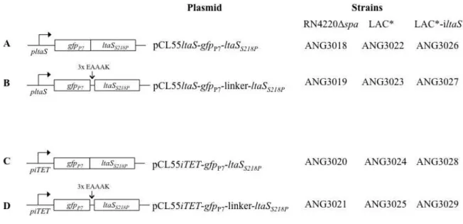
For the LtaS localisation studies, a fast folding version of GFP (GFPP7) was
fused to the N-terminal end of the non-cleavable LtaSS218P variant, with and without a
3x EAAAK amino acid linker region. These fusions were expressed either form the ltaS
native promoter or the Atet inducible iTET promoter.
For construction of pCL55pltaS-gfpP7-ltaSS218P, the promoter sequence of ltaS
was amplified from Newman chromosomal DNA using primer pair 1334/1697 and gfpP7
was amplified from pTrc99A-gfpP7 using primer pair 1698/1699. These PCR products
were fused by SOE PCR using primer pair 1334/317, resulting in the pltaS-gfpP7
fragment. The gene ltaSS218P was amplified from pOK-ltaSS218P using primer pair
1700/317 and fused to pltaS-gfpP7 using primer pair 1334/317, giving rise to pltaS
-gfpP7-ltaSS218P. The final PCR product and pCL55 were digested with EcoRI/KpnI and
ligated. The plasmid pCL55pltaS-gfpP7-ltaSS218P was initially obtained in E. coli XL1
Blue yielding the strain ANG2990 and subsequently introduced into S. aureus strains RN4220Δspa, LAC* and LAC*-iltaS yielding strains ANG3018, ANG3022 and ANG3026 respectively.
For construction of a pltaS-gfpP7-linker-ltaSS218P fusion, the promoter sequence
of ltaS was amplified from Newman chromosomal DNA using primer pair 1334/1697 and gfpP7 was amplified from pTrc99A-gfpP7 using primer pair 1698/1699. These PCR
products were fused by SOE PCR using primer pair 1334/1701, giving rise to pltaS
-gfpP7. The gene ltaSS218P was amplified from pOK-ltaSS218P using primer pair 1700/317
and both pltaS-gfpP7 and ltaSS218P were digested with SacII and ligated. The ligation
product was amplified using primer pair 1334/317 to yield the fusion pltaS-gfpP7
-linker-ltaSS218P. The final PCR product and pCL55 were digested with EcoRI/KpnI and ligated.
The plasmid pCL55pltaS-gfpP7-linker-ltaSS218P was initially obtained in E. coli XL1
Blue yielding the strain ANG2991 and subsequently introduced into S. aureus strains RN4220Δspa, LAC* and LAC*-iltaS yielding strains ANG3019, ANG3023 and ANG3027 respectively.
For construction of pCL55iTET-gfpP7-ltaSS218P fusion, gfpP7 was amplified from
pTrc99A-gfpP7 using primer pair 1702/1699 and ltaSS218P was amplified from
PCR using primer pair 1702/319. The final PCR product and the plasmid pCL55iTET
were digested with AvrII/BglII and ligated. The plasmid pCL55iTET-gfpP7-ltaSS218P was
initially obtained in E. coli XL1 Blue yielding strain ANG2992 and subsequently introduced into S. aureus strains RN4220Δspa, LAC* and LAC*-iltaS yielding strains ANG3020, ANG3024 and ANG3028 respectively.
For construction of pCL55iTET-gfpP7-linker-ltaSS218P fusion, gfpP7 was
amplified from pTrc99A-gfpP7 using primer pair 1702/1701 and ltaSS218P was amplified
from pOK- ltaSS218P using primer pair 1337/319. Both PCR products were digested with
SacII and ligated. The ligation product was amplified using primers 1702/319 to yield fusion iTET-gfpP7-linker-ltaSS218P. The final PCR product and pCL55iTET were
digested with AvrII/BglII and ligated. The plasmid pCL55iTET-gfpP7-linker-ltaSS218P
was initially obtained in E. coli XL1 Blue yielding strain ANG2993 and subsequently introduced into S. aureus strains RN4220Δspa, LAC* and LAC*-iltaS yielding strains ANG3020, ANG3024 and ANG3028 respectively.
2.4.
S. aureus
growth curves
RN4220 (ANG113), RN4220∆spa (ANG314), LAC* (ANG1575), RN4220-iltaS (ANG499) and LAC*-iltaS (ANG 2505) S. aureus strains and the remaining strains of interest for fluorescence microscopy were grown overnight at 37°C with shaking in TSB or LB supplemented with the appropriate antibiotics and 1 mM IPTG for the inducible strains. The next day, 1 ml culture was washed three times in TSB or LB and diluted 1:100 into 5 ml TSB or LB medium with and without IPTG, supplemented with the appropriate antibiotics. The cultures were grown at 37°C with shaking and bacterial growth was monitored by determining OD600nm every 2 h until the
2.5.
Live/Dead bacterial viability assay
The same S. aureus strains as used for growth curves (see section 2.4. S. aureus
growth curves) were grown overnight at 37°C with shaking in TSB supplemented with the appropriate antibiotics and 1 mM IPTG for the inducible strains. The next day 1 mL culture was washed three times in TSB and diluted 1:400 into 25 ml TSB medium with and without IPTG, supplemented with the appropriate antibiotics. Cultures were grown at 37°C with shaking and bacterial growth was monitored by determining OD600nm
every 2 h until the 6 h timepoint. From the overnight culture and at each time point, a 1 ml sample was centrifuged at 10,000 xg for 3 min and suspended in a volume of 0.85% NaCl normalised for OD600nm readings (where a culture with OD600nm of 0.5 was
suspended in 100 μl 0.85% NaCl).
In order to distinguish between living and dead cells, the Live/Dead Baclight Bacterial Viability Kit (Invitrogen) was used. Following the manufacturer’s instructions, equal volumes of component A (SYTO 9 dye) and component B (propidium iodide) were mixed and diluted 1:10 in DMSO (dimethyl sulfoxide). Of this mixture, 3 μl was added to 100 μl bacterial culture (suspended in 0.85% NaCl). Four μl was spotted on the slide and covered with a coverslip, before observation using a Nikon Eclipse E600 microscope equipped with a Nikon Digital DXM1200 camera and Digital Sight ACT-1 software. Cells were counted using the Image J software.
2.6.
Membrane potential assay
min, suspended in 1 ml buffer B (5mM HEPES pH 7.2, 5mM glucose, 100mM KCl) and diluted in the same buffer for a final OD600nm of 0.05. Two hundred μl of this
suspension were added to a black 96-well polystyrene fluorescence MaxiSorp plate (Thermo Scientific Nunc) plate. The membrane potential-sensitive fluorescent dye 3,3’ -dipropylthiadicarbocyanine iodide (DiSC3(5), Invitrogen) was added at a final concentration of 2 μM and the samples were incubated for 15 min to enable dye uptake and fluorescence quenching. Buffer B only and buffer B with dye was used as background and positive controls respectively. To one sample with wild-type bacteria, nisin, a pore forming peptide, was added at a final concentration of 10 μg/ml in order to guarantee fluorescence can be detected when there is no dye uptake. The fluorescence was measured at an excitation wavelength of 622 nm and emission wavelength of 670 nm.
2.7.
β
-Galactosidase activity assay in
S. aureus
S. aureus strains of interest were grown overnight in 4 ml TSB supplemented
with the appropriate antibiotics at 37°C with shaking. The following day cultures were diluted 1:100 into 5 ml fresh TSB medium and grown at 37°C with shaking and as negative control, S. aureus RN4220 pCL55iTET was grown in the presence of 200 ng/ml Atet.
Cultures were grown without inducer for 4 h and with inducer for 5 h. The OD600nm was recorded, 1 ml from each culture centrifuged at 17,000 xg for 10 min and
pellets were subsequently suspended in 1 ml ABT buffer (60 mM K2HPO4, 40 mM
KHPO4, 100 mM NaCl, pH 7.0, 1% Triton X-100) containing 20 μg/ml lysostaphin and
(MU) in ABT buffer at known concentrations were made and subsequently these values were used to determine the concentration (in μM) of product in each sample.
2.8.
Sodium Dodecyl Sulphate Polyacrylamide Gel Electrophoresis
(SDS-PAGE) and western immunoblot
In order to separate proteins and LTA prior to western immunoblot analysis, sodium dodecyl sulphate polyacrylamide gel electrophoresis (SDS-PAGE) was performed using 10 % or 15 % polyacrylamide gels produced as previously described (Sambrook et al., 1989). In order to estimate protein size, 5 µl of protein ladder (BenchMarkTM Prestained, Invitrogen or PageRuler Plus Prestained Protein Ladder, Thermo Scientific) was loaded beside 10 µl of the different samples, and separated by electrophoresis at 200 V (with the exception of LtaS samples that were separated at 100 V) using a Hoefer Mini Protein Electrophoresis system in 1X SDS buffer (14.4 g/L glycine, 3 g/L Tris-base, 1 g/L SDS).
using the Amersham ECL Prime Western Blotting Detection Kit (GE Healthcare), following the manufacturer’s instructions. The chemiluminescent signal was detected in an ECL Hyperfilm (GE Healthcare) and the film was developed using AGFA-Healthcare N. V. automated developer. Exposure time was variable depending on signal strength.
2.8.1. Preparation of samples and detection of LTA by western immunoblot
In order to obtain LTA samples for immunoblot analysis, the same S. aureus
strains as used for growth curves (see section 2.4. S. aureus growth curves) were grown overnight at 37°C with shaking in TSB supplemented with the appropriate antibiotics and 1 mM IPTG for the inducible strains. The following day 1 ml culture was washed three times in TSB and diluted 1:100 into 5 ml TSB medium with and without IPTG for the inducible strains and supplemented with the appropriate antibiotics. Cultures were grown for 4 h at 37°C with shaking and bacterial growth was monitored by determining OD600nm every 2 h. Cells were lysed by adding 1 ml culture to 0.5 ml of 0.1 mm glass
beads and vortexing upside down for 45 min at 4°C. In order to settle the beads, the samples were centrifuged for 1 min at 200 xg and the supernatant containing lysed bacteria was transferred into a fresh eppenford tube. Bacterial debris was collected by centrifuging for 15 min at 17,000 xg. The pellet containing LTA was suspended in 2 % SDS-containing sample buffer normalised for OD600nm readings (where a culture with
OD600nm of 3 was suspended in 90 μl buffer). Samples were boiled for 20 min and