Universidade Nova de Lisboa
Instituto de Higiene e Medicina Tropical
Different Models of DNA Immunization as Strategy
for Vaccine Development Against African
Trypanosomiasis
Adriana Beatriz Oliveira Temporão
DISSERTATION TO OBTAIN THE MASTER'S DEGREE IN BIOMEDICAL SCIENCES WITH SPECIALIZATION IN MOLECULAR BIOLOGY IN TROPICAL AND INTERNATIONAL MEDICINE
Universidade Nova de Lisboa
Instituto de Higiene e Medicina Tropical
Different Models of DNA Immunization as Strategy
for Vaccine Development Against African
Trypanosomiasis
Author: Adriana Beatriz Oliveira Temporão
Supervisor: Marcelo Sousa Silva, Assistant Researcher
Dissertation presented to fulfil the necessary requirements to obtain a Master's degree in Biomedical Sciences with specialization in Molecular Biology in Tropical and International Medicine
Acknowledgments
To my supervisor, Professor Marcelo Silva, for the patience, availability, optimism, for
all the support throughout this year and for all the transmitted knowledge.
To Diego Guerin, Ibai López-Marijuan and to Biofisika, for all the help in the VLP part
of the work and for making me feel welcomed.
To my lab colleagues, Jailson Querido, Cláudia Moreno, Joana Monteiro, Daniela
Portugal, Sónia Pestana, Janine Domingos, Joana Frazão, Filipa Ferreira and Ana Reis,
for the support, help and also for the great patience.
To Julie Dawes, for those two hours a week of fun and learning, for the advice and for
always pushing me to be better: I wouldn´t be writing this, if it wasn´t for you.
To Vânia Pereira, for being my sweetest friend.
To Aisén Vivas, for being the best tour guide ever.
To my Covilândia family: Mariana Nunes and Raquel Duarte. Thank you for adopting
me during that year, for all the good and memorable times, they will never be forgotten.
To Tiago Veiga, my dearest friend, for the advice, for the shared secrets and for being
always there, even miles and miles apart.
To my migos do coração, Tânia Rodrigues, Liliana Pires, David Migueis, Paulo
Carvalho and Ricardo Paulo, for always supporting me, for the fun times, for the
laughter, for the Saturday night coffees, and for showing me that friends are the family
we choose, and I chose the best one, ‘cause I will always love you more than chocolate.
To my miga, Catarina Azevedo, for all the coffees, serious (and not that serious) talks,
for being as mean as me, for the support and, of course, for putting up with me during
my panic attacks.
To my adoptive families: Cláudia, Henrique, Sara and Hugo; Alexandrina, Ismael and
Luis Miguel; Romy, Miguel and my little sister, Leonor; Maria José Brandão, João
To my sister from another mother, Helena Pereira, for the great times, the random
conversations, the long nights of study, the good and bad advice, for just being there, no
matter what, for the sarcasm and ironies, and for believing in me even when I didn’t.
To my parents, for making all this possible, for the support, for the constructive
criticism, for the laughter, for all the good and bad times, for making me what I am
today, because gosto de vocês até ao inxinito.
To my sister, for the things that just we understand, for being the most annoying person
in the world, for all the 5 minutes arguments, for all the crazy times and for always
being by my side, forever sisters metralha.
And to all the people who contributed directly or indirectly to my academic and
personal growth.
Index
1. INTRODUCTION 1
1.1 Pathology, Diagnosis and Treatment of Human African Trypanosomiasis 4
1.2 Trypanosoma brucei 7
1.2.1 Life cycle of Trypanosoma brucei 7 1.2.2 Cell structure of Trypanosoma brucei parasites 10 1.2.3 The Variant Surface Glycoprotein Coat of Trypanosoma brucei 11 1.2.4 Major Surface Protease and Phospholipase C from Trypanosoma brucei 12
1.3 Immunobiology of African Trypanosomiasis 14
1.4 DNA vaccination 16
1.5 The Virus-like Particles 19
2. WORK AIMS 22
3. MATERIALS AND METHODS 24
3.1 Animals and parasites 25
3.2 Infection protocol of mice and total protein extract from Trypanosoma brucei
brucei 25
3.3 Obtaining genomic DNA from Trypanosoma brucei 26
3.4 Obtaining the gene sequences of Trypanosoma brucei 26 3.5 Construction and acquisition of the primers used in the amplification of
Trypanosoma brucei genes of interest 26
3.6 Polymerase Chain Reaction (PCR) 27
3.7 Construction of DNA vaccine prototypes 28
3.7.1 Plasmid construction 28
3.7.2 Bacterial transformation 29
3.7.3 Plasmid purification 30
3.8 Subcloning the genes into plasmid pET28a 31
3.9 Immunization protocol 32
3.10 Harvesting the serum of the immunized mice 33
3.11 Determination of anti-Trypanosoma brucei antibodies by ELISA in mice
immunized by DNA vaccine 34
3.12 Expression of the recombinant MSP and PLC proteins in Escherichia coli 34 3.13 Immunological identification of native MSP and PLC proteins of bloodstream forms of T. b. brucei with serum from immunized mice 35
3.14 Parasite challenge of the immunized mice 36
3.15 Analysis in silico of MSP and PLC genes 36
4. RESULTS AND DISCUSSION 39 4.1 Restriction Analysis of the plasmids 40 4.1.1 Restriction Analysis of the plasmids cloned in pVAX1 40 4.1.2 Restriction Analysis of the plasmids sub-cloned in pET28a 41 4.2 Analysis by automated sequencing of the MSP and PLC genes in pVAX1 and
pET28a 43
4.3 Purification of the plasmids used in theimmunization protocol 44 4.4 Determination of anti-Trypanosoma brucei brucei antibodies in mice
immunized by DNA vaccine 44
4.5 Immunological identification of native and recombinant Trypanosoma brucei
brucei proteins from immunized mice 46
4.6 Challenge assays of immunized mice 49 4.7 Analysis in silico of MSP and PLC genes 52 4.8 Design of the antigenic candidates to the VLPs 53
5. CONCLUSION AND FUTURE PERSPECTIVES 63
6. REFERENCES 66
Figure Index
Figure 1: Distribution of human African Trypanosomiasis. 3
Figure 2: Schematic illustration of the life cycle of Trypanosoma brucei. 8
Figure 3: Life cycle stages in Trypanosoma brucei. 9
Figure 4: Schematic drawing of the T. brucei structure. 10
Figure 5: Schematic representation of the mechanism of action of DNA vaccines. 18
Figure 6: Schematic representation of the plasmid pVAX1. 29
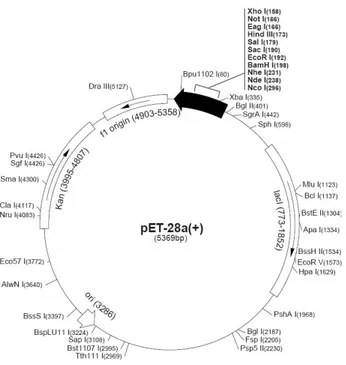
Figure 7: Schematic representation of the plasmid pET28a. 32
Figure 8: Immunization protocol. 33
Figure 9: Restriction analysis of the genes MSP and PLC cloned in pVAX1. 40
Figure 10: Restriction analysis of the genes MSP and PLC cloned in pET28a. 42
Figure 11: IgG humoral immune response elicited by DNA vaccination. 46
Figure 12: Results of the SDS-PAGE to test the expression of the recombinant
proteins. 47
Figure 13: Electrophoretic profile of total protein extract of Trypanosoma brucei brucei
in polyacrylamide gel electrophoresis and stained with Comassie Brilliant Blue. 48
Figure 14: Immunodetection of antibodies in pools of serum of the immunized
mice. 49
Figure 15: Parasite concentration at days 5, 8, 10, 12 and 15 post-infection with
Trypanosoma brucei brucei. 50
Figure 16: Survival of immunized mice after challenge with Trypanosoma brucei
brucei. 51
Figure 17: Representation of the capsid of TrV, with the capsid proteins. 53
Figure 18: Representation of the epitopes on the tertiary structure of the GP63. 55
Figure 19: Detailed view of the most exposed amino acids of Epi-1 in the GP63. 56
Figure 20: Detailed view of the most exposed amino acids of Epi-2 in the GP63. 56
Figure 21: Detailed view of the most exposed amino acids of Epi-3 in the GP63. 57
Figure 22: Representation of the TrV protomer with the mutation regions
Figure 23: Detailed view of the most exposed amino acids of Epi-1 inserted
in TrV. 58
Figure 24: Detailed view of the most exposed amino acids of Epi-2 inserted
in TrV. 59
Figure 25: Detailed view of the most exposed amino acids of Epi-3 inserted
in TrV. 59
Figure 26: Detailed view of the Epi-3 of three distinct protomers. 60
Figure 27: Detailed view of the disulfide bond in GP63, before mutation. 61
Table Index
Table 1: Nucleotide sequences used in the primer synthesis for the gene amplification
from Trypanosoma brucei. 27
Table 2: Mix constituents. 27
Table 3: Vaccine composition per group. 33
Table 4: Alignment of the sequencing results with the original sequence from the MSP
and PLC genes from Trypanosoma brucei brucei. 43
Table 5: Parameters of the plasmids used in the immunization protocol. 44
Table 6: Results of an analysis in silico of MSP and PLC. 52
Table 7: Relevant information about the three chosen epitopes. 55
Abbreviations List
APC - Antigen Presenting Cells
BBB - Blood-Brain Barrier
BGH - Bovine Growth Hormone
CMV - Cytomegalovirus
CNS - Central Nervous System
CRD - Cross-Reacting Determinant
CSF - Cerebrospinal Fluid
DC - Dendritic Cells
DNA - Desoxiribonucleic acid
EDTA - Ethylenediamine tetraacetic acid
ELISA - Enzyme-Linked Immunosorbent Assay
GPI - Glycosylphosphatidylinositol
HAT - Human African Trypanosomiasis
HBV - Hepatitis B virus
HIC - Hydrophobic Interaction Chromatography
HIV - Human Immunodeficiency Virus
HPV - Human Papillomavirus
HRP - horseradish peroxidase
IL - Interleukin
ISG - Invariant Surface Glycoprotein
mfVSG - membrane form VSG
MHC - Major Histocompatibility Complex
MSP - Major Surface Protease
NO - Nitric Oxide
OPD - o-phenylenediamine dihydrochloride
PAMP - pathogen- associated molecular pattern
PBS - phosphate buffered saline
PCR - Polymerase Chain Reaction
PLC - Phospholipase C
SDS-PAGE - SDS-polyacrylamide gel electrophoresis SIF - Stumpy Induction Factor
sVSG - soluble VSG TAE - Tris-Acetate-EDTA TLR - Toll-Like Receptors TNF - Tumor Necrosis Factor
TSA - Trans-sialidase
TrV - Triatoma virus VLP - Virus-Like Particles
Abstract
African Trypanosomiasis, also known as sleeping sickness, caused by the protozoan Trypanosoma brucei, is a neglected tropical disease. This disease can be successfully controlled, as has been proven in the past; nevertheless, the growing number of people affected and at risk makes the development of a vaccine a priority. T. brucei is capable of constantly evading the host immune system, due to a remarkable mechanism of defense, which provides a great antigenic variation. Due to this mechanism it has been very difficult to develop an effective vaccine. However, new approaches have been pursued, one of which, the vaccination strategy with plasmid DNA has revealed some promising results. Based on this, this work aims to use three immunization strategies: the first one, DNA vaccination, using two plasmids DNA, encoding antigenic candidates from Trypanosoma brucei; the second one, using these antigenic candidates together with a nanoformulation; and the third one, using VLPs (Virus-Like Particles). The three models used in the development of DNA vaccines against T. brucei use two important proteins of the parasite: MSP (Major Surface Protease) and PLC (Phospholipase C). MSP is a surface zinc metalloprotease that is believed to be responsible by the release of a VSG (Variable Surface Glycoprotein) fragment. PLC is a phospholipase anchored to a GPI (Glycosylphosphatidylinositol) residue that cleaves a full-length VSG protein from the cell surface. As we can see, both proteins are responsible by the VSG release, by the normal differentiation from bloodstream to procyclic form, and they participate synergistically in VSG loss during differentiation.
After immunization with the first two strategies, although the titres were low, mice produced antibodies anti-Trypanosoma brucei brucei. The animals that presented a better immune response were the ones immunized with the mix of plasmids together with the nanoformulation. Regarding the third model of immunization, the design of the VLPs was made, and the next step is evaluating them biologically.
Resumo
A tripanosomose Africana, também conhecida como Doença do Sono, causada pelo protozoário Trypanosoma brucei, é uma doença tropical negligenciada. Esta doença pode ser controlada, tal como foi provado no passado; no entanto, o crescente número de pessoas afectadas e em risco torna o desenvolvimento de uma vacina uma prioridade. T. brucei é capaz de evadir constantemente o sistema imunitário do hospedeiro, devido ao seu extraordinário mecanismo de defesa, que lhe proporciona uma grande variação antigénica. Devido a este mecanismo de defesa tem sido muito difícil de desenvolver uma vacina eficaz. Contudo, têm sido procuradas novas técnicas, entre elas, uma estratégia de vacinação com DNA plasmídico que têm revelado resultados promissores. Tendo em conta estes resultados, este trabalho tem como objectivo o uso de três estratégias de imunização: a primeira, recorrendo a vacinas de DNA, usando dois plasmídeos que codificam candidatos antigénicos de Trypanosoma brucei; a segunda, usando estes candidatos antigénicos conjugados com uma nano-formulação; e a terceira, usando VLPs (Vírus-Like Particles). Os três modelos usados no desenvolvimento de vacinas de DNA contra T. brucei recorreram ao uso de duas importantes proteínas do parasita: a MSP (Major Surface Protease) e a PLC (Phospholipase C). A MSP é uma metaloprotease de zinco de superfície, que se acredita ser responsável pela libertação de um fragmento de VSG (Variable Surface Glycoprotein). A PLC é uma fosfolipase, ancorada a um resíduo de GPI (Glycosylphosphatidylinositol), que cliva integralmente uma proteína de VSG da superfície da célula. Como se pode ver, ambas as proteínas são responsáveis pela libertação das VSG, pela normal diferenciação da forma procíclica para a forma de corrente, e participam de forma sinérgica para a perda de VSG durante a diferenciação.
Após a imunização com as duas primeiras estratégias, apesar de em baixos níveis, os murganhos produziram anticorpos anti-Trypanosoma brucei brucei. Os que apresentaram melhor resposta imunológica foram os imunizados com a mistura de plasmídeos conjugados com a nano-formulação. Em relação ao terceiro modelo de imunização, o desenho das VLPs foi efectuado, e o próximo passo é a avaliação biológica das mesmas.
1. Introduction
Human African Trypanosomiasis (HAT), also known as, sleeping sickness, is
caused by the protozoan parasite Trypanosoma brucei, and it is transmitted by the bite
of tsetse flies (Glossina spp.). There are two subspecies of T. brucei that are pathogenic
for humans and cause very distinct pathologies: T. b. gambiense and T. b. rhodesiense
(WHO 2015a). It is estimated that 69.3 million people, in 36 sub-Saharan Africa
countries, are at risk of having sleeping sickness, 57 million of which are at risk of
getting T. b. gambiense and 12.3 million T. b. rhodesiense (Simarro et al. 2012; WHO
2015a). The first descriptions of this disease date back to the colonial period and were
made by ship doctors and medical officers (WHO 2015b).
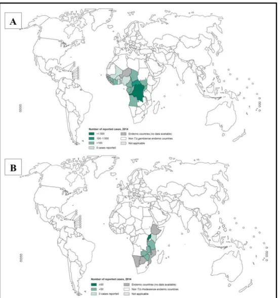
T. b. gambiense is found in Western and Central Africa (Figure 1A), and causes
a chronic form of HAT that can have a duration of about 3 years (Checchi et al. 2008).
On the other hand, T. b. rhodesiense, can be found in Eastern and Southern Africa
(Figure 1B), causes an acute and fulminant form of the disease, which can lead to death
within weeks or months (Odiit et al. 1997).
There is a third subspecies of Trypanosoma brucei parasites: Trypanosoma
brucei brucei, which causes African Animal Trypanosomiasis and does not infect
humans, just wild and domestic animals, such as donkeys, horses, goats, camels, mules,
sheep, antelope. This disease is chronic and sometimes fatal in cattle, however is rarely
fatal in swine. In animals, the first symptoms are lymphadenopathy, intermittent fever,
anemia and progressive emaciation. The animal disease can vary between acute or
chronic depending on the species, the age of the host and the parasite load (Acha &
Szyfres 2003). This subspecies is widely used in experimental models of animal and
human African Trypanosomiasis (de Sousa et al. 2010; Lança et al. 2011).
HAT was close to elimination during the 1960s, due to the establishment of
systematic screening, treatment, and follow-up of patients for the T. b. gambiense HAT,
and control of reservoir animals and vectors, in Eastern and Southern Africa, for the T.
b. rhodesiense HAT (Simarro et al. 2011). The rarity of cases resulted in a lack of
re-emergence in the 1980s. This re-re-emergence reached epidemic proportions, most of the
infections being caused by T. b. gambiense (Holmes 2014).
Figure 1: Distribution of Human African Trypanosomiasis: A- Distribution of Human African Trypanosomiasis (T. b. gambiense) (WHO 2015c); B- Distribution of Human African Trypanosomiasis (T. b. rhodesiense) (WHO 2015d).
Due to more health care facilities doing screening and the improvement of the
diagnostic tests, in 2009, the number of new cases of HAT fell below 10.000 and the
1.1 Pathology, diagnosis and treatment of Human African Trypanosomiasis
HAT has two different stages, being the clinical signs slightly different between
T. b. rhodesiense and T. b. gambiense. However, they have one feature in common, if
left untreated they can lead to coma and death.
After inoculation, the trypanosome multiplies at the site of infection, producing
a local skin reaction, called chancre. This reaction is more common in T. b. rhodesiense
than in T. b. gambiense, and is the first symptom of this disease (reviewed in Barrett et
al. 2003).
The first stage of the disease starts when the parasites reach the draining lymph
nodes and the bloodstream; this stage goes by the name of haemolymphatic stage. The
commonest characteristic symptom of this stage is the irregular fever, which reflects the
parasitemia levels in the blood. Other symptoms, like headache, pruritus and
lymphadenopathy are also seen. However, there are some differences between East and
West African trypanosomiasis. In the first one, the symptoms, even at the first stage of
the disease, can be severe, if there is no quick access to treatment, patients can die. On
the other hand, in the infection caused by T. b. gambiense the most common signs of the
disease are lymphoadenopathy (Winterbottom’s sign), hepatosplenomegaly and faint
rash (reviewed in Stich et al. 2002; Barrett et al. 2003; Brun et al. 2010).
As the disease progress to the second stage, also known as, meningoencephalitic
stage, the parasites cross the blood-brain barrier (BBB), invading the central nervous
system, causing progressive neurological damage. In T. b. rhodesiense, this happens
within a few weeks of infection, unlike T. b. gambiense, which takes between several
months to years. The main symptom of the second stage is the sleep disorder; this is
characterized by deregulation of the circadian rhythm of the sleep cycle and
fragmentation of the sleeping pattern. Other neurological symptoms are tremor,
fasciculation, general motor weakness, paralysis of a limb, hemiparesis, akinesia, and
abnormal movements such as dyskinesia or chorea-athetosis. Difficulty in
concentration, reduced higher mental functions, are other symptoms which culminate in
a final state of sleepiness (reviewed in Stich et al. 2002; Barrett et al. 2003; Brun et al.
This disease needs an accurate and early diagnosis. Control programmes use a
three-step approach: screening, diagnostic confirmation and staging. The diagnostic
method may vary between the two forms of the disease.
The card agglutination test for trypanosomiasis - T. b. gambiense, used in the
screening step, is a specific and, most of all, fast and practical serological test, that
allows hundreds of people to be tested daily. Nevertheless, this method has limitations:
high frequency of equivocal results and a limited sensitivity. Other serological tests
used to screen, are immunofluorescence and enzyme-like immunosorbent assays
(ELISA) (Noireau et al. 1988; Lejon et al. 1998). However, in addition to all necessary
equipment, these techniques are time-consuming, making them mainly used in
non-endemic countries, where the number of samples are lower and the equipment already
exists. Even though the serologic test comes back negative, in patients who have had
recent infection or a clinical suspicion of sleeping sickness, health professionals, are
advised to search for parasites in the blood. For T. b. rhodesiense, there is no serological
screening test. In this case, screening step has to be done, based on non-specific clinical
presentation and history of exposure (reviewed in Brun et al. 2010).
For diagnostic confirmation, a microscopic examination of lymph node aspirate
and blood, or both, is needed. It might be needed a concentration step, to increase the
sensitivity of this method (Lutumba et al. 2007). PCR (Polymerase Chain Reaction) on
blood has 99% sensitivity and 97.7% specificity, but, usually, is impracticable in
endemic regions, because of the advanced facilities required to perform this method
(Mugasa et al. 2012).
In the diagnosis process it is essential to differentiate the stage of the disease, by
examination of the cerebrospinal fluid (CSF) after lumbar puncture, due to the
pharmacological differences between the two stages. According to the WHO, the
presence of trypanosomes in the CSF or a white blood cell count of more than 5 cells
per µL, or both, defines the late-stage disease (Noireau et al. 1991). The measurement
of IgM concentrations, in the CSF, is a useful adjunct to the second-stage diagnosis
There are some non-invasive staging methods, like, polysomnography and
actigraphy; however, they are not used as a basis for guiding the initial treatment.
Polysomnography identifies alterations of sleep structure that occur in the late-stage
HAT (Buguet et al. 2005). Actigraphy uses an actigraph on the wrist to measure body
movement, and can detect the sleep/wake cycle abnormalities (Njamnshi et al. 2012).
The treatment of sleeping sickness is based on the disease stage and on the
subspecies of trypanosome. There are few drugs available to treat this condition.
First-stage T. b. gambiense HAT is treated with intramuscular pentamidine. This
drug, although effective has many possible complications, as hyperglycaemia or
hypoglycaemia, prolongation of the QT interval on electrocardiogram, hypotension and
gastrointestinal features. The treatment for early-stage T. b. rhodesiense HAT is
suramin, usually administered intravenously, which has as potential complications renal
failure, skin lesions, anaphylactic shock, bone marrow toxicity and neurological
complications (reviewed in Babokhov et al. 2013).
There is only one drug effective in treating second-stage Eastern HAT,
melarsoprol. Nevertheless, this drug is very toxic, injections are exceedingly painful,
and there are records of treatment failures due to resistance. Melarsoprol treatment can
result in reactive encephalopathy, agranulocytosis, skin rashes, peripheral neuropathy
and cardiac arrhythmias. Eflornithine is the drug used to treat late-stage T. b.
gambiense. This drug can cause bone marrow toxicity, alopecia, seizures and
gastrointestinal symptoms, and there is a potential for drug resistance in the affected
areas in Africa (reviewed in Kennedy 2013).
Two new drugs for second-stage gambiense HAT, Fexinidazole and
SCYX-7158, are being evaluated in clinical trials (Jacobs et al. 2011; Tarral et al. 2014). There
is a study being conducted using melarsoprol, where it was changed the delivery
method by constructing melarsoprol-cyclodextrin inclusion complexes, and it is a
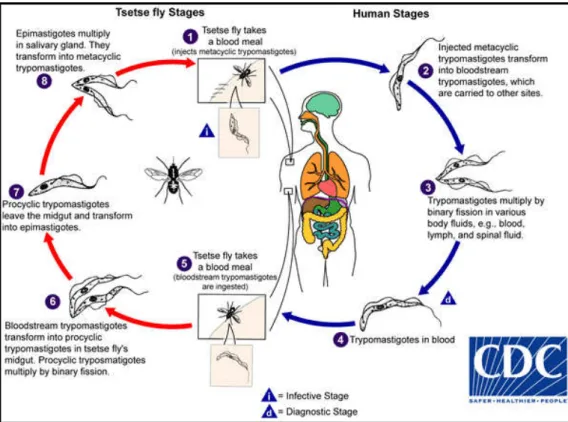
1.2 Trypanosoma brucei
1.2.1 Life cycle of Trypanosoma brucei
Trypanosoma brucei is a heteroxenic protozoan parasite from the
Trypanosomatidae family and the Kinetoplastida order (reviewed in Simpson et al.
2006). There are four main developmental stages in the entire life cycle of T. brucei:
epimastigotes, procyclic forms, slender metacyclic trypomastigotes, and stumpy
metacyclic trypomastigotes (Figure 2).
Once an infected tsetse fly takes a blood meal it injects stumpy metacyclic
trypomastigotes into the mammalian host. The metacyclic trypomastigotes differ into
proliferative, slender, trypomastigotes that can multiply themselves by binary fission
throughout the body fluids, such as lymph and cerebrospinal fluid. During the whole
life cycle in the mammalian host, T. brucei is in the extracellular space. When a tsetse
fly bites an infected mammal, ingests bloodstream trypomastigotes that differentiate
into proliferative procyclic trypomastigotes in the tsetse midgut and transform into
epimastigotes. Then, in the salivary glands, they multiply and transform into
non-proliferative, stumpy, metacyclic trypomastigotes. Only then, the trypanosomes are
ready to infect another host, once the Glossina takes a blood meal (D’Avila-Levy et al.
Figure 2: Schematic illustration of the life cycle of Trypanosoma brucei (CDC 2015).
As the parasitemia increases, in the vertebrate host, so does the density (Figure
3), which induces the parasites to produce a trypanosome factor, the stumpy induction
factor (SIF). SIF stimulates the transition from slender forms to stumpy forms. These
forms differ in terms of capacity to proliferate – slender forms proliferate, while stumpy
forms are arrested; nevertheless, both express the bloodstream stage-specific antigen,
variant surface glycoprotein (VSG). The fact that stumpy forms are cell-cycle arrested
combined with an elaborated mitochondrial activity, makes them pre-adapted to life in
the tsetse vector (Vassella et al. 1997; Tyler et al. 1997). The establishment of parasite
numbers in the blood and the immune evasion, through antigen variation, is due to the
proliferation of slender forms. On the contrary, stumpy forms control the expansion of
parasite numbers in the mammalian host bloodstream and consequently prolonging the
host survival, increasing the probability of disease transmission (Tyler et al. 2001;
Figure 3: Life cycle stages in Trypanosoma brucei (Matthews et al. 2004).
African trypanosomes have at least three different survival strategies that are
responsible for the persistent infection. The first one, is the antigenic variation of the
surface coat (Cross 1996); second one, it is observed a repression of stimulatory
components that results in immunosuppression by unknown mechanisms (reviewed in
Donelson et al. 1998); and at least, clearance of surface-bound antibodies by the
parasite motility. However, this clearance is, only effective when the antibody
concentrations are low, it is not sufficient to protect the cells at high antibody titers
(Engstler et al. 2007). It is believed, that the antigenic variation and the
immunosuppression are the reason for the long-term persistence of an infecting
population, and that the clearance of surface-bound immunoglobulin may contribute for
the survival of individual cells during the emergence of a specific humoral immune
1.2.2 Cell structure of Trypanosoma brucei parasites
The elongated trypanosome cell (Figure 4) is composed of a highly polarized
microtubule cytoskeleton, which gives the cell the characteristic shape and maintains it
intact throughout the cell cycle (Sherwin & Gull 1989). The flagellar pocket is
positioned at the posterior end of the cell. This structure is the exit point for the
flagellum and is responsible by all the processes of endo and exocytosis of the parasite
(reviewed by Overath & Engstler 2004). The motility of the T. brucei depends of its
single flagellum, a semi-rigid structure found in the Kinetoplastids (Bastin et al. 1998;
Vaughan & Gull 2003). Nowadays, the flagellum is recognized as the main contributor
to the pathogenicity of these protozoa. In addition to the motility, it contributes to the
recognition of the host/vector environment and promotes the recognition and attachment
necessary for immobilizing the parasite at the vector surfaces at some life-cycle stages
(Vaughan & Gull 2003). The flagellum begins in the basal body, which in turn, is
connected through the mitochondrial membrane to the mitochondrial genome which is
contained by the kinetoplast. A tripartite attachment complex links the basal body and
the kinetoplast, and has to cross the cell and the mitochondrial membranes (Ogbadoyi et
al. 2003).
1.2.3 The Variant Surface Glycoprotein Coat of Trypanosoma brucei
One of the main features of the T. brucei that raises interest between the
scientific communities, is his capability of evading the host immune system, and
thereby remains in circulation for months to years. This is achieved by antigenic
variation of the parasite coat. The Trypanosoma brucei has a 15 nm Variant Surface
Glycoprotein (VSG) coat that covers the entire cell, during the bloodstream form
(Vickerman 1969). The VSG coat, as the name suggests, is composed by,
approximately, 10 million copies of VSG molecules, and represents up to 10% of the
total cell protein (Cross 1996).
The VSG monomer consists of an exposed N-terminal domain, comprising
about ! 350 residues, that contains the biologically relevant epitopes (Hsia et al. 1996)
and a membrane-proximal C-terminal domain, of 50 to 100 residues, that is inaccessible
to the antibodies (Johnson & Cross 1979; Schwede et al. 2011). The C-terminal is
attached to the plasma membrane by a glycosylphosphatidylinositol (GPI) anchor
(Ferguson et al. 1988).
Over the course of the infection, the host immune system develops a response
against the VSG expressed at that time, however this is inefficient in eradicating the
entire parasite population, as some individuals have already switched their VSG
(Vickerman 1978). The VSG coat also serves to protect less variable or even invariant
surface proteins from immune effectors (Hutchinson et al. 2003).
There are two more life cycle steps where the VSG is shed from the parasite
surface: when all cells exchange the metacyclic VSG for a bloodstream form VSG,
within a few days after entry into a mammalian host (Esser & Schoenbechler 1985); and
when the trypanosome enters the tsetse midgut. The procyclic form stops expressing
any VSG and starts to express another GPI-anchored glycoprotein, yet invariant, the
procyclin (Figure 3). When the parasites re-enter the vertebrate host as metacyclic
trypomastigotes, the expression of VSG is activated once again (Bülow et al. 1989).
When the VSG coat is shed in the differentiation from stumpy bloodstream
vitro, within hours of the initiation of differentiation: one is full-length VSG protein that
is cleaved from the cell surface by a GPI-phospholipase C (GPI-PLC) (Ziegelbauer et
al. 1993) and the other, is a truncated VSG fragment released by proteolysis, by the
major surface protease (MSP) (Bangs et al. 1997; Gruszynski et al. 2006).
1.2.4 Major Surface Protease and Phospholipase C from Trypanosoma
brucei
There is a parasitic protozoa evolutionary related with T. brucei, that occurs as
an extracellular form in the sand fly vector and as an intracellular form in their
mammalian hosts macrophage, the Leishmania spp. These parasites contain a major
surface zinc metalloprotease (MSP, also called GP63), which has been extensively
studied. Among other function, this metalloprotease provides resistance to
complement-mediated lysis before Leishmania enters the macrophage, participates in the attachment
and entry into the macrophage supporting survival in the macrophage after entry
(Brittingham et al. 1995; reviewed in Yao et al. 2003).
Previously, LaCount et al. reported the presence in the genome of the African
trypanosome, of at least, three gene families (TbMSP-A, -B, and -C) encoding
homologues of the GP63 found in Leishmania spp. The TbMSPs share about 33%
sequence identity, positional conservation of 20 cysteines and 10 prolines, and a
metalloprotease catalytic site motif, HEXXH, with Leishmania GP63, which suggests
common three-dimensional features. All of these three families are expressed in
bloodstream stage trypanosomes, but only TbMSP-B is found in procyclic stage.
Although, they all are expressed in the bloodstream stage, the TbMSP-A mRNA is
about twice as abundant as the TbMSP-B mRNA, which is four times more than
TbMSP-C mRNA. The sequences from the three TbMSP share about 33% identity, and
the major difference is in their termini. The TbMSP-A has an extended C-terminal
region, rich in serines and glutamates, that was not seen in the other two, and finishes in
a short hydrophobic segment. TbMSP-B has a hydrophobic tail in the C-terminal.
prolines, which indicates that it is not linked to a membrane via GPI anchor, unlike
TbMSP-A and -B (LaCount et al. 2003).
It has been shown that TbMSP is a surface-localized zinc metalloprotease, with
60 kDa, that is expressed, mainly, in differentiating bloodstream to procyclic parasites
and in established procyclic cells. This MSP, in concert with PLC, participates in the
removal of the VSG coat when bloodstream stage trypanosomes differentiate to
procyclic trypanosomes. A TbMSP-B-/-PLC-/- double mutant cell line, failed to
differentiate into the procyclic form, most VSG remaining in the cell surface. This cell
line also exhibited an altered morphology: enlarged with multiple nuclei and
kinetoplast, and detached flagellae. However, the single mutant cell line, TbMSP-/- and
PLC-/-, showed a delay in VSG loss, but they were still able to differentiate, which leads
to the conclusion, that MSP and PLC act synergistically in the VSG loss during
differentiation (Grandgenett et al. 2007).
A further detailed analysis provides evidence that TbMSP-B and GPI-PLC
expression is coordinately and inversely regulated during differentiation (Gruszynski et
al. 2006).
Another possible function of the MSP, it is related to the capacity of the
Trypanosoma brucei in disrupting the blood-brain barrier and invade the central
nervous system (CNS). It has been shown that the metalloproteases expressed on T. b.
brucei bloodstream forms display hydrolytic activity on proteins like, gelatin, casein,
and matrix proteins such as collagen (de Sousa et al. 2010).
Trypanosoma brucei has also an endogenous PLC of 39-40 kDa anchored to
GPI residue, this protein does not have a N-terminal signal peptide nor a
trans-membrane domain, it behaves as an integral trans-membrane protein (Bülow & Overath
1986; Fox et al. 1986; Carrington et al. 1989). It is capable of hydrolysing the GPI
anchor of the VSG, releasing dimyristyl glycerol (Ferguson et al. 1985). This hydrolysis
results in the conversion of the hydrophobic membrane form of VSG (mfVSG) to a
water soluble VSG (sVSG), resulting then in the shedding of the VSG from the parasite
contained in the residue of the anchor attached to the VSG, the cross-reacting
determinant (CRD) (de Almeida & Turner 1983; Zamze et al. 1988).
GPI-PLC concentrates in the flagellar membrane, disposed in a patchy, linear
array along the flagellar attachment zone and extending into the free flagellum and it
remains in the same location, both before and after activation, which means that to
cleave the GPI anchor of VSG it does not move from its location (Hanrahan et al. 2009;
Sunter et al. 2013).
The GPI-PLC is only present in metacyclic and bloodstream stages of the life
cycle, procyclic trypanosomes are resistant to this protein, because it contains GPI
anchors bearing an extra acylation on the inositol ring (Field et al. 1991).
Although GPI-PLC does not act alone in the VSG release, as explained before, it
is described as a virulence factor. In a cell line with the PLC gene deleted, the infection
was able to proliferate, however the levels of parasitemia were lower when compared to
a control cell line, and the survival times of the mice were longer (Webb 1997).
GPI-PLC is unable to release VSG of another parasite, when expressed by one
trypanosome only targets its own plasma membrane. Also, VSG is not the only protein
cleaved by GPI-PLC, this protein also acts in other proteins of lower and higher
molecular mass, like other GPI biosynthetic intermediates (Cardoso De Almeida et al.
1999).
The action of GPI-PLC can be inhibited by some sulphydryl reagents, like Zn2+
and p-chloromercurylphenylsulphonic (Carnall et al. 1997).
1.3 Immunobiology of African Trypanosomiasis
Being an extracellular parasite, T. brucei encounters both innate and adaptive
immune response from the host. As soon as the parasite enters in the host bloodstream it
encounters the innate immune system as first barrier. The first response of the host to
control the first peak of parasitemia consists of classical activated macrophages which
and trypanotoxic molecules, such as Tumoral Necrosis Factor - alpha (TNF)-" and
nitric oxide (NO) (Mabbott et al. 1994; Sternberg & Mabbott 1996; Kaushik et al. 2000;
Pan et al. 2006). In addition, these macrophages can be activated by the trypanosome
DNA (Desoxiribonucleic acid) (Harris et al. 2006) or by the GPI anchor of the VSG
(Coller et al. 2003).
Although initial inflammatory response is beneficial to the host at an early stage,
a sustained inflammation can cause pathological disorders. Therefore, it is essential to
reduce the inflammation, reducing the classical activated macrophages and,
consequently, reduce the pro-inflammatory cytokines. In order to accomplish this, type
II cytokines, like IL-4, IL-10 and IL-13 have to be secreted, so they can induce
alternatively activated macrophages. These macrophages are involved in a longer
survival of the host because they are more anti-inflammatory (Namangala et al. 2001).
Thus, to facilitate the control of the parasitemia and the pathology, it is needed a shift
from a type I inflammatory response, in an early stage, to a type II inflammatory
response, in a later stage (Namangala et al. 2009).
Another mediator responsible, not just, for the control of the parasite levels, but
also for the development of the pathology, is TNF-" (Magez et al. 1999). During
trypanosome infections, TNF is involved both in parasitemia control and infection
associated pathology such as anaemia, neurological disorders, fever and cachexia during
both human and animal trypanosomiasis (reviewed in Taylor & Mertens 1999). VSG
was identified as major TNF inducing component in trypanosome-soluble extract
(Magez et al. 1998). In addition, protection from neuroinflammatory pathology of
sleeping sickness has been associated with the levels of IL-10 and IL-6 in the brain
(Sternberg et al. 2005).
Since this parasite is extracellular there is a dominant humoral response of the
host. The activation of polyclonal B cells results in an increase in the number of B cells
and an elevation in plasma immunoglobulin, mainly IgM (Hudson et al. 1976; Diffley
1983). The successive parasitemia waves are a result of the specific antibodies directed
against the epitopes of the VSG coat. These antibodies are able to opsonize the parasites
making possible the efficient phagocytosis and destruction of the immune complexes, in
Trypanosome infections are characterized by a drastic suppression of the
immune responses, which makes the host more susceptible to opportunistic infections.
During the AT/HAT infections there is an inhibition of the T cell proliferation due to
down regulation of IL-2 production and expression of IL-2 receptor, which leads to the
host immunosuppression (Sileghem et al. 1989; Darji et al. 1992). It is believed, that
TNF also plays a role in the process of suppressing the immune system (Schleifer &
Mansfield 1993).
1.4 DNA vaccination
Since the first description where it was shown for the first time that an
intramuscular injection of a plasmid was able to induce protein expression in muscle
cells (Wolff et al. 1990), DNA vaccines have been object of many interest.
Followed this initial report on DNA vaccines there was a proliferation of studies
from cancer (Buchan et al. 2005), to infectious virus diseases, most prominently HIV
(Human Immunodeficiency Virus) (Hammer et al. 2013), hepatitis C virus (Frelin et al.
2004) and cytomegalovirus (Kharfan-Dabaja et al. 2013), but also parasitic diseases are
potential targets to DNA vaccination, like malaria (Wang et al. 1998), Chagas disease
(Quijano-Hernández et al. 2013) and African trypanosomiasis (Silva et al. 2009; Lança
et al. 2011).
In order to create the plasmid, first the gene sequence of interest has to be
generated, synthetically or by PCR, and then is enzymatically inserted into the multiple
cloning region of the plasmid backbone, purified and delivered to the inoculation site.
DNA vaccination is capable of inducing both, humoral and cellular immune responses
(Figure 5). It starts when the optimized gene sequence is delivered, and the plasmid
enters the nucleus of transfected local cells, such as myocytes (1) and antigen presenting
cells (APCs) (2). Once this entry is completed, foreign antigens are generated as
proteins, as a result of the expression of the plasmid-gene. These antigens become the
subject of immune surveillance by the Major Histocompatibility Complex (MHC) class-
MHC-I molecules, which is followed either by direct transfection by the plasmid
vaccine (2) or by cross-presentation of cell-associated exogenous antigens (3).
Additionally, after the secretion of protein antigens that have been shed from
transfected cells are captured and processed within the endocytic pathway, APCs
mediate the display of peptides on MHC II molecules (4). Antigen-loaded APCs travel,
via the afferent lymphatic vessel (5), to the draining lymph node, where they present
antigenic peptide to naïve T cells, providing the necessary secondary signals to initiate
an immune response and expansion of T cells (6), or instead, activation of B cells and
antibody production (7). This activation is due to cytokines secreted by the CD4 T
helper cells during cell-to-cell interaction with B cells, which is a response to
peptide-bound MHC molecules and co-stimulatory secondary signals. Once T cells are primed
in the draining lymph node they can be restimulated and expanded, by the presentation
of the peptide-MHC complexes, at the immunization site. The activation of both T and
B cells, elicit specific immunity against the antigen encoded by the plasmid. These cells
travel through the efferent lymphatic system and provide a surveillance system (8)
(reviewed in Kutzler & Weiner 2008).
Over the years there were some safety concerns about DNA vaccination: the
potential to be integrated into cellular DNA, the development of autoimmunity and the
possibility of antibiotic resistance. However, studies conform the safety of this class of
vaccines. DNA vaccines tested did not shown relevant levels of integration into host
cellular DNA, it typically occurs at lower rates than spontaneous mutation frequencies
(Temin 1998; Manam et al. 2000; Ledwith et al. 2000). Considering the possible
development of autoimmunity induced by DNA vaccines, studies made did not detect
convincing evidence of autoimmunity (Le et al. 2000). With regards to antibiotic
resistance, it is improbable that resistances emerge because the resistance genes
contained in the plasmids are not commonly used to treat human infections. Although
all of these concerns, DNA vaccination has been proven to be safe. And in order to
respond to the concerns, the European Medicines Agency (EMA) and the US Food and
Drug Administration developed guidelines on safety and testing DNA vaccines
In addition to this advantage, DNA vaccines are able to encode several types of
genes and proteins, they are stable, ease to store and can be manufactured on a large
scale (reviewed in Kutzler & Weiner 2008).
Figure 5: Schematic representation of the mechanism of action of DNA vaccines (Kutzler & Weiner
2008).
One of the disadvantages of DNA vaccination is the poor immunogenicity. In
order to solve this problem, immunization techniques, nowadays, are changing and
being improved. Electroporation is considered a promise as a second generation DNA
vaccine technology for humans (Otten et al. 2004). This method consists on the
this way to an increase of cell permeability (Satkauskas et al. 2005). This approach
expands DNA distribution in the injected muscles and uptake of DNA by myocytes
which results in a higher efficiency of DNA vaccines (Dupuis et al. 2000), leading to an
increase in the immune response and in the levels of expression (Hirao et al. 2008;
Rosati et al. 2008; Vandermeulen et al. 2014). Other technique is the particle-mediated
epidermal delivery, or particle bombardment, a needle-free DNA delivery technology
that was developed as a physical gene transfer method to deliver DNA vaccines coated
onto 1-3 µm gold beads into skin cells (reviewed in Yager et al. 2009; Wang & Lu
2013). Unlike routine intramuscular injection, this technique does not need larger
amounts of DNA to have the same, or even higher, cellular and humoral immune
response (Deng et al. 2014). This, may be due to the fact that DNA is delivered directly
into the host’s skin cells instead of in extracellular spaces (Roy et al. 2000). Another
needle-free technique is high-pressure mediated delivery, it is similar to particle
bombardment, forces a liquid through a small orifice under pressure to deliver the
vaccine parentally (Mumper 2003; Graham et al. 2013). This technique, showed a
higher immune response, when compared with approaches using needles or syringes
(Graham et al. 2013), and it is being used in clinical trials of DNA vaccines to
Ebolavirus and Marburgvirus, with positive results (Sarwar et al. 2014).
One example of a noninvasive technique is a dermal patch, like DermaVir
(Genetic Immunity). It has the vaccine antigens encoded in a plasmid DNA formulated
into a nanoparticle that target specialized dendritic cells under the skin. A single
DermaVir immunization is able to produce a specific immune response (Cristillo et al.
2007; Jin & Kim 2014).
1.5 The Virus-like particles
Virus-like particles (VLP) are multimeric, sometimes multiprotein,
supramolecular assemblages, geometrically well-defined, with diameters in the range of
25-100 nm, lacking genetic material, that mimic the overall structure of the native
virions. The fact that recombinant VLPs do not contain viral genetic material, offers a
idea of using non-infectious virus particles to develop prophylactic human vaccines has
been attractive, since the discovery of non-infectious hepatitis B virus (HBV) particles
(Millman et al. 1969).
Commonly, VLPs are more immunogenic than subunit or recombinant vaccines,
and are able to stimulate the humoral and cellular immune system. VLPs have a
multivalent display and a highly ordered structure that constitute the
pathogen-associated molecular pattern motifs (PAMPs). Since these motifs are, by and large,
unique to microbial antigens, the mammalian immune system has evolved to respond
vigorously to this arrangement of antigens. PAMPs are able to trigger the innate
immune sensing mechanisms and can be recognized by Toll-like receptors (TLRs)
(reviewed in Plummer & Manchester 2011). VLPs are able to induce a strong B-cell
response by efficiently cross-linking the membrane associated immunoglobulin
molecules that constitute the B-cell receptor, thanks to the correct display of the
antigenic epitopes and the highly repetitive structure (reviewed in Grgacic & Anderson
2006). VLPs are efficiently taken up by dendritic cells (DCs) for processing and
presentation by MHC-II and for promoting DC maturation and migration, stimulating
the innate immune system (Fifis et al. 2004; Gamvrellis et al. 2004). As native virus,
VLPs can also be processed in the cytosol of DCs and are presented by MHC-I
molecules to cytotoxic CD8+ T cells by the cross-presentation mechanism, which is
essential for the clearance of intracellular pathogens and for the induction of a potent
cytotoxic immune response (Murata et al. 2003; Win et al. 2011).
Several VLP-based vaccines are already licensed. The first recombinant vaccine
based on VLPs was the RECOMBIVAXHB® (Merck), against HBV (Krugman 1982).
The next recombinant VLP-based vaccine took 20 years to be licensed: Gardasil®
(Merck) against human papillomavirus (HPV). According to a study by the US Centre
for Disease Control and Prevention, thanks to this vaccine there was a reduction of over
50% in HPV infection among teenagers (Markowitz et al. 2013). A second VLP-based
HPV vaccine, Cervarix® (GlaxoSmithKline), was licensed in the USA, almost 3 years
after Gardasil® (Deschuyteneer et al. 2014).
Based on the structure of their parental viruses, VLPs can be divided in two
licensed HPV VLP vaccines, consist of one or more components of a vaccine target
antigens displayed on the VLP surface as a fusion to a heterologous viral protein with
the ability to self-assemble, and they do not include any host components. These VLPs
can be produced in prokaryotic and eukaryotic cells. On the contrary, the enveloped
VLPs consist in the host cell membrane with the integrated target antigens displayed in
the outer surface (reviewed in Lua et al. 2014).
VLPs are not just suitable as vaccines for homologous virus from which they are
derived. Additionally, they can be used for the display of foreign antigens, in order to
enhance their immunogenicity. Many soluble antigens have a poor immune response;
this can be overcome by rendering them highly repetitive in a single particle. In this
case, VLPs have two roles: they serve as scaffolds for presenting antigens derived from
other pathogens in a suitable repetitive configuration, and as adjuvants to boost the
immune response. An example of this kind of VLPs, is the new VLP-based vaccine for
malaria, that includes a portion of the circumsporozoite protein of Plasmodium
falciparum fused to the N-terminus of hepatitis B surface antigen (Rutgers et al. 1988)
and is, already, in phase III of the clinical trial (Agnandji et al. 2012).
It was recently described a VLP based on the Triatoma virus (TrV) (Querido et
al. 2013; Sánchez-Eugenia et al. 2015; Rodrígues Aguirre & Guérin 2015). This virus
infects Triatomine bugs, the vector of Trypanosoma cruzi, the ethiological agent of
Chagas Disease (Grayson 2010). The native TrV particles are composed by a 30 nm
icosaedral capsid, with no lipid envelop. Its capsid is made up of 60 copies of a
protomer, consisting of the structural proteins VP1 (29.7 kDa), VP2 (28.4 kDa) and
VP3 (31.8 kDa), and VP4 (5.5 kDa) lying within the capsid and in contact with the
2. Work Aims
The main aims of the present work are:
1- Cloning the genes MSP and PLC of the bloodstream forms of Trypanosoma
brucei brucei in pVAX1 and pET28a;
2- Use the plasmids MSPpET28a and PLCpET28a to express the recombinant
protein of MSP and PLC in Escherichia coli;
3- In silico analysis and design the antigenic epitopes as candidates to the VLPs.
4- Use the plasmids MSPpVAX1 and PLCpVAX1 as model to the development of
DNA vaccines against Trypanosoma brucei, determine anti-Trypanosoma brucei
antibodies and identify native proteins with serum from immunized mice by DNA
3. Materials and Methods
3.1 Animals and parasites
All animals used in this work were obtained and maintained in the bioterium of
the Instituto de Higiene e Medicina Tropical, Lisbon – Portugal. Female Balb/C mice,
between 5 to 8 weeks old, were used in the experimental infection with T. brucei and in
the DNA immunization protocols. The strain of Trypanosoma brucei brucei used in the
experimental model was a GVR35 obtained from the Unidade de Clínica Tropical of
the IHMT.
3.2 Infection protocol of mice and total protein extract from Trypanosoma brucei
brucei
Mice were inoculated intraperitoneally with trypanosomes prepared by dilution
of the frozen stabilate T. b. brucei GVR35 with PBS-glucose 20 mM pH 7.4. In order to
obtain protein extract of T. b. brucei, blood from infected Balb/C was collected, by
cardiac puncture, in syringes with heparin (B. Braun - Germany) as anti-coagulant.
Then, 200 µL of PBS-glucose 20 mM pH 7.4, was added to the collected blood and
submitted to purify bloodstream forms of the parasite using a gravity-operated
DEAE-cellulose chromatography column, previously equilibrated with PBS-glucose 20 mM
pH 7.4 (Lanham & Godfrey 1970). To evaluate the presence of bloodstream forms in
the resultant fractions of the chromatography procedure, they were monitored by
conventional optical microscopy. The fractions containing parasites were centrifuged,
during 10 minutes at 380g, the pellet was suspended with PBS, and then frozen at
-20ºC. When the samples were defrosted, a protease inhibitor was added. In order to
determine the protein concentration of the extract, the Bicinchoninic Acid Protein Assay
3.3 Obtaining genomic DNA from Trypanosoma brucei
The blood from the infected mice was used to purify genomic DNA from T.
brucei, using a commercial kit QIAamp® DNA Blood Mini Kit (Qiagen – USA). After
this step, the DNA concentration was determined by spectrophotometry at a wavelength
of 260 nm. The DNA samples were used as template for the amplification assays of
Trypanosoma brucei brucei genes of interest.
3.4 Obtaining the gene sequences of Trypanosoma brucei
Two T. brucei genes of interest were used as possible antigenic targets for the
construction of DNA vaccine prototypes. The nucleotide sequences of these genes,
named Major Surface Protease (MSP) and Phospholipase C (PLC), were obtained from
GenBank® from National Library of Medicine – National Center for Biotechnology
Information (NCBI), Bethesda – USA, under the gene identification (GI) AY230807
and XM_946646.1, respectively.
3.5 Construction and acquisition of the primers used in the amplification of
Trypanosoma brucei genes of interest
To each gene of interest (MSP and PLC) a pair of primers were design to the
terminal regions 5’ and 3’. Subsequent to this, these primers, commercially acquired
from Thermo Electron Corporation (Germany), were used in the amplification assays
with Polymerase Chain Reaction (PCR).
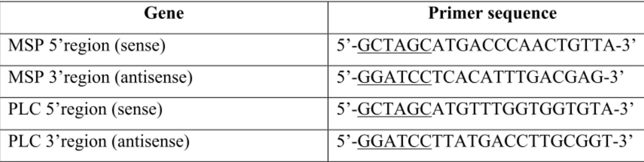
As cloning strategy, was added to the extremities of each pair of primers the
restriction sites to the enzymes NheI (GCTAGC) (Promega - USA) and BamHI
genes. The following table (Table 1) shows nucleotide sequences of the primers used in
the PCR.
Table 1: Nucleotide sequences used in the primer synthesis for the gene amplification from Trypanosoma brucei.
Gene Primer sequence
MSP 5’region (sense) 5’-GCTAGCATGACCCAACTGTTA-3’
MSP 3’region (antisense) 5’-GGATCCTCACATTTGACGAG-3’
PLC 5’region (sense) 5’-GCTAGCATGTTTGGTGGTGTA-3’
PLC 3’region (antisense) 5’-GGATCCTTATGACCTTGCGGT-3’
3.6 Polymerase Chain Reaction (PCR)
The PCR assays were used for the amplification of the gene of interest from
Trypanosoma brucei. To this end, the amplification reaction was optimized under the
conditions described below (Table 2):
Table 2: Mix constituents.
Constituents Quantity
Genomic DNA from Trypanosoma brucei
brucei 100 ng
Primers 100 pmol each
dNTPs
25 mM of each nucleotide dATP, dCTP,
dTTP and dGTP)(Stratagene, California –
USA)
MgCl2 (Bioline – Germany) 3 mM
DNA Polimerase (BIOTAQTM, Bioline –
Germany) 5 U
Reaction buffer NH4 (Bioline – Germany) 5 µL
After mixing the above constituents, the parameters used for the reaction were
established in a thermocycler (Bio-Rad - USA): an amplification cycle with a
denaturation temperature of 94ºC for 2 minutes, the annealing temperature of the
primers of 55ºC for 1 minute and 30 seconds, and a extension temperature of 72ºC for 2
minutes, in a total of 40 amplification cycles.
After the amplification reaction, the samples were mixed with a Loading buffer
(Bioline – Germany), and then submitted to a 0,8% (w/v) agarose gel electrophoresis
(Bioline – Germany) with a Tris-Acetate-EDTA (TAE) with ethidium bromide (Sigma-
USA).
The amplification products were then extracted from the gel and purified with a
commercial kit, QIAquick Gel Extration Kit (Qiagen - USA). In order to determine the
size of the amplified fragments, was used the molecular weight marker Hiperladder ITM
(Bioline – Germany).
3.7 Construction of DNA vaccine prototypes
3.7.1 Plasmid construction
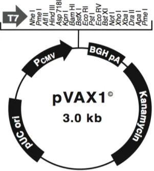
All the vectors used in the immunization process were derived from the
commercially available plasmid vector pVAX1 (2999 bp, Invitrogen - USA) (Figure 6),
designed to be use in the development of DNA vaccines. This vector contains the
human cytomegalovirus (CMV) immediate-early promoter, the bovine growth hormone
(BGH) polyadenylation signal and a kanamycin resistance gene.
The genes amplified by PCR were submitted to a cloning strategy in the plasmid
pVAX1. In order to accomplish this, both, fragments and the plasmid vector pVAX1,
were submitted to a digestion step (37ºC for 3 hours) with restriction enzymes (NheI
and BamHI), followed by a dephosphorylation step of the plasmid, using the enzyme
alkaline phophatase (Promega - USA), with the purpose of removing the phosphate
to a 0.8 % (w/v) agarose gel electrophoresis and the fragments were extracted from the
gel and purified by QIAquick Gel Extraction Kit (Qiagen – USA). The products of this
purification were cloned. To accomplish this, the enzyme DNA ligase (Promega - USA)
was used, and the manufacturer's instructions were followed.
Figure 6: Schematic representation of the plasmid pVAX1 (Invitrogen 2002).
3.7.2 Bacterial transformation
Plasmid DNA was added to competent cells, Escherichia coli DH5" cells, and
submitted to a heat shock. First, the mixture was kept on ice for 30 minutes, then heated
at 42ºC during 1 minute, and, immediately after, cooled down on ice for 2 minutes. The
mixture was placed in 1 mL of LB Broth (Sigma - USA) medium and incubated for 40
minutes to one hour at 37ºC. Then, the culture was centrifuged at 855g for 5 minutes,
the resultant pellet was suspended and placed on LB/Agar (Sigma – USA) plates
containing 30 #g/mL(w/v) kanamycin for selection of transformed E. coli colonies and
Plasmid DNA used in the immunization process was prepared from 250 mL of
E. coli DH5" in 20 g/L of LB medium with 30 #g/mL(w/v) of kanamycin (Bioline -
UK), overnight at 37ºC in an orbital shaker.
3.7.3 Plasmid Purification
Plasmid purification was performed by two different techniques, taking into
account the quantity of plasmid needed: by standard alkaline lysis followed by
hydrophobic interaction chromatography (HIC) (Diogo et al. 2000), when higher
amounts of plasmid were needed, for example for the immunizations; and when lesser
amounts were needed, was used the ISOLATE II Plasmid Mini Kit (Bioline - UK).
Initially, to perform the alkaline lysis, the inoculum was centrifuge at 3000g for
15 minutes at 5ºC. After discard the supernatant, was added 8 mL of solution P1 (in
section 7), 8 mL of solution P2 (in section 7) and incubated for 5 minutes at room
temperature, and then 8 mL of solution P3 (in section 7) and incubated for 5 minutes in
ice. After all the solutions were added, the mixture was centrifuged for 30 minutes, at
18000g. The supernatant was transferred to a new tube, and centrifuged one more time,
in order to remove all the floating material. The clean supernatant, was then passed to a
new tube and was added 0.8 of the final volume of isopropanol (Panreac – Germany),
and cooled down at 4ºC overnight.
The hydrophobic interaction chromatography, started with a centrifugation at
18000g, during 30 minutes, at 5ºC. After the centrifugation, the pellet was washed with
2 mL of ethanol at 70%, in a way, that just the DNA stays undissolved. Another step of
centrifugation, in the previous conditions was performed, and the supernatant was
discarded and the pellet was left to dry. Then it was suspended in 500 µL of Solution 1
(in section 7) and placed in microtubes of 1.5 ml with 0.16 g of ammonium sulphate,
and centrifuged at 9503g for 5 minutes. The 500 µL of sample were placed in the
column, previously equilibrated with Solution 2 (in section 7). After the column stopped
dripping, first, 3 mL of Solution 2 was added, and then 2 mL. So, the final volume of