DOI: 10.14260/jemds/2014/2577
CASE REPORT
J of Evolution of Med and Dent Sci/ eISSN- 2278-4802, pISSN- 2278-4748/ Vol. 3/ Issue 19/May 12, 2014 Page 5215
HEMANGIOPERICYTOMA OF NASAL CAVITY
P. Narmadha1, D. Premcharles2, P. Viswanathan3, V. U. Shanmugam4, B. Krishnasamy5
HOW TO CITE THIS ARTICLE:
P. Narmadha, D. Premcharles, P. Viswanathan, V. U. Shanmugam, B. Krishnasamy. Hemangiopericytoma of Nasal Cavity. Journal of Evolution of Medical and Dental Sciences 2014; Vol. 3, Issue 19, May 12;
Page: 5215-5218, DOI: 10.14260/jemds/2014/2577
ABSTRACT: A 9 Years old female came with complaints of mass in the right nasal cavity with nasal bleed. Biopsy was taken and sent for histopathological examination which was diagnosed as Hemangiopericytoma involving the nasal cavity. Hemangiopericytoma is an uncommon vascular mesenchymal neoplasm of the nasal cavity.
KEYWORDS: Hemangiopericytoma, sinonasal type.
INTRODUCTION: The term Hemangiopericytoma otherwise called as glomangiopericytoma is the term applied for neoplasms with true pericytic differentiation and appears to be related to glomus tumor. 15%-25% of these occur in the head and neck equally affecting both sexes in sixth and seventh decades of life.
CASE REPORT: A 9 Years old female came to ENT OP with complaints of mass in right nasal cavity for 10 days with on and off nasal bleed. On examination there was a grayish white fleshy mass in right nasal cavity found to be attached to the lateral wall. Cold spatula test and cotton wool test were positive. Probe test was done and there was no bleeding. Hematological investigations were unremarkable.
Radiologic findings:
CT scan was done and it showed following features:
1. Right inferior turbinate appeared grossly edematous.
2. Mucosal opacity in right maxillary, frontal, ethmoidal sinus with occlusion of osteomeatal complex.
3. Bilateral partial conchobullosa.
Mass was completely excised and was sent for histopathological examination.
Post operatively bleeding was present on and off. Nasal cavity was explored and remaining tissue was excised and packed. Further period was uneventful.
Macroscopic Examination: Multiple grey white grey brown soft tissue pieces altogether measuring 5ml in aggregate.
DOI: 10.14260/jemds/2014/2577
CASE REPORT
J of Evolution of Med and Dent Sci/ eISSN- 2278-4802, pISSN- 2278-4748/ Vol. 3/ Issue 19/May 12, 2014 Page 5216 DISCUSSION: It is an uncommon neoplasm occurring primarily over lower extremities, pelvis. Sinonasal type hemangiopericytoma can occur virtually in any site, but most frequent site are nasal cavity and paranasal sinuses.1, 2 Typically presents with obstruction and epistaxis. The radiologic appearance is opacification of involved sinus. Bony erosion can be seen due to pressure effects. Arteriographic finding reveal a richly vascular neoplasm. There are no specific etiologic factors. Hemangiopericytoma is thought to arise from pericytic cells which are normally found in capillary wall and function as baroreceptors to regulate the caliber of capillary lumen.
Sinonasal type of hemangiopericytoma varies from soft tissue type, being more myogenic in appearance. This finding coupled with overall benign behavior of sinonasal type of hemangiopericytoma contrasts with hemangiopericytoma of soft tissue origin.3,4 Hence it led to the consideration that sinonasal type of hemangiopericytoma are a related entity but separate from their soft tissue counterparts. This gave a designation of these sinonasal tumors as hemangiopericytoma -like.
The gross appearance is that of red to tan-gray soft to firm polypoid mass of varying size. On histologic examination lesion is seen in submucosa delineated but unencapsulated cellular tumor. It has a diffuse growth pattern and composed of a single cell type that is situated around endothelium lined vascular spaces. The tumor cells are uniform and have round to oval nuclei with vesicular to hyperchromatic chromatin and an indistinct cytoplasm, occasionally spindle cells are found5,6 An occasional mitotic figure can be seen. The vascular spaces range from capillary size to sinusoidal spaces that may have a staghorn or anter like pattern. The characteristic feature is perivascular hyalinization. Collagen is usually absent. Heterologous elements like bone and cartilage can also be seen.
DIFFERENTIAL DIAGNOSIS:
1. Lobular capillary hemangioma-vascular proliferation in arranged in lobules or clusters composed of central capillaries and small ramifying small tributaries.
2. Glomus tumour-glomus cells are rounded with round nuclei.
3. Angiofibroma- variable sized vascular spaces and a fibrocollagenous stroma.
CONCLUSION: There are various vascular tumors that arise from nasal cavity. Hemangiopericytoma is distinguished from them by its typical histopathological characteristics and also with the help of immunohistochemistry.
REFERENCES:
1. Gorenstein A, facer G.W, Weiland L.H. Hemangiopericytoma of the nasal cavity. ORL J Otorhinolaryngol Relatspec 86: 405-415, 1978.
2. Thompson LDR, Miettinen M, Weing B.M. Sinonasal Hemangiopericytoma: clinicopathologic and immunophenotypic analysis of 104 cases. MOD Pathol 15: 224a, 2002.
3. Compagno J, Hyams V.J. Hemangiopericytoma like intranasal tumours. A clinicopathological study of 23 cases. Am J Clinicopathol 66:672-683, 1976.
DOI: 10.14260/jemds/2014/2577
CASE REPORT
J of Evolution of Med and Dent Sci/ eISSN- 2278-4802, pISSN- 2278-4748/ Vol. 3/ Issue 19/May 12, 2014 Page 5217
5. Eichorn JH, Dickerson GR, Bhan AK, et al. Sinonasal Hemangiopericytoma: a reassessment with electron microscopy, immunohistochemistry and long term follow up. Am J Surg Path 14: 856-866, 1990.
6. El-Naggar A, Batsakis JG, Garcia GM, et al. Sinonasal Hemangiopericytomas: A Clinicopathologic and DNA content study. Arch Otolaryngol Head and Neck Surg 118:134- 137, 1992.
MICROSCOPIC PICTURES
Deeper to respiratory epithelium tumors composed of vascular channels are seen.
Multiplevascular channels showing staghorn pattern lined by endothelial cells surrounded by pericytes which are spindle shaped
Fig. 2: 20x Fig. 1: 10x
DOI: 10.14260/jemds/2014/2577
CASE REPORT
J of Evolution of Med and Dent Sci/ eISSN- 2278-4802, pISSN- 2278-4748/ Vol. 3/ Issue 19/May 12, 2014 Page 5218
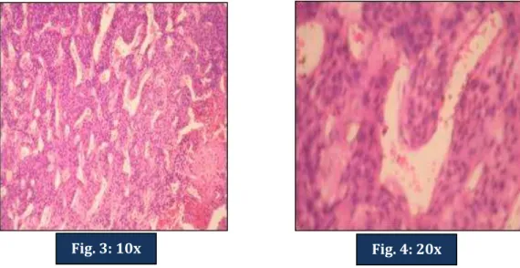
Showing areas of hemorrhage and necrosis.
AUTHORS:
1. P. Narmadha 2. D. Premcharles 3. P. Viswanathan 4. V. U. Shanmugam 5. B. Krishnasamy
PARTICULARS OF CONTRIBUTORS:
1. 2nd Year Post Graduate, Department of
Pathology, Rajah Muthiah Medical College, Annamalai University.
2. 2nd Year Post Graduate, Department of
Pathology, Rajah Muthiah Medical College, Annamalai University.
3. Professor, Department of Pathology, Rajah Muthiah Medical College, Annamalai University.
4. Professor, Department of E.N.T, Rajah Muthiah Medical College, Annamalai University.
5. Professor, Department of Pathology, Rajah Muthiah Medical College, Annamalai University.
NAME ADDRESS EMAIL ID OF THE CORRESPONDING AUTHOR:
Dr. P. Viswanathan,
Professor, Department of Pathology, Faculty of Medicine,
Rajah Muthiah Medical College,
Annamalai University, Chidambaram-608002, Tamilnadu, India.
E-mail: [email protected] Date of Submission: 12/04/2014. Date of Peer Review: 13/04/2014. Date of Acceptance: 25/04/2014. Date of Publishing: 10/05/2014.