International Journal of Pharmaceutical Sciences and Drug Research 2009; 1(2): 74-79
74
Research Article
ISSN 0975-248X
Mucoadhesive Microcapsules of Indomethacin: Evaluation for
Controlled Release and Ulcerogenic Activity
K.P.R. Chowdary
1, K. Sree Deepthi
2, Y. Srinivasa Rao
2*2
Pharmaceutical Technology Division, Vignan Institute of Pharmaceutical Technology, Duvvada, Visakhapatnam, India
1
University College of Pharmaceutical Sciences, Andhra University, Visakhapatnam, India
ABSTRACT
Mucoadhesive microcapsules of indomethacin were prepared by an emulsification-ionic gelation process employing sodium carboxy methylcellulose, methylcellulose, Carbopol and hydroxy propyl methyl cellulose along with alginate and the microcapsules were evaluated for release kinetics and ulcerogenic activity. The resulting microcapsules were discrete, free flowing, multinucleate, monolithic and spherical. Microencapsulation efficiency was 41-70 % and relatively high with alginate-sodium carboxymethylcellulose. Indomethacin release from these mucoadhesive microcapsules was found to be slow and extended over longer periods of time and depended on the composition of coat and size of the microcapsules. Drug release was diffusion controlled and followed first order kinetics. Alginate-methyl cellulose and alginate-sodium carboxymethylcellulose microcapsules were found suitable for oral controlled release. The microcapsules exhibited good
mucoadhesive property in the in vitro wash-off test. Release from some microcapsules fulfilled the official (USP 23) drug
release test-2 requirement of indomethacin extended release capsules. A 62-80 % reduction in ulcerogenic activity was observed with these microcapsules when compared to pure drug indomethacin.
Keywords: mucoadhesive microcapsules, indomethacin, controlled release, ulcerogenic activity.
INTRODUCTION
In recent years considerable attention has been focused on the development of new drug delivery systems known as controlled release drug delivery systems. Controlled release
drug delivery systems [1] are those dosage formulations
designed to release an active ingredient at rates, which differ significantly from their corresponding conventional dosage forms. The controlled release drug delivery systems are aimed at controlling the rate of drug delivery, sustaining the duration of therapeutic activity and/or targeting the delivery of the drug to a tissue. Drug release from these systems should be at a desired rate, predictable and reproducible. Among the various approaches for controlled systems, microencapsulation process and microcapsules have gained good acceptance as a process to achieve controlled release and drug targeting. Microencapsulation by various polymers
*Corresponding author: Dr. Y. Srinivasa Rao Professor of Pharmaceutical Technology and
Principal of the Vignan Institute of Pharmaceutical Technology, Duvvada, Visakhapatnam-530 046, Andhra Pradesh, India
Ph: +91-891-2511222, Fax: +91-891-2752333 E-mail: [email protected]
and their applications are described in standard textbooks.
[2-3]
Mucoadhesion is a topic of current interest in the design of controlled release drug delivery systems to prolong the residence time of the dosage form at the site of application or absorption and to improve and enhance the bioavailability
of drugs. [4-6] Though several studies [7] reported
mucoadhesive drug delivery systems in the form of tablets, films, patches and gels for oral, buccal, nasal, occular and topical routes, however, very few reports on mucoadhesive
microcapsules are available. [8-14] The hydrophilic polymers
are reported [15] to have excellent mucoadhesive properties.
Indomethacin, which requires controlled release owing to its
short biological half-life [16] of 2.4 ± 0.4 h and
MATERIALS AND METHODS Materials
Indomethacin was a gift sample from M/s Micro Labs, Pondicherry. Sodium carboxymethylcellulose (sodium CMC, having a viscosity of 1500-3000 cps of 1 % wt/vol aqueous
solution at 25°C), methylcellulose (having a methoxyl
content of 28.32 % wt/vol and a viscosity of 65 cps in 0.5%
wt/vol aqueous solution at 25°C), and hydroxypropyl
methylcellulose (HPMC, having a viscosity of 50 cps in a 2%
by wt/vol aqueous solution at 20°C) were gift samples from
M/s Natco Pharma Ltd (Hyderabad, India). Carbopol 934P was a gift sample from M/s SmithKline Beecham Pharmaceuticals (Bangalore, India). Sodium alginate (SD Fine Chem, Mumbai, India) and calcium chloride (Qualigens, Mumbai) were procured from commercial sources. All other reagents used were of analytical grade.
Preparation of Microcapsules
Microcapsules containing indomethacin were prepared employing sodium alginate in combination with sodium CMC, methyl cellulose, Carbopol and HPMC as coat materials. No methods are reported for microencapsulation
by these polymers. The ionic gelation processes [17-18] which
has been extensively used to prepare large sized alginate beads, was used to prepare the microcapsules.
Sodium alginate (1.0 g) and the mucoadhesive polymer (1.0 g) were dissolved in purified water (32 ml) to form a homogeneous polymer solution. Core material, indomethacin (-120+200 mesh) (2.0 g) was added to the polymer solution and mixed thoroughly to form a smooth viscous dispersion. The resulting dispersion was then added in a thin stream to about 300 ml of groundnut oil contained in a 600 ml beaker while stirring at 400 rpm. A Remi medium duty stirrer with speed meter (Model RQT 124) was used for stirring. The stirring was continued for 5 min to emulsify the add dispersion as fine droplets. Calcium chloride (10 % w/v) solution (40 ml) was then added slowly while stirring for ionic gelation (or curing) reaction. Stirring was continued for 15 min to complete the curing reaction and to produce spherical microcapsules .The mixture was then centrifuged and the product thus separated was washed repeatedly with water and dried at 45°C for 12 h.
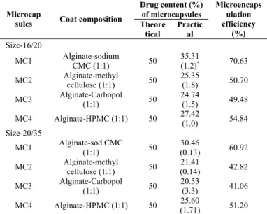
Table 1: Coat Composition, Drug Content and Microencapsulation Efficiency of the Microcapsules Prepared
Drug content (%) of microcapsules Microcap
sules Coat composition Theore tical Practic al Microencaps ulation efficiency (%) Size-16/20
MC1 Alginate-sodium
CMC (1:1) 50
35.31
(1.2)* 70.63
MC2 Alginate-methyl
cellulose (1:1) 50
25.35
(1.8) 50.70
MC3 Alginate-Carbopol
(1:1) 50
24.74
(1.5) 49.48
MC4 Alginate-HPMC (1:1) 50 27.42
(1.0) 54.84
Size-20/35
MC1 Alginate-sod CMC
(1:1) 50
30.46
(0.13) 60.92
MC2 Alginate-methyl
cellulose (1:1) 50
21.41
(0.14) 42.82
MC3 Alginate-Carbopol
(1:1) 50
20.53
(3.3) 41.06
MC4 Alginate-HPMC (1:1) 50 25.60
(1.71) 51.20 *Figures in parentheses are Coefficient of Variation (CV) values
Evaluation of Microcapsules
Indomethacin content in the microcapsules was estimated by
using UV spectrophotometric method [19] based on the
measurement of absorbance at 318 nm in phosphate buffer of pH 6.2. The method was validated for linearity, accuracy and precision. The method obeyed Beer’s law in concentration
range 1-40 μg/ml. When a standard drug solution was
assayed repeatedly (n=6), the mean error (accuracy) and relative standard deviation (precision) were found to be 1.2 % and 2 %, respectively.
Microencapsulation efficiency was determined by estimating the drug content in the microcapsules.
The formula, microencapsulation efficiency = (estimated percent drug content / theoretical percent drug content) × 100.
For size distribution analysis, different sizes in a batch were separated by sieving using a range of standard sieves. The amounts retained on different sieves were weighed. The microcapsules prepared along with their coat composition indomethacin content and microencapsulation efficiency are listed in Table 1.
Scanning electron microscopy
The microcapsules were observed under a scanning electron
microscope (SEM-LEICA, S430, London, UK). They were
mounted directly onto the SEM sample stub using double-sided sticking tape and coated with gold film (thickness 200 nm) under reduced pressure (0.001 mm of Hg).
Drug release study
Release of indomethacin from the microcapsules of size 16/20, and 20/35 was studied in phosphate buffer of pH 6.2 (900 ml) using an USP XXIII three-station Dissolution Rate Test Apparatus (Model DR-3, M/s Campbell Electronics, Bombay, India) with a basket stirrer at 75 rpm as per USP XXIII drug release test prescribed for indomethacin extended
release capsules. [18] A sample of microcapsules equivalent to
75 mg of indomethacin was used in each test. Samples were
withdrawn through a filter (0.45 μm) at different time
intervals and were assayed at 318 nm for indomethacin using a Shimadzu UV-150 double-beam spectrophotometer (Shimadzu Corporation, Japan). The drug release experiments were conducted in triplicate.
Mucoadhesion testing byin vitrowash-off test
The mucoadhesive property of the microcapsules was
evaluated by an in vitro adhesion testing method known as
the wash-off method. [20] The mucoadhesiveness of these
microcapsules was compared with that of a nonbioadhesive material, ethylene vinyl acetate microcapsules. Freshly excised pieces of intestinal mucosa (2 × 2 cm) from sheep were mounted onto glass slides (3 × 1 inch) with cyanoacrylate glue. Two glass slides were connected with a suitable support. About 50 microcapsules were spread onto each wet rinsed tissue specimen, and immediately thereafter the support was hung onto the arm of a USP tablet disintegrating test machine. When the disintegrating test machine was operated, the tissue specimen was given a slow,
regular up-and-down movement in the test fluid at 37°C
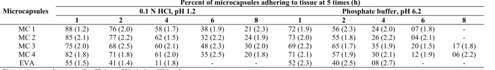
Table 2: Results of In Vitro Wash-Off Test To Assess Mucoadhesive Property of the Microcapsules
Percent of microcapsules adhering to tissue at 5 times (h)
0.1 N HCl, pH 1.2 Phosphate buffer, pH 6.2 Microcapsules
1 2 4 6 8 1 2 4 6 8
MC 1 88 (1.2) 76 (2.0) 58 (1.7) 38 (1.9) 21 (2.3) 72 (1.9) 56 (2.3) 24 (2.0) 07 (1.8) - MC 2 85 (2.1) 77 (2.2) 62 (1.5) 32 (2.2) 24 (1.9) 73 (2.0) 55 (1.8) 26 (2.2) 04 (2.1) - MC 3 75 (2.0) 68 (2.5) 60 (2.1) 48 (2.3) 30 (2.0) 69 (2.2) 65 (1.7) 35 (1.9) 20 (1.5) 17 (1.8) MC 4 82 (1.8) 71 (1.8) 61 (2.0) 35 (2.5) 20 (1.8) 71 (2.1) 57 (1.9) 30 (2.1) 12 (1.9) 06 (2.2)
EVA 55 (1.5) 41 (1.4) 11 (1.8) - - 52 (2.3) 40 (2.5) 08 (2.7) - -
Figures in parentheses are Coefficient of Variation (CV) values
Table 3: Release Characteristics of Mucoadhesive Microcapsules Prepared
Percent Indomethacin Released at 5 times (h) (
x
± s.d) Microcapsule1.0 2.0 4.0 8.0 12.0
T50 (h) K1×102 (h-1)
Size-16+20
MC1 33.6±4.20 48.6±4.90 63.1±5.40 79.8±10.00 87.7±7.03 3.3 17.38
MC2 8.8±0.08 22.5±5.10 44.5±8.80 66.6±1.21 75.7±1.43 5.0 12.50
MC3 16.6±0.90 34.9±0.19 63.1±0.11 90.8±0.87 97.1±0.21 2.4 29.14
MC4 25.2±0.21 45.5±1.25 74.2±0.97 89.5±2.50 98.2±0.11 2.9 36.89
Size-20+35
MC1 47.6±5.90 65.5±5.00 77.1±3.90 93.1±3.71 100.0±0.27 1.1 29.96
MC2 27.6±0.93 45.1±1.79 60.8±0.67 76.4±0.94 92.2±0.45 2.5 17.63
MC3 48.8±0.39 65.0±1.26 79.9±0.64 94.7±0.64 99.9±2.84 1.6 30.51
MC4 30.9±1.00 51.8±1.76 78.2±1.33 96.3±2.31 99.9±0.16 1.2 43.06
T50 is time for 50% release and K1 is first order release rate constant
Table 4: Ulcerogenic Activity Of Indomethacin And Its Mucoadhesive Microcapsules Description
Product
Pylorus Fundus
Ulcer index Mean±S.E
Percentage reduction in U.A.
t-test differences
Indomethacin Badly redden, 3-4 big ulcers, 6 very very big ulcer patches
Reddened. 20-25 big ulcers
5-6 big ulcer patches 4.00±0.26 - -
MC1 Slightly reddened No ulcers Normal, 5 microscopic ulcers,6 small
ulcers, 1 big ulcer 1.17±0.31 70.75 S
MC2 Slightly reddened No ulcers Normal,4-5 small ulcers 1 big ulcer 0.83±0.17 79.25 S
MC3 Moderate reddened, 1 big ulcer patch 2 ulcers, 5-10 haemorrhagic spots 1.50±0.22 62.50 S
MC4 Slightly reddened, 8 fine ulcers Slightly reddened, 1 big ulcer 1.00±0.26 75.00 S
S: Significant
Evaluation of Ulcerogenic Activity
The mucoadhesive microcapsules MC1, MC2, MC3 and MC4 of size 20/35 were evaluated for ulcerogenic activity in comparison with pure drug indomethacin. The approval of the Institutional Animal Ethics Committee was obtained before starting the study. The ulcergenic studies were carried
out by the method of Okabe. [21] Wistar rats of either sex
weighing between 120-150 g were used. The animals were starved for 24 h prior to experimentation. The pylorus was ligated under light ether anesthesia. After the recovery of animal, the preparation was administered orally at a dose equivalent to 5 mg of indomethacin per kg of body weight. The animals were sacrificed after 8 h and the stomach mucosa was collected for the observation of ulceration. The mucosa of the fundus and the pyloric part of the stomach was observed with magnifying lens for ulcers and perforations. The rating of ulcer formation (ulcer index) was done according to scoring system described by Anderson and
Soman [22] as follows,
Score Condition
1 A few small ulcers up to 4 2 Several small ulcers 5 to 8 3 Many small ulcers
4 Large areas of ulceration with confluence or more small ulcers (16-30) or impending perforations.
5 More than 30 small ulcers or large areas of ulcers with confluence or impending perforations
Each preparation was tested in six rats. The average of the individual scores in each group was calculated. The results are given in Table 4. The photomicrographs of stomach mucosa collected in the ulcerogenic studies are shown in Fig. 1.
RESULTS AND DISCUSSION
Mucoadhesive microcapsules of indomethacin with a coat consisting of alginate and a mucoadhesive polymer (1:1) namely sodium CMC, or methylcellulose, or Carbopol or HPMC could be prepared by an emulsification and ionic gelation process. The microcapsules (Fig. 2) were found to be discrete, spherical and free flowing. Regarding the internal structure, the nature of the method indicates that the microcapsules produced are of multinucleate monolithic type. The sizes could be separated and more uniform size range of microcapsules could readily be obtained by sieving. The size analysis of different microcapsules showed that the size distributions were normal in each case with a large proportion, 55-70 % in the size range of –16+20 mesh. The average size was found to be 888.7, 852.2, 831.9 and 885.7
μm respectively in the case of microcapsules MC1, MC2,
MC3 and MC4.
Low C.V. (< 2.0 %) in percent drug content indicated uniformity of drug content in each batch of microcapsules (Table 1). Drug content of the microcapsules was same in different sieve fractions. The microencapsulation efficiency was in the range of 41-70 %. The microencapsulation efficiency was relatively high with alginate-sodium CMC combination. The variations in the microencapsulation efficiency observed with various polymers are due to the possible differences in the porosity of the coat formed. High porosity of the coat leads to less microencapsulation efficiency.
A B
C D
E
Fig. 1: Photomicrograph of stomach mucosa of rat following oral administration of indomethacin (A) and its mucoadhesive microcapsules, MC1 (B), MC2 (C), MC3 (D) and MC4 (E).
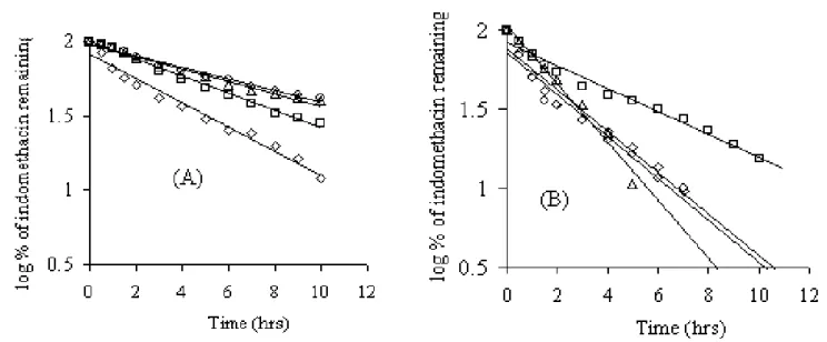
Fig. 3: First order plots of drug release from the mucoadhesive microcapsules prepared; size 16/20 (A) and size 20/35 (B). Mucoadhesive polymer in the coat: Sodium CMC (◊), methyl cellulose ( ), carbopol (○) and HPMC (∆).
Fig. 4: Percent released Vs t plots of drug release from the mucoadhesive microcapsules prepared; size 16/20 (A) and size 20/35 (B). Mucoadhesive polymer in the coat: Sodium CMC (∆), methyl cellulose ( ), carbopol (○) and HPMC (◊ ).
property in the in vitro wash-off test when compared to
non-mucoadhesive material, ethylene vinyl acetate microcapsules. The wash-off was slow in the case of microcapsules containing alginate-mucoadhesive polymer as coat when compared to that of EVA microcapsules (Table 2). The wash off was relatively rapid in phosphate buffer than in acid fluid. The results of wash-off test indicated fairly good mucoadhesive property of the microcapsules.
Drug Release from the Microcapsules
Indomethacin release from the microcapsules was studied in phosphate buffer (pH: 6.2) for a period of 12 h as prescribed in the drug release test-2 of indomethacin extended release capsules in USP XXIII. Indomethacin release from the microcapsules was slow and spread over extended periods of time (Table 3). Plots of log percent drug remaining Vs time (Fig. 3) were found to be linear (r >0.98) with all the microcapsules indicating that the drug release from these microcapsules was according to the first order kinetics. The
release was depended on the composition of the coat and size of the microcapsules. The release increased as the size of the microcapsules decreased due to large surface area of smaller microcapsules. Microcapsules of alginate-carbopol and alginate-HPMC gave relatively fast release when compared to alginate-sodium CMC and alginate-methylcellulose. The order of increasing release rate observed with various microcapsules was alginate-methylcellulose<alginate-sodiumCMC<alginate-carbopol<alginate-HPMC in both the sizes studied. The differences in the drug release characteristics of various microcapsules are due to the differences in the porosity of the coat formed and its solubility in the dissolution fluid. The drug release from the microcapsules was diffusion controlled as plots of amount
released Vs√t (Fig. 4) were found to be linear (r >0.97).
Indomethacin release from microcapsules MC2 (size 20/35) also fulfilled the official (USP XXIII) drug release test-2 requirement of indomethacin extended release capsules.
Ulcerogenic Activity of the Mucoadhesive Microcapsules Ulcer formation and the degree of its severity were
significantly (p <0.01) reduced in the rats, which received the
mucoadhesive microcapsules than those received the pure drug, indomethacin. About 62-80 % reduction in ulcerogenic activity was observed with all the mucoadhesive microcapsules and the microcapsules were found to have negligible ulcerogenic activity. The reduced ulcerogenic activity of mucoadhesive microcapsules is due to the slow release of indomethacin from the microcapsules and also due to the possible protective nature of the mucoadhesive polymer present in the microcapsules. Thus, microencapsulation of indomethacin with mucoadhesive polymers offers an effective method to avoid the undesirable ulcergenic effects of indomethacin besides achieving oral controlled release.
Mucoadhesive microcapsules of indomethacin with a coat consisting of alginate and a mucoadhesive polymer such as sodium carboxymethylcellulose, methyl cellulose, Carbopol and hydroxypropylmethylcellulose could be prepared by an emulsification-ionic gelation process. The microcapsules
exhibited good mucoadhesive property in vitro tests. The
resulting microcapsules were discrete, multinucleate, monolithic, spherical and free flowing. Indomethacin release from these mucoadhesive microcapsules were slow, diffusion controlled and followed first order kinetics. Alginate-methyl cellulose and alginate-sodium carboxymethylcellulose microcapsules were found suitable for oral controlled release. Release from some microcapsules fulfilled the official (USP 23) drug release test-2 requirement of indomethacin extended release capsules. A 62-80 % reduction in ulcerogenic activity was observed with these microcapsules when compared to pure drug indomethacin.
REFERENCES
1. Ballard BE, In: Robinson JR. eds., An overview of prolonged action drug dosage form, in sustained and controlled release drug delivery systems. Inc. New York, NY: Marcel Dekker, 1978, 22. 2. Kondo A, ed. Microcapsule Processing and Technology. New
York, NY: Marcel Dekker; 1979, 18.
3. Gutcho MH, ed. Microcapsules and Microencapsulation Techniques. Park Ridge, NJ: Noyes Data Corporation; 1976, 236.
4. Ikeda K, Murata K, Kobayashi M, Noda K. Enhancement of bioavailability of dopamine via nasal route in beagle dogs. Chem Pharm Bull 1992; 40(8): 2155-2158.
5. Nagai T, Nishimoto Y, Nambu N, Suzuki Y, Sekine K. Powder dosage forms of insulin for nasal administration. J Control Release 1984; 1: 15-22.
6. Illum L, Farraj NF, Critcheley H, Davis SS. Nasal administration of gentamicin using a novel microsphere delivery system. Int J Pharm 1988; 46: 261-265.
7. Gavini E, Sanna V, Juliano C, Bonferoni MS, Giunchedi P. Mucoadhesive vaginal tablets as veterinary delivery system for the controlled release of an antimicrobial drug, acriflavine. AAPS PharmSci Tech 2002; 3: 20.
8. Chowdary KPR, Srinivas L. Mucoadhesive drug delivery systems: a status of current review. Indian Drugs 2000; 37: 400-406. 9. Chowdary KPR, Srinivasa Rao Y. Preparation and evaluation of
mucoadhesive microcapsules of indomethacin. Indian J Pharm Sci 2003; 65: 49-52.
10. Sakagami M, Kinoshita W, Sakon K, Sato JI, Makino Y. Mucoadhesive beclomethasone microspheres for powder inhalation: their pharmacokinetics and pharmacodynamics evaluation. J Control Release 2002; 80: 207-218.
11. Takishima J, Onishi H, Machida Y. Prolonged intestinal absorption of cephradine with chitosan-coated ethylcellulose microparticles in rats. Biol Pharm Bull 2002; 25: 1498-1502.
12. Lim ST, Forbes B, Berry DJ, Martin GP, Brown MB. In vivo evaluation of novel hyaluronan/chitosan microparticulate delivery systems for the nasal delivery of gentamicin in rabbits. Int J Pharm 2002; 231: 73-82.
13. Cuna M, Alonso MJ, Torres D. Preparation and in vivo evaluation of mucoadhesive microparticles containing amoxicillin-resin complexes for drug delivery to the gastric mucosa. Eur J Pharm Biopharm 2001; 51: 199-205.
14. Chowdary KPR, Srinivasa Rao Y. Design and in vitro and in vivo evaluation of mucoadhesive microcapsules of glipizide for oral controlled release. AAPS PharmSci Tech 2003; 4:39.
15. Longer M A. Robinson J R. Fundamentals of bioadhesion. Pharmacy International 1986; 7: 114-117.
16. Insel PA. In: Hardman JG, Limbard LE, Molinoff PB, Ruddon RW, Gilman AG, eds. Goodman and Gilman’s The Pharmacological Basis of Therapeutics. 9th Ed. New York, NY: McGraw- Hill; 1996, 633.
17. Kim CK, Lee EJ. The controlled release of blue dextran from alginate beads. Int J Pharm 1992; 79: 11-19.
18. Hari PC, Chandy T, Sharma CP. Chitosan/calcium alginate microcapsules for intestinal delivery of nitrofurantoin. J Microencapsul 1996; 13: 319-329.
19. The United States Pharmacopeia, XXIII. Rockville, MD: United States Pharmacopeial Convention, Inc; 1995: 801.
20. Lehr CM, Bowstra JA, Tukker JJ, Junginer HE. Intestinal transit of bioadhesive microspheres in an in situ loop in the rat. J Control Release 1990; 13: 51-62.
21. Okabe S, Takeuchi K, Kakamura K, Takagi K. Evaluation of ulcerogenic activity of substances. Jap J Pharmacol 1974; 24: 363-365.