*Correspondence: Sarwar Beg. Department of Pharmaceutics, Faculty of Pharmacy, Hamdard University, Hamdard Nagar, New Delhi - 110062 - India. E-mail: [email protected]
A
vol. 48, n. 2, apr./jun., 2012
Antimicrobial activity assessment of time-dependent release
bilayer tablets of amoxicillin trihydrate
Sarwar Beg
1*, Amit Kumar Nayak
2, Kanchan Kohli
1,
Suryakanta Swain
3, MS Hasnain
21Department of Pharmaceutics, Faculty of Pharmacy, Hamdard University, New Delhi, India, 2Department of Pharmaceutics, Seemanta Institute of Pharmaceutical Sciences, Odisha India, 3Department of Pharmaceutics, Roland Institute of
Pharmaceutical Sciences, Berhampur, Odisha, India
The aim of present study was the assessment of antimicrobial activity of prepared time-dependent release bilayer tablets of amoxicillin trihydrate and in vitro evaluation of drug release by antimicrobial assay using agar plate diffusion method. The bilayer tablets comprised of a delayed and sustained release layer. Direct compression method was used for the preparation of bilayer tablets containing Eudragit-L100 D55 as delayed release polymer, and HPMCK4M and HPMCK15 as sustained release polymers. The prepared bilayer tablets containing amoxicillin trihydrate were evaluated for hardness, thickness, friability, weight variation and drug content. Further, in vitro drug release was assessed by antimicrobial assay using S. aureus and E. coli as test microorganisms. The aliquot samples of in vitro drug release study were found to be effective against both microorganismsfor 16 hours due to sustained action. The in vitro drug release study and antimicrobial assay showed that bilayer tablets have sustained release proile of drug delivery with time-dependent burst release after a lag-time of 2 hours. The lower MIC value (2 µg/mL) of prepared bilayer tablets vis-à-vis marketed preparation (5 µg/mL) represented its good antimicrobial activity.
Uniterms: Chronotherapeutics. Time-dependent release. In vitro dissolution. Minimum inhibitory concentration. Zone of inhibition.
O objetivo do presente estudo foi avaliar a atividade antimicrobiana de formulações de comprimidos de dupla camada contendo amoxicilina triidratada para liberação tempo dependente e avaliação da liberação in vitro do fármaco pelo ensaio de atividade antimicrobiana utilizando o método de difusão em placa de ágar. Os comprimidos de dupla camada consistem em uma camada para liberação retardada e outra sustentada. O método de compressão direta foi usado para a preparação dos comprimidos de dupla camada contendo Eudragit-L 100 D55 como polímero para liberação retardada e HPMCK4M ou HPMCK15 como polímeros para liberação sustentada. As formulações de comprimidos de dupla camada contendo amoxicilina triidratada foram avaliadas quanto a dureza, espessura, friabilidade, variação de peso e conteúdo de fármaco. Além disso, a liberação do fármaco in vitro foi avaliada por ensaio de atividade antimicrobiana usando S. aureus e E. coli como microrganismos teste. A alíquota das amostras do estudo de liberação do fármaco in vitro demonstrou ser efetiva contra ambos os microrganismos por um período de 16 horas devido à ação sustentada. O estudo de liberação do fármaco in vitro e o ensaio de atividade antimicrobiana mostraram que os comprimidos de dupla camada tiveram um peril de liberação sustentada do fármaco com um pico de liberação após 2 horas de ensaio. O menor valor de MIC (2 ug/mL) dos comprimidos de dupla camada quando comparados à formulação comercial (5 ug/ mL) representa uma boa atividade antimicrobiana.
INTRODUCTION
Time-dependent release systems are designed to deliver drugs after a particular lag-time period. Lag-time equates to the time taken for the drug to release from a dosage form at the absorption site (Aurora, Talwar, Pathak, 2006; Singh et al. 2010). Over the past few years, pharmaceutical formulators and scientists have shown increasing interest in the development of time-dependent release systems for controlled release delivery of drugs (Akhgari, Sadeghi, Garekani, 2006, 2009).
Bacterial infections are the chronobiological dis-eases whose progression depends upon the circadian rhythm of the body. Growth cycle of bacteria involves four different phases such as lag phase, exponential phase, stationary phase and decline phase. The bacterial growth is
higher during reproductive phase speciically in the early
daytime, which is a chronobiology-mediated phenomenon. The time-dependent release systems of antibiotics are useful as they provide drug release from the dosage form by mimicking the bacterial reproduction cycle to achieve higher Cmax, AUC and to reduce the bacterial population (Cha, Rybak, 2004; Sun, Lee, Banevicius, Du, Maglio, Nicolau, 2005; Leuthner, Cheung, Rybak, 2006).
Amoxicillin trihydrate (AMT), chemically α-amino
hydroxyl benzyl penicillin, is a broad spectrum
semi-syn-thetic penicillin belonging to the β-lactam family (Hervey,
1991). It has been found to be highly effective against gram-positive and gram-negative bacteria especially for
Helicobacter pylori by inhibiting their cell wall synthesis (Donowitz, Mandell, 1988; Sahasathian et al., 2007). It binds topenicillin-binding proteins of the inner membrane of thebacterial cell wall. In actively growing cells, the binding ofamoxicillin within the cell wall leads to inter-ference withproduction of peptidoglycan, and subsequent lysis ofthe cell in an iso-osmotic environment (Papich, 1987; Yellanki, Singh, Ali, 2010).AMT issusceptible
to degradation by β-lactamase producingbacteria. Fur-ther, low plasma protein binding (20%) and very low t1/2 (1-1.5 hours) and lack of stability in gastric acidic pH calls for the design of novel pharmaceutical formulations (Wilson, Lee, Mukherji, 2002). Several formulation ap-proaches have been explored to develop sustained release dosage forms of AMT to address the above-mentioned problems (Bonev, Hopper, Parisot, 2008; Hilton, Deasy, 1992; Patel, Amiji, 1996; Risbud, Hardikar, Bhat, 2000). The novel time-dependent release bilayer tablets com-prised of a delayed release layer (i.e., Eudragit-L100 D55 as pH dependent enteric polymer which releases the drug
after speciic lag-time), and a sustained release layer (i.e.,
HPMCK4M and HPMCK15). Thus, the prime objectives
behind designing the once-a-day bilayer tablet formulation of AMT were to provide gastric protection, lower the mini-mum inhibitory concentration (MIC) and subsequently improve the patient compliance.
The present studies, therefore, entails the method of preparation of time-dependent release bilayer tablets of amoxicillin trihydrate and evaluation of its antimicrobial activity using agar plate diffusion method.
MATERIAL AND METHODS
Material
AMT was generous gift sample from M/s Ranbaxy Laboratories Ltd., Gurgaon, India. Eudragit-L100 D55, HPMCK4M, and HPMCK15 were purchased from M/s
Colorcon Asia Ltd., (Mumbai, India). Aerosil-200, siliciied
microcrystalline cellulose (Prosolv-HD60) and magnesium stearate were obtained from M/s FMC Biopolymer (Mum-bai, India). All other reagents and chemicals used were of analytical grade. Deionized double-distilled water (Milipore Corporation, USA) was used throughout the study.
Method of preparation of bilayer tablets
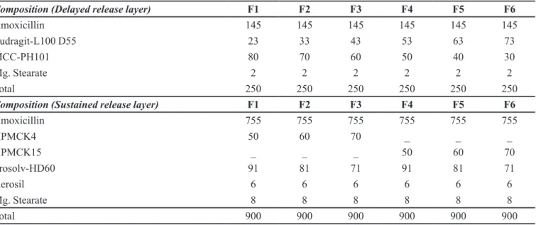
The time-dependent release bilayer tablets containing divided dose of amoxicillin trihydrate in delayed release layer and sustained release layer were prepared by direct compression method. The total dose of AMT in the matrix tablet was kept as 900 mg, which was equivalent to 775 mg of amoxicillin, i.e., once-daily dose for AMT in treatment of bacterial infections. The delayed release layer contained 145 mg of AMT and sustained release layer contained 755 mg of AMT. The delayed release granules were pre-pared by wet granulation method. Drug was granulated with Eudragit-L100 D55 dissolved in acetone. The wet mass was passed through sieve no. 18 (#BSS), and dried in hot air oven at 40 oC for 20 min. Finally magnesium stearate was mixed with dried granules for lubrication. Similarly, sustained release granules were prepared by dry blending of drug with excipients. Finally both the layers were com-pressed into a single bilayer tablet by direct compression technique using (20 x 9 mm) capsule-shaped punch in a rotary tablet compression machine (Cadmach India Ltd., In-dia). The compositions of prepared time-dependent release bilayer tablets of AMT are shown in Table I.
Characterization
Evaluation of delayed release granules
evalu-TABLE I - Composition of time-dependent bilayer tablet of amoxicillin trihydrate
Composition (Delayed release layer) F1 F2 F3 F4 F5 F6
Amoxicillin 145 145 145 145 145 145
Eudragit-L100 D55 23 33 43 53 63 73
MCC-PH101 80 70 60 50 40 30
Mg. Stearate 2 2 2 2 2 2
Total 250 250 250 250 250 250
Composition (Sustained release layer) F1 F2 F3 F4 F5 F6
Amoxicillin 755 755 755 755 755 755
HPMCK4 50 60 70 _ _ _
HPMCK15 _ _ _ 50 60 70
Prosolv-HD60 91 81 71 91 81 71
Aerosil 6 6 6 6 6 6
Mg. Stearate 8 8 8 8 8 8
Total 900 900 900 900 900 900
*All the quantities expressed are in mg
ated for micromeritic properties like percentage loss on drying (%LOD), angle of repose, bulk density and tapped density, Carr’s compressibility index and Hausner’s ratio.
Evaluation of bilayer tablets
The different formulations of time-dependent release bilayer tablets were evaluated for hardness, thickness, fri-ability, weight variation and drug content as per USPXXI
speciications. For drug content evaluation, 20 tablets were
weighed and crushed into powder. An accurately weighed quantity of powder was suitably dissolved in phosphate buffer (pH 7.4), appropriate dilutions were made and analysed by UV-Visible spectrophotometer to calculate the percentage drug content. The acceptance criteria of
all these tests were based on the USPXXI speciications.
In vitro drug release
In vitro drug release from the prepared bilayer tablets was performed using USP Type-1 dissolution apparatus
using SGF (pH 1.2) (900 mL) for irst 2 hours followed by
phosphate buffer (pH 7.4) (900 mL) for upto 16 hours at 50 rpm/37±0.5 0C. Aliquot samples (5 mL) were taken at various time intervals (0.5, 1, 1.5, 2, 2.5, 3, 4, 6, 8, 10, 12
and 16 hours), replaced with fresh media, iltered through 0.45 µm nylon ilter (Millipore, Mumbai, India) and absor -bance was determined spectrophotometrically at 273 nm. Further, the %drug release was determined and a plot was constructed between time (h) versus cumulative %drug release. Similarly, dissolution study was performed for marketed preparation Amoxil® (Dabur, India), containing AMT equivalent to 500 mg, using USP Type-I apparatus
with phosphate buffer (pH 7.4, 900 mL, 50 rpm/37±0.5 oC)
for 4 hours. At speciic time intervals (0.5, 0.75, 1, 1.5, 2,
2.5, 3, 3.5 and 4 hours) aliquots (5 mL) were collected, filtered and analysed spectrophotometrically. The in vitro drug release samples were further subjected to
anti-microbial activity evaluation using speciic test organisms.
Assay of AMT and antibacterial activity assessment
The tablet formulations which showed optimum drug release were taken for further evaluation using antimi crobial activity. The antimicrobial assay of bilayer tablets of AMT was performed using agar plate diffusion method. The different dilutions of standard were prepared in distilled water with concentrations ranging from 1-250 µg/mL. The
aliquots obtained after dissolution were iltered through
0.45 µm nylon filter (Millipore, Mumbai, India). Each
1 mL of the iltered samples was carefully transferred into the wells prepared with sterile borer on solidiied nutrient
agar (Himedia, India) plate in petridishes inoculated with test gram positive cocci, S. aureus (ATCC29213), and gram negative bacilli, E. coli (ATCC25922). After inoculation, petri-dishes were kept in an incubator (Remi Corporation, India) at 37 oC for 24 hours. After incubation the zone of inhibition (ZOI) for time-dependent release bilayer tablet, marketed preparation and standard dilution of antibiotic were measured with the help of slide calliper scale in mm. The concentration of AMT in aliquot samples was calcu-lated using the following equation(1):
Where, MIC is the minimum inhibitory concentra-tion, x is the zone of inhibition (mm), c is the concentration
of antibiotic (µg/mL), D is the diffusion coeficient, and
t is the time required for antibiotic diffusion. A plot was made between x2 vs. ln (c) graphs for standard dilutions of the antibiotic using each test organism. From this graph, the unknown concentration of AMT present in samples ob-tained after dissolution of bilayer tablets can be determined by extrapolating zone of inhibition (x) with respect to the
concentration (c) for each microbial strain to ind out the
%drug release. This helps in establishing the correlation between the in vitro drug release and anti-microbial assay.
RESULTS
Evaluation of delayed release granules
The micromeritic properties of the granules are given in the Table II. The %LOD was found to be less than 13%, while other parameters like angle of repose (25-30%), Carr’s index (12-16%) and Hausner’s ratio (<1.25) for all the formulations are within the acceptable
range, indicated good low property and compressibility.
Evaluation of bilayer tablets
The prepared bilayer tablets were capsular in shape with good physical appearance. Thickness, hardness, friability, weight variation and drug content of all formu-lations were found to be satisfactory as shown in Table III. Results indicated that all batches of prepared tablet
formulations met the USPXXI speciications with thick -ness <5%, hard-ness 18 kg/cm2, friability <1% and weight variation ±10%. Drug content uniformity was within 98.9±0.35 to 102.4±0.16%, respectively.
In vitro drug release and antibacterial activity
assessment
The in vitro drug release proile of different batches
of time-dependent release bilayer tablet formulations are depicted in Figure 1. The formulation F2 showed optimum time-dependent release with more than 85% drug release in 16 h. This optimized formulation was subjected to an-timicrobial activity evaluation using agar plate diffusion method as discussed above. According to equation (1), x2
vs. ln (c) plots were drawn using zone of inhibition data of control against S. aureus and E. coli as obtained from agar plate diffusion assay in Figure 2 and Figure 3. The
correlation coeficient values for these plots were found
to be 0.9983 and 0.9876 indicated good linearity. From these plots, the concentration of AMT from in vitro drug release samples of bilayer tablets was determined by com-paring their zones of inhibition (mm) data with respective concentrations (µg/mL) for each test organism (Table IV).
The in vitro drug release proile of time-dependent
release bilayer tablets using S. aureus and E. coli as test organisms is shown in Figure 4. Further, the in vitro drug
release proile using S. aureus and E. coli were compared by plotting y-axis and x-axis, respectively. The R2 value was found to be 0.9939 (Figure 5), showing a positive correlation between the assay results of two different
TABLE II - Micromeritic properties of delayed release granules of AMT
Formulation code % LOD
(Loss on drying)
Angle of repose
(θ) Bulk density (g/mL)
Tapped density (g/mL) Carr’s Index (%) Hausner’s Ratio F1 F2 F3 F4 F5 F6 13.0 12.9 13.1 13.0 12.9 13.2 26.9 28.2 29.9 28.6 26.4 27.1 1.14 1.14 1.13 1.14 1.14 1.13 1.32 1.30 1.31 1.31 1.32 1.33 13.6 12.3 15.9 12.9 13.6 15.0 1.15 1.14 1.15 1.14 1.15 1.17
TABLE III - Technological characterization of sustained release tablets of AMT (Mean± S.D, n=6)
Formulation code Thickness (mm) Hardness (kg/cm2) Friability (%) Weight variation (mg) Drug content (%)
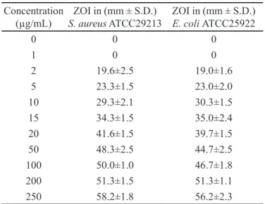
TABLE IV - Antibacterial activity of control samples against S. aureus ATCC29213 and E. coli ATCC25922
Concentration (µg/mL)
ZOI in (mm ± S.D.)
S. aureus ATCC29213
ZOI in (mm ± S.D.)
E. coli ATCC25922
0 0 0
1 0 0
2 19.6±2.5 19.0±1.6
5 23.3±1.5 23.0±2.0
10 29.3±2.1 30.3±1.5
15 34.3±1.5 35.0±2.4
20 41.6±1.5 39.7±1.5
50 48.3±2.5 44.7±2.5
100 50.0±1.0 46.7±1.8
200 51.3±1.5 51.3±1.1
250 58.2±1.8 56.2±2.3
ZOI- Zone of inhibition; n=6
FIGURE 1 -In vitro drug release profile of time-dependent release bilayer tablet containing AMT by dissolution study.
FIGURE 2 - x2vs. ln (c) plot of control (standard amoxicillin
trihydrate) using S. aureus. x = zone of inhibition (mm); c =
concentration of control samples (µg/mL)
FIGURE 3 - x2vs. ln (c) plot of control (standard amoxicillin
trihydrate) using E. coli. x = zone of inhibition (mm); c =
concentration of control samples (µg/mL)
organisms and also indicated that AMT estimation from the drug release samples was not affected by bacterial strains used in this investigation. Figure 6 and Figure 7 depicts the zone of inhibition of standard dilutions of AMT against S. aureus and E. coli. It was observed that zones of inhibition for these two different bacteria were almost the same. In vitro drug release data showed that AMT release from time-dependent release bilayer tablet was sustained up to 16 hours and delayed release occurred after 2 hours. Figure 8 and Figure 9 depicts the zone of inhibition of samples obtained from in vitro drug release study of time-dependent release tablet of amoxicillin trihydrate against
S. aureus and E. coli.
Table IV, V represented the zone of inhibition of time-dependent release bilayer tablet and marketed preparation (Amoxil®) of AMT performed using agar
FIGURE 4 - Comparative in vitro drug release profiles of optimized time-dependent release bilayer tablet containing AMT using S. aureus and E. coli as test organisms.
of pure drug at 5 µg/mL concentration. This indicated that bilayer tablets have lower value of MIC as compared to marketed preparation in the test organisms used.
The optimized time-dependent release bilayer tablet formulation showed maximum zone of inhibition of 76.1±2.05 mm in gram-positive and 74.0±1.07 mm in gram-negative bacteria within 16 hours, while marketed immediate release tablets showed maximum zone of in-hibition of 49.4±2.00 mm in gram-positive and 49.9±1.05 in gram-negative bacteria within 4 hours. Further, a prominent increase in zone of inhibition was observed with dissolution after 2 hours in time-dependent release formu-lation due to burst release of antibiotic to kill the viable
growth of microorganisms and showed good correlation with in vitro drug release data. This indicated the higher
ef-icacy of time-dependent release bilayer tablet formulation
FIGURE 5 - Comparative in vitro drug release profiles of optimized time-dependent release bilayer tablet containing AMT using S. aureus and E. coli as test organisms.
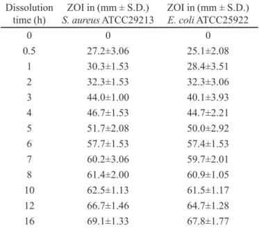
TABLE V - Antibacterial activity indicating the ZOI of samples
obtained from in vitro drug release study of time-dependent
bilayer release matrix tablet of amoxicillin trihydrate against S.
aureus ATCC29213 and E. coli ATCC25922
Dissolution time (h)
ZOI in (mm ± S.D.)
S. aureus ATCC29213
ZOI in (mm ± S.D.)
E. coli ATCC25922
0 0 0
0.5 27.2±3.06 25.1±2.08
1 30.3±1.53 28.4±3.51
2 32.3±1.53 32.3±3.06
3 44.0±1.00 40.1±3.93
4 46.7±1.53 44.7±2.21
5 51.7±2.08 50.0±2.92
6 57.7±1.53 57.4±1.53
7 60.2±3.06 59.7±2.01
8 61.4±2.00 60.9±1.05
10 62.5±1.13 61.5±1.17
12 66.7±1.46 64.7±1.28
16 69.1±1.33 67.8±1.77
ZOI- Zone of inhibition; n=6
FIGURE 9 - The pictures depicting zone of inhibition of samples
obtained from in vitro drug release study of time-dependent
release bilayer tablet formulation (F2) of AMT in E. coli.
FIGURE 8 - The pictures depicting zone of inhibition of samples
obtained from in vitro drug release study of time-dependent
release bilayer tablet formulation (F2) of AMT in S. aureus.
FIGURE 7 - The pictures depicting zone of inhibition of different
standard dilutions of (1-250 µg/mL) against E. coli.
FIGURE 6 - The pictures depicting zone of inhibition of different
over conventional immediate release marketed preparation due to the low MIC and enhanced antimicrobial action.
CONCLUSION
The time-dependent release bilayer tablets of AMT were prepared and evaluated for its antibacterial activity. The in vitro drug release from these tablets was estimated by antimicrobial assay using agar plate diffusion method. The aliquot dissolution samples of bilayer tablet formu-lation were found to be sensitive against both the test organisms, S. aureus and E. coli over 24 hours study. The drug release from bilayer tablet was found to be sustained up to 16 hours with burst release achieved after 2 hours. Furthermore, the MIC of prepared time-dependent release bilayer tablets was found to be 2 µg/mL, while marketed conventional tablet preparation was found to be 5 µg/mL. The lower MIC indicated higher anti-microbial activity of the prepared bilayer tablet formulation to arrest the growth of microorganisms. Thus, chronotherapeutics by time-dependent release bilayer tablets containing amoxicillin trihydrate may be more effective alternative over conventional dosage forms for the management of bacterial infections.
ACKNOWLEDGEMENTS
The authors are thankful to the Department of Microbiology, Majeedia Hospital, Hamdard University, New Delhi, for conducting the present work. Further, the authors are thankful to Mr. Md Irfanuddin, London School of Commerce, London, UK for overall correction of the
manuscript for good lair of English.
CONFLICT OF INTEREST
Authors have no conlict(s) of interest.
REFERENCES
AKHGARI, A.; GAREKANI, H.A.; SADEGHI, F. Combination of inulin and time dependent polymethacrylates as a coating
system to achieve colonic delivery of indomethacin. DARU
J., v.17, p.199-208, 2009.
AKHGARI, A.; SADEGHI, F.; GAREKANI, H.A. Combination of time-dependent and pH-dependent polymethacrylates as a single coating formulation for colonic delivery of
indomethacin pellets. Int. J. Pharm., v.320, p.137-142,
2006.
AURORA, J.; TALWAR, N.; PATHAK, V. Colonic drug
delivery challenges and opportunities - an overview. Eur.
Gastroenterol. Hepatol. Rev., v.1, p.1-6, 2006.
BONEV, B.; HOPPER, J.; PARISOT, J. Principles of assessing bacterial susceptibility to antibiotics using the agar diffusion
method. J. Antimicrob. Chemother., v.61, p.1-7, 2008.
CHA, R.; RYBAK, M.J. Pulsatile delivery of amoxicillin against
Streptococcus pneumonia. J.Antimicrob. Chemother., v.54, p.1067-1071, 2004.
DONOWITZ, D.R.; MANDELL, G.L. Beta-lactam antibiotic.
N. Engl. J. Med., v.318, p.419-426, 1988.
HERVEY, S.C. Antimicrobial drugs. In: GENNARO A.R. (Ed.).
Remington’s Pharmaceutical Sciences. 18.ed. Eston: Mack Publishing Company, 1991. cap.62, p.1163-1241.
HILTON, A.K.; DEASY, P.B. In vitro and in vivo evaluation of
an oral sustained-release loating dosage form of amoxicillin
trihydrate. Int. J. Pharm., v.86, p.79-88, 1992.
LEUTHNER, K.D.; CHEUNG, C.M.; RYBAK, M.J. Pulsatile delivery of clarithromycin alone or in combination with
amoxicillin against Streptococcus pneumoniae. Antimicrob
Agents Chemother., v.50, p.813-816, 2006.
PAPICH, M.G. The beta-lactam antibiotics: clinical
pharmacology and recent developments. Compend. Contin.
Educ. Pract. Vet., v.9, p.68-74, 1987.
PATEL, V.J.; AMIJI, M.M. Preparation and characterization of freeze-dried chitosan-poly (ethylene oxide) hydrogels for
site-speciic antibiotic delivery in stomach. Pharm. Res.,
v.13, p.588-593, 1996.
RISBUD, M.B.; HARDIKAR, A.A.; BHAT, S.V. pH sensitive freeze-dried chitosan-polyvinyl pyrrolidone hydrogels as
controlled release system for antibiotic delivery. J. Control.
Release, v.68, p.23-30, 2000.
S A H A S AT H I A N , T . ; K E R D C H O L P E T C H , T . ; C H A N W E R O C H , A . ; P R A P H A I R A K S I T, N . ; SUWONJANDEE, N.; MUANGSIN, N. Sustained release
of amoxicillin from chitosan tablets. Arch. Pharm. Res.,
SINGH, S.K.; CHIDRAWAR, V.R.; USHIR, Y.V.; VADALIA, K. R . ; SHE T H, N. R . ; SI NGH, S. Ph a r m a c e u t i c a l characterization of amoxicillin trihydrate as mucoadhesive
microspheres in management of H. pylori. Int. J. PharmTech.
Res., v.2, p.348-358, 2010.
SUN, H.K.; LEE, S.Y.; BANEVICIUS, M.A.; DU, X.; MAGLIO, D.; NICOLAU, D.P. Efficacy of pulsatile amoxicillin and clarithromycin dosing alone and in
combination in a murine pneumonia model. J. Antimicrob.
Chemother. v.56, p.559-565, 2005.
WILSON, C.G.; LEE, W.W.; MUKHERJI, G. Time-dependent systems for colonic delivery. In: RATHBONE, M.J.; HADGRAFT, J.; ROBERTS, M.S. (Eds.). Modiied-release
drug delivery technology. New York: Informa Healthcare, 2002. chapt.20, p.243-248.
YELLANKI, S.K.; SINGH, J.; ALI, S.J. Design and characterization of amoxicillin trihydrate mucoadhesive
microspheres for prolonged gastric retention. Int. J. Pharm.
Sci. Drug Res., v.2, p.112-114, 2010.
Received for publication on 27th July 2011