189
Effects of radiotherapy on bone tissues
Radiol Bras 2007;40(3):189–192
Original Article
EFFECTS OF RADIOTHERAPY ON BONE TISSUE*
Samantha Seara Da Cunha1
, Viviane Almeida Sarmento2
, Luciana Maria Pedreira Ramalho2 , André Carlos de Freitas2
, Darcy de Almeida1
, Maria Eulina Tavares3
, Jaílton Caetano Souza4 , Elaine Bauer Veeck5
, Nilza Pereira da Costa6
OBJECTIVE: To investigate the effects of radiotherapy on bone tissues and the accuracy of gray level mea-surements on radiographic images. MATERIALS AND METHODS: Four Wistar rats were submitted to
exter-nal radiotherapy (single 3000 cGy dose) on an area of 2 cm × 2 cm of their right legs. The animals were
sacrificed six weeks after radiotherapy, and both irradiated and contralateral (non-irradiated) legs were re-moved, dissected, evaluated for thickness, x-rayed in a standardized form and histologically processed (stained with hematoxylin-eosin and picrosirius red). The radiographs were digitalized and the gray level average was measured with the ImageTool® software. RESULTS: The femur thickness of non-irradiated legs was greater than that of the irradiated legs (p < 0.05). Radiographically, the findings indicated a higher bone density in
the non-irradiated legs, although with no statistically significant difference (p > 0.05). Histological analysis
of the irradiated legs demonstrated a decrease in the number of osteocytes and Haversian canals, although with no statistically significance (p > 0.05). On the other hand, a significant increase in adipocytes was
observed, resulting in a reduction of medullary tissue in the irradiated legs (p < 0.05), besides a higher
osteoblastic activity in the non-irradiated legs (p < 0.05). CONCLUSION: Radiotherapy within the above
mentioned parameters determined a decrease in activity of bone remodeling, which could be radiographi-cally detected in the majority of the evaluated specimens.
Keywords: Radiotherapy; Ionizing radiation; Bone tissue.
Efeitos da radioterapia no tecido ósseo.
OBJETIVO: Avaliar os efeitos da radioterapia e a acurácia da mensuração do nível de cinza do tecido ósseo em imagens radiográficas. MATERIAIS E MÉTODOS: Quatro ratos Wistar foram submetidos a radioterapia
externa (dose única de 3.000 cGy) em uma área de 2 cm × 2 cm na perna direita. Os animais foram
sacri-ficados seis semanas após a radioterapia, e tanto as pernas irradiadas quanto as contralaterais (não-irradia-das) foram avaliadas na sua espessura, radiografadas de forma padronizada e processadas histologicamente (hematoxilina-eosina e picrossírius). As radiografias foram digitalizadas e a média dos níveis de cinza foi men-surada no programa Image Tool®. RESULTADOS: A espessura do fêmur foi maior na perna contralateral do
que na irradiada (p < 0,05). Radiograficamente, observou-se maior quantidade de tecido ósseo na perna
contralateral em relação à perna irradiada, porém sem diferença estatística significante (p > 0,05).
Histolo-gicamente, foi possível observar, na perna irradiada, diminuição do número de osteócitos e dos canais de
Havers, porém sem diferença estatística significante (p > 0,05). Por outro lado, foi observado aumento
significante de adipócitos, com conseqüente diminuição de tecido medular na perna irradiada (p < 0,05) e
maior atividade osteoblástica na perna contralateral (p < 0,05). CONCLUSÃO: A radioterapia, na dose
apli-cada, determinou diminuição da atividade de remodelação óssea, que pôde ser detectada radiograficamente na maioria dos espécimes avaliados.
Unitermos: Radioterapia; Radiação ionizante; Tecido ósseo. Abstract
Resumo
* Study developed at Universidade Federal da Bahia (UFBA), Hospital Santa Izabel (HSI) and União Metropolitana de Educa-ção e Cultura (Unime), Salvador, BA, and at Pontifícia Universi-dade Católica do Rio Grande do Sul (PUCRS), Porto Alegre, RS, Brazil.
1. PhD, Universidade Federal da Paraíba (UFPB), João Pes-soa, PB, and Universidade Federal da Bahia (UFBA), Salvador, BA, Brazil.
2. PhD, Pontifícia Universidade Católica do Rio Grande do Sul (PUCRS), Porto Alegre, RS, Associate Professors at Department of Complementary Propedeutics and Integrated Clinic, Faculda-de Faculda-de Odontologia da UniversidaFaculda-de FeFaculda-deral da Bahia (UFBA), Salvador, BA, Brazil.
3. Radiotherapist at Hospital Santa Izabel (HSI), Salvador, BA, Brazil.
4. Physicist at Hospital Santa Izabel (HSI), Salvador, BA, Bra-zil.
5. PhD, Associate Professor at Pontifícia Universidade Católica do Rio Grande do Sul (PUCRS), Porto Alegre, RS, Brazil.
6. PhD, Universidade de São Paulo, Bauru, SP, Titular
Profes-INTRODUCTION
According to data from The World Health Organization (WHO)(1), more than 11 million people are diagnosed with can-cer each year, with estimated 16 million new cases per year as from 2020. Besides, cancer annually causes seven million
deaths, that is to say, 12.5% of deaths in the world. Amongst therapeutic modalities, radiotherapy represents a well established method for treatment of head and neck cancer. Approximately 50% of patients with cancer are submitted to radiotherapy at some phase of their treatment, either isolatedly or in association with other forms of oncologic therapy(2).
Although being frequently observed in the treatment of malignant tumors, the use of high radiation doses may generate unde-sirable side effects, since ionizing radiation cannot differentiate tumor cells from healthy
sor at Pontifícia Universidade Católica do Rio Grande do Sul (PUCRS), Porto Alegre, RS, Brazil.
Mailing address: Dra. Samantha Seara Da Cunha. Alameda Praia de Jaguaripe, 76, Vilas do Atlântico. Lauro de Freitas, BA, Brazil, 42700-000. E-mail: [email protected]
190
Álvares BR et al.
Radiol Bras 2007;40(3):189–192 cells(3,4). Consequently, the destruction of
healthy tissues is a factor limiting the com-prehensive utilization of radiation therapy. As with other tissues, bones also are affected by the radiation effects, resulting in a significant change of the bone regen-eration capacity when it is injured(4,5). One of these alterations would be a disorder in the balance between the osteoblastic and osteoclastic activities, leading to a bone destructive process. Also, a decrease in the number of osteocytes and osteoblasts may be observed following the tissue irradia-tion. Significant post-radiation alterations of the bone matrix develop slowly, the ini-tial changes being a result from an injury to the bone remodeling system, i.e., osteo-blasts, osteocytes and osteoclasts. Osteo-blasts tend to be more sensitive than osteo-clasts, therefore an increase in the cellular lysis may occur(6). As a result, the process of bone matrix formation stops, hindering the mineralization process, which may lead to a spontaneous bone fractures and osteo-radionecrosis(7–9). Endothelial cells also are heavily affected, and the vascular fibrosis results in a decrease in the vascularization, affecting the bone and medullary cells vi-tality, so the area remains susceptible to infection and necrosis, even after a small trauma(5,9). For this reason, dental extrac-tions are contraindicated during an one-year period following radiotherapy(10).
The severity of the tissue lesion will depend on the total radiotherapy dose, the effective biological dose, the size of the radiation field, the number of radiotherapy sessions and the interval between them, the dose fractioning, and the surgical and/or traumatic aggression to the irradiated tis-sue. Severe cases of tissue destruction usu-ally are associated with doses > 7,000 cGy, although 6,000 cGy may result in mandible osteoradionecrosis(11,12).
The present study was aimed at evalu-ating the radiotherapy effects on bone tis-sues, and the accuracy of the measurement of gray levels on radiographic images as a predictor of histological alterations of bone tissues in animal models.
MATERIALS AND METHODS
The present study utilized four male, adult, clinically healthy rats Rattus
norve-gicus albinus, Rodentia, Mammalia, of the lineage Wistar, weighing about 210–260 g. These animals were kept in individual cages measuring 20 cm × 30 cm × 13 cm, at 22°C room temperature, luminosity (12-hour day/12-(12-hour night cycle), 50% relative humidity, and received commercial rodents diet (Nuvilab® CR 1) and water ad libitum. All of the animals were submitted to general anesthesia by intraperitoneally in-jected sodic thiopental in the dosage 0.2 ml/100 g. Then the rats underwent tri-chotomy on their back legs coxofemoral region, and were immobilized during the irradiation procedure by means of an espe-cially developed acrylic device based on an experiment performed by Machado(13). The animals were submitted to a single session of ionizing radiation from a cobalt-60 source, with a total of 3,000 cGy delivered to a 2 cm × 2 cm area of their right legs, utilizing only one irradiation field from the top to the bottom. The rest of the animal body was protected by lead blocks inserted into the radiotherapy device. The animals were sacrificed six weeks after radio-therapy, and both irradiated and non-irra-diated legs were dissected.
The animals’ legs were placed with their ventral surfaces directly onto an imaging plate for cephalometric radiography of a DenOptix® (300 dpi, pixel de 85 µm) digi-tal radiographic system. A five-step alu-minium wedge with 1 mm increment was added to the apparatus. The radiographic device (Timex® – 70 kV e 7 mA) was set for 0.06 s exposure time, 1.20 cm focal distance and perpendicular beam.
After the radiographic exposure, the plate was read by the DenOptix® system, allowing the generation of the correspond-ing digital images which were exported and stored as bitmap files. These files are opened in the Photoshop® application for bright-ness correction with basis on the penetrom-eter and saved again. After that, the images were opened with the Image Tool® software, and a polygon was traced on the irradiated region of each rat. The mean gray level was measured by the “histogram” tool. So the mean gray levels measured for each speci-men were compared (Student-t test).
Then the specimens were longitudinally incised to expose the whole femur whose thickness was measured with a digital
Starrett 727 series pachymeter and a Zeiss® magnifying glass.
The femurs were sent for histological processing and hematoxylin-eosin and picrosirius red staining. The following pa-rameters were analyzed on the histological slides: presence of collagen fibers, level of periosteal osteoblastic activity, degree of bone reabsorption near the medulla, and amount of adipous tissue. The latest three items were evaluated as for their intensity degree as follows: mild intensity, grade 1; moderate intensity, grade 2; severe inten-sity, grade 3. These results were evaluated by means of the non-parametric chi-square test (error probability, 5%). Additionally, a counting of the number of osteocytes and Haversian canal was performed in 10 fields. Such results were analyzed by means of the non-parametric Kruskal-Wallis test (error probability, 5%).
RESULTS
After the femurs thickness measure-ment, a lower thickness was observed on the irradiated legs (4.20 mm × 2.84 mm) as compared with the non-irradiated legs (4.67 mm × 3.11 mm). This value was sta-tistically significant (p < 0.05).
Regarding the digital radiographic analysis, it was possible to observe that, in the irradiated legs, the bone tissue amount was smaller (mean gray levels, 118) as compared with the non-irradiated legs (mean gray level, 123.5), but this difference was not statistically significant (p > 0.05) (Figure 1).
be-191
Effects of radiotherapy on bone tissues
Radiol Bras 2007;40(3):189–192 Figure 1. Mean gray
level of the evaluated bone tissue.
tween the irradiated and non-irradiated legs (p > 0.05) was not statistically significant. The same has occurred with the counting of Haversian canals (p > 0.05). However, as regards the adipous/medullary tissues ratio, a mild presence of fat cells, and a moderate presence of medullary tissue were observed (Figure 3). This ratio was statistically significant as compared with the irradiated legs (p < 0.05). As regards the osteoblastic activity, the non-irradiated leg presented moderate to intense activity (Fig-ure 4), and this difference was statistically significant (p < 0.05). The presence of col-lagen fibers has not been observed, but this difference has not been statistically signifi-cant as compared with the irradiated leg (p
> 0.05). Mild to moderate bone reabsorp-tion has been observed, although with no statistically significant difference (p > 0.05).
DISCUSSION
The animal model chosen for the present study was the rat, since it is the most uti-lized for evaluating secondary effects of radiotherapy in studies developed by sev-eral authors (4,9,14,15).
Because of difficulties inherent to ani-mal model studies, the rats were exposed to a single radiation dose enough to cause trabecular changes; besides, this single dose was based on protocols reported by previous studies(4,9,14–16), ranging between 25 Gy and 35 Gy. The cobalt-60 apparatus was chosen because of its higher accessi-bility and for knowingly causing more ad-verse effects than other radiation sources(17). Based on the results of digital radio-graphic evaluation, it is possible to infer that the method has presented a good
sen-sitivity because, even in the absence of a statistically significant mean gray level between irradiated and contralateral legs, these results are similar to those of the his-tological analysis where significant differ-ences also have not been found among some structures evaluated. The sensitivity of the evaluation of gray levels on digital radiographic images of bone tissues has already been analyzed both in other in vivo
studies(18) and animal model studies(19). The literature has demonstrated that osseous alterations observed on irradiated bone are visible and are directly related to cellular scarcity in the bone structure(15). However, this assertion has been just par-tially evidenced with the present study, since although the irradiated bone has pre-sented less cellularized, it cannot be af-firmed that this has occurred as a result of radiotherapy, since the contralateral leg has not demonstrated statistically significant difference as regards the number of osteo-cytes.
The same has occurred with regard to the number of Haversian canals. On the irradiated leg, a little number of Haversian canals was observed, but this number was not statistically significant in comparison with the contralateral leg. Studies devel-oped by Morales et al.(20) have found a decreased local vascularization after expo-sure of rabbits mandibles to ionizing radia-tion. However, the animals mandibles uti-lized by these researchers as a comparison media, had not been submitted to any type of radiotherapy, as a comparison media.
Besides these effects, a low osteoblas-tic activity could be observed in the irradi-ated leg. These findings are similar in stud-ies developed by Matsumura et al.(21), Dare et al.(22) and Dudziak et al.(23), who have
found a decrease in osteoblastic prolifera-tion, leading to the idea that ionizing radia-tion implies the terminal differentiaradia-tion between precursor bone cells and osteo-blasts. The same fact has not occurred in the non-irradiated leg. The collagen produc-tion, however, was not affected, being ob-Figure 3. Photomicrograph of contralateral leg specimen, six weeks after radiotherapy. Note the medullary tissue/adipous tissue ratio. (approximate 100× increase of hematoxylin-eosin).
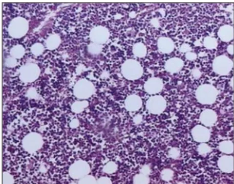
Figure 2. Photomicrograph of the irradiated leg specimen, six weeks after radiotherapy. Note the exuberant presence of adipous tissue in the bone marrow (approximate 100× increase of hematoxy-lin-eosin).
Figure 4. Photomicrograph of contralateral leg specimen, six weeks after radiotherapy. Note the presence of osteoblastic activity, with osseous neo-formation (approximate 200× increase of hema-toxylin-eosin).
Non-irradiated leg Irradiated leg
Specimens
G
ra
y
l
e
v
e
192
Álvares BR et al.
Radiol Bras 2007;40(3):189–192 served in 50% of the irradiated legs
al-though this finding has not been statisti-cally significant as compared with the con-tralateral leg which has not presented col-lagen.
Zones of bone reabsorption were vis-ible, although not with the expected inten-sity. Once more, there was no significant difference between irradiated and non-irra-diated legs. These findings go against those of Kiyohara et al.(4), who have found bone reabsorption in irradiated legs as from the fourth week after radiotherapy, with thin-ning of bone trabeculae. However, the bone thickness observed in the irradiated leg was statistically smaller than in the non-irradi-ated leg, contradicting the histological find-ings. This suggests that, in some moment, a possible bone reabsorption process has occurred. Also, an expressive increase in fat tissue was found, with the consequent reduction of bone marrow tissue. This fact had already been previously docu-mented(4,15).
CONCLUSION
It may be concluded that radiotherapy in the dose utilized in the present study, has determined a decrease in the bone remod-eling activity in the majority of specimens evaluated, which could be radiographically detected by measuring the gray levels of the bone tissue on digital radiographic images.
Acknowledgment
We thank to the Hospital Santa Izabel, for lending the hospital premises and for the devices operation.
REFERENCES
1. World Health Organization. Cancer home. Aces-sado em: 5/7/2005. Disponível em: http://www. who.int/cancer/en/
2. Hyderley LJ, Maddock PG. Noções gerais da ra-dioterapia. In: Manual de enfermagem oncoló-gica. São Paulo: Fundação Oncocentro de São Paulo, 1996;91–97.
3. Calhoun KH, Shapiro RD, Stiernberg CM, Calhoun JH, Mader JT. Osteomyelitis of the man-dible. Arch Otolaryngol Head Neck Surg 1988; 114:1157–1162.
4. Kiyohara S, Sakurai T, Kashima I. Early detection of radiation-induced structural changes in rat tra-becular bone. Dentomaxillofac Radiol 2003;32: 30–38.
5. Bras J, De Jonge HK, van Merkesteyn JP. Osteo-radionecrosis of the mandible: pathogenesis. Am J Otolaryngol 1990;11:244–250.
6. Vissink A, Jansma J, Spijkervet FKL, Burlage FR, Coppes RP. Oral sequelae of head and neck ra-diotherapy. Crit Rev Oral Biol Med 2003;14:199– 212.
7. Aitasalo K. Bone tissue response to irradiation and treatment model of mandibular irradiation injury. An experimental and clinical study. Acta Otolaryngol Suppl 1986;428:1–54.
8. Mitchell MJ, Logan PM. Radiation-induced changes in bone. RadioGraphics 1998;18:1125– 1136.
9. Würzler KK, DeWeese TL, Sebald W, Reddi AH. Radiation-induced impairment of bone healing can be overcome by recombinant human bone morphogenetic protein-2. J Craniofac Surg 1998; 9:131–137.
10. Németh Z, Somogyi A, Takácsi-Nagy Z, Barabás J, Németh G, Szabó G. Possibilities of preventing osteoradionecrosis during complex therapy of tumors of the oral cavity. Pathol Oncol Res 2000; 6:53–58.
11. Carlson ER, Zak MJ. Osteoradionecrosis and hyperbaric oxygen. Proceedings of the First In-ternational Congress on Maxillofacial Prosthet-ics 1996;184–191.
12. Jereczek-Fossa BA, Orecchia R. Radiotherapy-induced mandibular bone complications. Cancer Treat Rev 2002;28:65–74.
13. Machado RCL. Avaliação do efeito protetor da glutamina na resposta inflamatória do intestino
delgado à radioterapia abdominal: estudo expe-rimental em ratos. (Dissertação de Mestrado em Farmácia). Salvador, BA: Universidade Federal da Bahia, 2002.
14. Arnold M, Stas P, Kummermehr J, Schultz-Hec-tor S, Trott KR. Radiation-induced impairment of bone healing in the rat femur: effects of radiation dose, sequence and interval between surgery and irradiation. Radiother Oncol 1998;48:259–265. 15. Maeda M, Bryant MH, Yamagata M, Li G, Earle JD, Chao EY. Effects of irradiation on cortical bone and their time-related changes. A biome-chanical and histomorphological study. J Bone Joint Surg Am 1988;70:392–399.
16. Jacobsson M, Jonsson A, Albrektsson T, Turesson I. Dose-response for bone regeneration after single doses of 60Co irradiation. Int J Radiat Oncol Biol Phys 1985;11:1963–1969.
17. Pachigolla R, Pou A, Quinn F. The principles of radiation oncology. Grand Rounds Presentation, UTMB, Dept. of Otolaryngology, 2000. Acessado em: 10/11/2005. Disponível em: http://www. utmb.edu/otoref/Grnds/Radiation-Oncology-200001/Radiation-Oncology-200001.htm 18. Sarmento VA, Rubira IRF. Mensuração da
den-sidade óptica apical – uma proposta para diagnós-tico diferencial em endodontia. J Bras Odontol Clín, Curitiba 1998;12:65–68.
19. Sarmento VA, Pretto SM. Diagnóstico radiográ-fico de alterações periapicais de origem endodôn-tica através da determinação do nível de cinza em imagens digitais – estudo experimental em ratos. Rev Pós-Grad Fac Odontol Univ São Paulo 2003; 10:333–345.
20. Morales MJ, Marx RE, Gottlieb CF. Effects of pre-and postoperative irradiation on the healing of bone grafts in the rabbit. J Oral Maxillofac Surg 1987;45:34–41.
21. Matsumura S, Jikko A, Hiranuma H, Deguchi A, Fuchihata H. Effect of x-ray irradiation on pro-liferation and differentiation of osteoblast. Calcif Tissue Int 1996;59:307–308.
22. Dare A, Hachisu R, Yamaguchi A, Yokose S, Yoshiki S, Okano T. Effects of ionizing radiation on proliferation and differentiation of osteoblast-like cells. J Dent Res 1997;76:658–664. 23. Dudziak ME, Saadeh PB, Mehrara BJ, et al. The