*Corresponding author.
E-mail: jffweber@puncakalam.uitm.edu.my (J.F.F. Weber) A R T I C L E I N F O
Article history: Received 20 July 2013 Accepted 20 September 2013
Keywords Ajisamat
Salacia macrophylla Prismatomeris glabra Pharmacognostic features Anthraquinones
Oxidized triterpenes
A B S T R A C T
Ajisamat, an herb commonly used as an aphrodisiac in the Malaysian traditional medicine, corresponds to two different species from different families - Salacia macrophylla Blume, Celastraceae, and Prismatomeris glabra (Korth.) Valeton, Rubiaceae. Macromorphological inspection of the vegetative parts both plants reveals only a slight difference in the arrangement of the petioles. Microscopic investigation of the plants roots used as crude drugs revealed however distinctive anatomical features. Prismatic calcium oxalate crystals and banded paratracheal parenchyma are characteristics of S. macrophylla while P. glabra displays an abundance as crystals. Other features such as vessels diameters and arrangements are also of diagnostic importance. Some of these characters were also identified in the powder of thes e plant materials and proposed for diagnostic purpose. The values for extraction of ethanol and water as well as total ash, acid-insoluble ash, water-soluble ash and sulfated ash were determined for both plants. Phytochemical studies were carried out on hexane and chloroform extracts of S. macrophylla and methanolic extract of P. glabra. S. macrophylla was shown to contain highly oxidized pentacyclic triterpenes while P. glabra contains anthraquinones. The pharmacognostical and phytochemical information can be utilised as the identificationtools for Salacia macrophylla and Prismatomeris glabra
© 2013 Elsevier Editora Ltda. All rights reserved.
Original Article
Chemical and pharmacognostical characterization of two
Malaysian plants both known as Ajisamat
Tengku Azlan S. Tengku Mohamad
a,b, Humera Naz
a,b, Ratni S. Jalal
a,b, Khatijah Hussin
c,
Mohd R. Abd Rahman
c, Aishah Adam
b, Jean-Frédéric F. Weber
a,b,*aAtta-ur-Rahman Institute for Natural Product Discovery, Universiti Teknologi MARA, Puncak Alam, Selangor, Malaysia
bDepartment of Pharmacology and Chemistry, Faculty of Pharmacy, Universiti Teknologi MARA, Puncak Alam, Selangor, Malaysia cSchool of Environmental Science and Natural Resources, Universiti Kebangsaan Malaysia, Bangi, Selangor, Malaysia
Introduction
Ajisamat, Haji Samat or Tongkat Haji Samat is a commonly used plant in Malaysian traditional plant medicines. The aqueous extract of the dried roots of the plant have been traditionally used by the aborigines and certain rural Malays for wellness,
enhancing stamina and for its ergogenic effect. This plant has been used for generations to an aphrodisiac in Kelantan and Terengganu (Azmi et al., 2011). The names Ajisamat, Haji Samat
that the name referred to other two plants of different species and family i.e.Prismatomeris glabra (Korth.) Valeton, Rubiaceae, and Salacia macrophylla Blume, Celastraceae. The appearance of the vegetative parts including roots of both plants are nearly similar. Therefore, this study was carried out in an attempt to characterize the morphological, microscopic and chemical profiles of both types of Ajisamat plants.
Salacia macrophylla is a woody liana or climber found in swamps, lowland forest, on the mountain slopes as well as in old rubber estates. Usually it climbs up trees to several meters in height (Metcalfe and Chalk, 1965). Plants of this genus are known to possess pentacyclic triterpenes with highly oxidized A/B rings, as well as thiosugars, salacinol and kotalanol (Dhanabalasingham et al., 1996; Muraoka et al., 2008). These compounds have been reported to show a variety of biological activities such as cytotoxicity, antimicrobial, antifertility, anti-inflammatory, antimalarial and α-glucosidase inhibitory properties (Jeller et al., 2004; Maregesi et al., 2010).
Prismatomeris glabra is a slender tree and usually found in keranga and undisturbed mixed dipterocarp forests up to 700 m altitude in Peninsular Malaysia, Sumatra and Borneo (Slik, 2006). Usually it is found on hillsides and ridges, along rivers and streams. It can grow on sandy to clay soils. The genus
Prismatomeris usually contains anthraquinones and iridoids, compounds having cytotoxic (Hao et al., 2011) and antitumor activities (Krohn et al., 2007).
Materials and methods
Plant materials
Salacia macrophylla Blume, Celastraceae, was collected from a swamp forest near Taiping, Perak, Malaysia, while Prismatomeris glabra (Korth.) Valeton, Rubiaceae, was collected in Puchong, the dry lowland rain forest reserve, Selangor, Malaysia.
Life specimens of both plants were brought to Forest Research Institute Malaysia, Kepong, Selangor, Malaysia, and were identified by a botanist of specimens of both plants were deposited in the Herbarium of Faculty of Pharmacy, UiTM, Puncak Alam Campus under accession number UiTM191/HAB28 (P. glabra) and UiTM 278/TAS003 (S. macrophylla).
Macromorphological characterization
The macromorphological examination includes visual observation of the size, shape, color and was carried out following the method outlined by World Health Organization (WHO, 1998).
Micromorphological characterization
Micromorphological inspection was carried out on samples of the dried roots of the plants following the botanical microtechnique procedures described by Teh (1996). The dried roots were boiled and sectioned (25 µm in thickness) with a Reichert Jung sliding microtome slider. The obtained sections were bleached, stained with safranin, alcian green/blue and phloroglucinol, dehydrated, mounted with Canada balsam and
then studied under microscope. Microscopical examinations of the powdered roots of both plants were carried out by using the method modified from WHO (1998). The root powders of both plants were thoroughly mixed with two drops of glycerol on a glass slide and observed under the microscope.
Chemical characterization
Chemical characterization of both plant material included determination of solvent extractive values, ash values and color reaction tests.
Ethanol and water extraction amounts were determined following the methods described in WHO (1998) and Malaysian Herbal Monograph (2009). Briefly, the root powder S. macrophylla
was used to calculate the values of the hot and cold extraction in water and ethanol, while the powder of root of P. glabra
was extracted with only cold water and cold ethanol due to insufficient sample.
Measurements of total, acid-insoluble and water-soluble ashes were made according to the procedures discribed in Wiart and Kumar (2001) and in WHO (1998), while determination of sulfated ash was carried out following the method described in British Pharmacopoeia (2010).
Color reaction tests were done following the method prescribed by the Malaysian Herbal Monograph (2009). One gram sample of powdered root was treated with each of the reagents from a specific list at room temperature and the observation on the colors produced was done under normal light. The reagents were concentrated sulfuric acid, concentrated hydrochloric acid, sodium hydroxide 5%, potassium hydroxide 5%, ammonium hydroxide 25% and ferric chloride 5%.
Phytochemical analysis
The powder of the roots of S. macrophylla (170 g) was successively extracted with n-hexane, chloroform and ethyl acetate, while the powder roots of P. glabra (400 g) using hexane, ethyl acetate and methanol.
TLC analyses were performed on silica gel 60 F254 plates (Merck, Germany). Spots were detected at 254 and 366 nm; cerric sulfate was used as a spray reagent for the detection of triterpenes, while the characterization of anthraquinones was achieved with Bornträger reaction (5% KOH in ethanol). Column chromatography was carried out using silica gel (40-63 μm, Merck, Germany). The 1H-NMR, 13C-NMR, 1H-1H-COSY, HMQC
and HMBC spectra were recorded on Bruker Avance II 400 and Bruker Avance III 500 MHz NMR spectrometers; chemical shifts are in ppm (δ) relative to the SiMe4 as internal standard and coupling constants are in Hz. MS were measured on Jeol HX 110 mass spectrometers (m/z) and Agilent 6220 LC/MS TOF.
Results and discussion
Macromorphological characterization
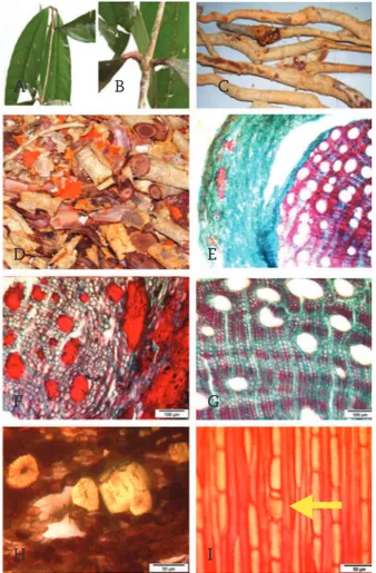
are simple, broadly elliptic in shape and glabrous. The leaf base is symmetrical (Fig. 1A) and arrangement of the petioles is subopposite (Fig. 1B). The roots of S. macrophylla are hard taproots, yellowish-brown in color and some with lateral roots. The root bark exfoliates easily. The outer layer is brown and the internal layer appears orange (Fig. 1C-D).
The second type of Ajisamat plant, Prismatomeris glabra, is a small plant, similar in appearance to the coffee plant. The leaves are simple, broadly elliptical in shape with an entire margin, and are oppositely arranged (Fig. 2A). Their scars or stipules are present and interpetiolar (Fig. 2B). Flowers are white with five petals, are black drupes when mature (Fig. 2C). The roots are hard woody taproots, yellowish-orange
color, with lateral roots (Fig. 2D). The roots of this species also show the unusual feature of an orange inner bark.
Generally both plants show a similar physical appearance. The leaf shape is similar with acute to acuminate apices. They are truly opposite for P. glabra, while slightly subopposite for S. macrophylla. However the two species can be distinguished by the presence of stipules or stipule scars and the raised midvein in P. glabra leaves.
Micromorphological characterization
The micromorphological inspection of both types of Ajisamat
plant revealed distinctive differences in the anatomical structures between the plants.
The vessel elements of S. macrophylla are mostly solitary, while some are in pairs, lack tyloses (Fig. 1E). Following the category of vessel characteristics by Menon (1967), the vessel size of S. macrophylla is moderately small (43-84 µm) and the vessels are numerous (21 to 40 vessels per mm2). The axial
parenchyma of S. macrophylla appears banded paratracheal of the 2- to 4-cells wide as well as confluent (Fig. 1G). In the tangential sections, (Muraoka et al., 2008), of root bark, brachysclereids are abundant within the phelloderm layer of the young and old roots (Fig. 1F). These are thick-walled with narrow pit canals isolated or arranged in groups (Fig. 1H). Uniseriate rays are of different heights, some with solitary prismatic crystals of calcium oxalate (Fig. 1I).
In P. glabra, the vessels elements are solitary, few are in pairs and lacking tyloses (Figure 2E). The vessel size for P. glabra can be categorized as extremely small (12-25 µm) and the number
Rp
Wp
p
Fig. 1 - Photograph of Salacia macrophylla. A, leaf blades; B, arrangement of the petioles; C, whole roots; D, root pieces with exfoliated orange barks; E, TS of root; F, TS of root bark showing phelloderm region; G, TS of root secondary xylem showing banded paratracheal axial parenchyma; H, brachysclereids (unstained); I, TLS root of secondary xylem showing an inclusion of prismatic calcium oxalate crystal in a ray cell (arrow).
Fig. 2 - Photograph of Prismatomeris glabra. A, leaf blades; B, stipule; C, drupes; D, whole root; E, TS of root (stained with phloroglucinol); F, TLS of root showing a bundle of raphides of calcium oxalate crystals (Rp) included in an axial parenchyma cell; G, TS of root showing uniseriate rays, multiseriate rays thick-walled fibres; H, TS of root bark and cork showing phelloderm (double-stained with safranin and alcian blue); I, TS of root showing distribution of vessels, fibers, axial parenchyma paratracheal and apotracheal (double-stained with safranin and alcian blue); J, TLS root of showing imperforated tracheids (T), vessels with alternate pitting wall (Wp) and simple perforation plate (P).
B
B
A
A
D
F
F
I
J
T
H
I
G
G
H
E
D
E
C
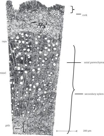
Fig. 3 - Anatomical characters of Salacia macrophylla root (TS).
Fig. 5 - Diagnostic features Salacia macrophylla powdered root; 1, corks cells; 2, fiber Bundle ; 3, vessel element; 4, group brachyscelereids; 5, fragment of vessel element; 6, tracheary elements; 7, unknown orange crystals; 8, prismatic crystals of calcium oxalate. (The scale marked * is for feature 7 and 8).
Fig. 4 - Anatomical characters of Prismatomeris glabra root (TS).
of vessels as very numerous (more than 40 vessels per mm2).
The parenchyma of P. glabra appears as both paratracheal and apotracheal axial. The paratracheal type is mostly of 2-cells wide, forming short tangential lines, while the apotracheal type includes uniseriate short tangential lines extending from rays to rays (Fig. 2 G-I). No brachysclereids were observed in P. glabra root bark (Fig. 2H).
Tangential longitudinal section (TLS) of P. glabra root reveals tracheids, vessels with alternate wall pitting and simple perforation plates (Fig. 2J). The parenchyma is irregularly distributed among fibers, some with inclusion of calcium oxalate crystals in the form of bundles of raphides (Fig. 2F).
The anatomical structure of tangential sections of the roots of both types of Ajisamat plants are presented in drawings in Fig. 3 and Fig. 4. The summary of the distinctive anatomical features between the two plants is presented in Chart 1.
Microscopical studies of root powder of both plants were carried out. The diagnostic features of powdered roots of S. macrophylla
and P. glabra are presented in Figs. 5 and 6 respectively. Their differential identification is therefore relatively easy. S. macrophylla
is characterized by abundant brachysclereids and solitary calcium oxalate crystals, while P. glabra contains numerous raphides and no sclereids.
secondary xylem
200 µm axial parenchyma cork
rays
vessel
pith
pith
cork
phellogen
brachysclereids
vessel with deposit
secondary xylem vessel
ray
axial
parenchyma
50 µm 50 µm
50 µm*
1 2
3 4
5
7
6
8
S. macrophylla P. glabra
Cork
Brick shaped with orange content
Brick shaped with content becoming blue or pink after staining with safranin-alcian blue
Parenchyma Banded paratracheal parenchyma
Both apotracheal and paratracheal parenchyma Xylem
vessels
Mainly solitary and some in pairs, moderately small in size and numerous, lack tyloses, with content that stains red with safranin
Solitary, extremely small in size and very numerous, lack tyloses, without vessel content
Sclereids Brachysclereids, mostly in groups
None
Calcium oxalate
Prismatic crystals in ray cells, abundant
Raphide crystals in bundles, in axial parenchyma, abundant
Rays Uniseriate Uniseriate to multiseriate
Method and solvent Mean extractive values (%)
S. macrophylla P. glabra
Cold method ethanol 1.4 1.1
water 1.4 6.2
Hot method ethanol 5.6 n.d.a
water 5.3 n.d.a
aNot determined due to lack of sample.
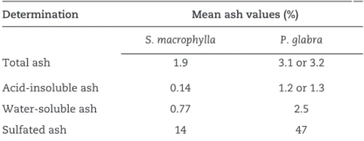
Determination Mean ash values (%)
S. macrophylla P. glabra
Total ash 1.9 3.1 or 3.2
Acid-insoluble ash 0.14 1.2 or 1.3
Water-soluble ash 0.77 2.5
Sulfated ash 14 47
Chart 1
Summary of anatomical characteristics of the roots for S. macrophylla and P. glabra.
Table 1
Extractive values for Salacia macrophylla and
Prismatomeris glabra.
Table 2
Ash values for Salacia macrophylla and Prismatomeris glabra.
Fig. 6 - Diagnostic features Prismatomeris glabra root powder. 1, starch grains; 2, raphide crystals; 3, oil globules; 4, fiber; 5, fragment of vessel element.
Chemical characterization
For S. macrophylla, both ethanol and water extractives were of similar value when produced by the cold method. With the hot method, the extractives are about four times greater than those produced by cold method (Table 1). In contrast, by cold method P. glabra produces a water extractive almost six times more abundant than the ethanol extractive.
The ash values obtained for S. macrophylla and P. glabra are presented in Table 2. Regardless of the type of the ashes, the values for P. glabra appear to be consistently higher than that of
S. macrophylla. Prabhu et al. (2009) reported that the presence of abundant calcium oxalate might contribute to high amounts of
1 2 3
4 5
20 µm
ash, especially for the sulfated ash values. Similarly, Shinkar (2007) reported that high values of acid-insoluble ash are related to the high content of calcium oxalate in plants. For P. glabra, calcium oxalate crystals were found to be abundantly present in the form of raphide needles. Therefore, the high ash values for this plant are likely due to the high content of calcium oxalate.
Chart 2 summarizes the color changes resulting from the reactions of root powders with reagents prescribed by the Malaysian Herbal Monograph (2009) for color reaction tests. Some distinctive differences in colors were produced were observed between the two types of Ajisamat plants that indeed could be used for fast discrimination between them.
Phytochemical analysis
Reagent Color
S. macrophylla P. glabra
Distilled water yellowish orange yellowish orange
Concentrated sulfuric acid
Black Green
Concentrated hydrochloric acid
No color changes Dark green
Sodium hydroxide (5%) Brown Dark red
Potassium hydroxide (5%) Brown Dark red Ammonium hydroxide
(25%)
Light brown Red
Ferric chloride Light green Brown
Chart 2
Color reaction tests for powdered roots of both Ajisamat
Repeated column chromatography (silica gel) of the n-hexane extract afforded S. macrophylla afforded β-sitosterol Patra et al., 2010 and four pentacyclic triterpenes namely, pristimerin (1) (Khalid et al., 2007), netzahualocoyene (2) (Dhanabalasingham et al., 1996) and netzahualocoyonol (3) (Jeller et al., 2004). The chloroform extract, when subjected to a similar fractionation process, produceded 2,3,7-trihydroxy-6-oxo-1,3,5(10),7-tetraene-24-nor-friedelane-29-oic acid methyl ester (4) (Ankli et al., 2000). Structure elucidations of compounds (1-4) were carried out by comparison of spectral data with literature. Triterpenes 1-4 belong to the D:A-friedo-nor-oleanane group and show the rare quinone-methide moiety. Such derivatives are significant chemotaxonomic characteristic of the family Celastraceae (Gamlath et al., 1990).
Chemical studies on the methanolic Prismatomeris glabra
extract led to the isolation of four anthraquinones. These compounds were isolated by MPLC (Medium Pressure Liquid
of anthraquinones warrant caution on the use of this plant. To the best of our knowledge, no pharmacological/toxicological study was carried out on S. macrophylla. The above mentioned triterpenes are also known for significant cytotoxicity (Ankli et al., 2000; Setzer et al., 1998). Yet, due to their low water solubility, it cannot be ascertained that a water extract of this plant is toxic without further tests. S. macrophylla therefore needs to be fully investigated. This is an endeavour undertaken by our group and results will be reported in due time.
Authorship
TASTM (MSc student) contributed in collection and identification of plant samples, confection of herbarium specimens, running laboratory work, analysis of date and drafted the paper. RSJ (MSc student) contributed to the phytochemical studies. MRAR (MSc student) and KH contributed in microscopic analysis. HN and JFFW designed the study, supervised the laboratory work. HN, JFFW and KH contributed to critical reading of the manuscript. AA conceptualized the research problem and facilitated administrative procedures. All the authors have read the final manuscript and approved the submission.
Acknowledgment
The authors wish to express their gratitude to Mr. Richard Chung and Mr. Kamarudin Salleh of Forest Research Institute Malaysia (FRIM) to identifying the plant species. This work was supported by Dana Kecemerlangan UiTM 600-RMI/ST/DANA 5/3/DST (225/200).
R E F E R E N C E S
Ankli, A., Heilmann, J., Heinrich, M., Sticher, O., 2000. Cytotoxic cardenolides and antibacterial terpenoids from Crossopetalum gaumeri. Phytochemistry. 54, 531-537.
Azmi, N., Loh, W.T., Omar, S.S., Jalil, J., Adam, A., 2011. Effects of aqueous extract of Prismatomeris glabra root on non-spatial memory in rats using object discrimination test. Sains Malaysiana. 40, 1097-1103.
Burnett, A.R., Thomson, R.H., 1968. Anthraquinones in two Digitalis species. Phytochemistry. 7, 1423-1423.
British Pharmacopoeia, 2010. British Pharmacopoeia-complete Edition CD, standard version 14.0. TSO information and publishing solution, London.
Dhanabalasingham, B., Karunaratne, V., Tezuka, Y., Kikuchi, T., Gunatilak, A.A.L., 1996. Biogenetically important quinonemethides and other triterpenoid constituents of Salacia reticulata. Phytochemistry. 42, 1377-1385.
Fraga, B.M., Quintana, N., Diaz, C.E., 2009. Anthraquinones from natural and transformed roots of Plocama pendula. Chem. Biodivers. 6, 182-192.
Gamlath, C.B., Gunatilaka, A.A.L., Tezuka, Y., Kikuchi, T., Balasubramanium, S., 1990. Quinone-methide, phenolic and related triterpenoids of plants of Celastraceae: future evidence for the structure of celastranhydride. Phytochemistry. 29, 3189-3192.
Chromatography) and preparative thin layer chromatographic techniques. They were identified as 1,3-dihydroxy-2 methoxymethyl-9,10-anthraquinone (5) (Reanmongkol et al., 2003), 1-hydroxy-3-methoxy-2-(methoxymethyl)-9,10-anthraquinone (6) (Joshi et al., 1955), 2-methyl-3-methoxy-9,10-anthraquinone (7) (Burnett and Thomson, 1968; Sartori et al., 1995) and 1,3-dimethoxy-2-methyl-9,10-anthraquione (8) (Fraga et al., 2009).
Hao, J., Feng, S.X., Qiu, S.X., Chen, T., 2011. Anthraquinone glycosides from the roots of Prismatomeris connata. Chin. J. Nat. Med. 9, 42-45.
Jeller, A.H., Silva, D.H.S., Liao, L.M., Bolzani V. S., Furlan, M., 2004. Antioxidant and phenolic triterpenes from quinomethide Cheiloclinium cognatum. Phytochemistry 65, 1977-1982. Joshi, B. S., Parkash, N., Venkataraman, K., 1955. Anthraquinone
and anthrone series. XVIII. A synthesis of lucidin. J. Sci. Ind. Res. 14B, 87-92.
Khalid, S.A., Friedrichsen, G.M., Christensen, S.B., El Tahir, A., Sattic, G.M., 2007. Isolation and characterization of pristimerin as the antiplasmodial and antileishmanial agent of Maytenus senegalensis (Lam.) Exell. Arkivoc. 129-134. Krohn, K., Gehle, D., Dey, S.K., Nahar, N., Mosihuzzaman, M.,
Sultana, N., Sohrab, M.H., Stephens, P.J., Pan, J.J., Sasse, F., 2007. Prismatomerin, a new iridoid from Prismatomeris tetrandra. Structure elucidation, determination of absolute configuration, and cytotoxicity. J. Nat. Prod. 70, 1339-1343. Department of Forestry Malaysia, 2005. Herbal Information, 2005.
Retrieved September 2005 from http://www.forestry.gov.my/ aji_samat.html.
Malaysian Herbal Monograph, 2009. Vol. 2. Malaysian Herbal Monograph Committee, Kuala Lumpur, Malaysia.
Maregesi, S.M., Hermans, N., Dhooghe, L., Cimanga, K., Ferreira, D., Pannecouque, C., Vanden Berghe, D. A., Cos, P., Maes, L., Vlietinck, A.J., Apers, S., Pieters, L., 2010. Phytochemical and biological investigations of Elaeodendron schlechteranum. J. Ethnopharmacol. 129, 319-326.
Menon, P.K.B., 1967. Structure and identification of Malayan woods. In: Sulaiman, A., Lim, S.C. (Eds.), 1993, Malayan Forest Records No. 25. Forest Research Institute Malaysia, Kepong, Malaysia.
Metcalfe, C.R., Chalk, L., 1965. Anatomy of the dicotyledons: leaves, stem and woods in relation to taxonomy with notes on economic uses. Clarendon Press, Oxford, UK.
Muraoka, O., Xie, W., Tanabe, G., Amer, M.F.A., Minematsu, T., Yoshikawa, M., 2008. On the structure of the bioactive constituent from ayurvedic medicine Salacia reticulata: revision of the literature. Tetrahedron Lett. 49, 7315-7317.
Patra, A., Jha, S., Murthy, P.N. A., Sharone, 2010., Isolation and characterization of stigmast-5-en-3b-ol (b-sitosterol) from the leaves of Hygrophila spinosa T. Anders. Int. J. Pharm. Sci. Res. 1, 95-100.
Prabhu, K., Ponnudurai, K., Hemalatha, S., Karar, P.K., 2009. Pharmacognostic investigations on the leaves of Viburnum coriaceum Blume. Nat. Prod. Radiance. 8, 520-524.
Reanmongkol, W., Subhadhirasakul, S., Panichayupakaranant, P., Kim, K.M., 2003. Anti-allergic and antioxidative activities of some compounds from Thai medicinal plants. Pharm. Biol. 41, 592-597.
Sartori, G., Bigi, F., Tao, X., Porta, C., Maggi, R., Predieri, G., Lanfranchi, M., Pellinghelli, M.A., 1995. An investigation of the reaction-mechanism of the bis-acylation of aromatics with o-phthaloyl dichlorides - regioselective synthesis of anthraquinones. J. Org. Chem. 60, 6588-6591.
Setzer, W.N., Setzer, M.C., Hopper, A.L., Moriarity, D.M., Lehrman, G.K., Niekamp, K.L., Morcomb, S.M., Bates, R.B., McClure, K.J., Stessman, C.C., Haber, W.A., 1998. The cytotoxic activity of a Salacia liana species from Monteverde, Costa Rica, is due to a high concentration of tingenone. Planta Medica. 64, 583-583. Shinkar, A.S., 2007. Phytochemical investigation and antiulcer
activity of Aphanamixis polystachya (Wall.) Parker., M. Pharm Dissertation. Rajiv Gandhi University of Health Sciences, Bangalore, India.
Slik, J.W.F., 2006. Trees of Sungai Wain. Retrieved April 2013 from http://www.nationaalherbarium.nl/sungaiwain/
Teh, C.P., 1996. Studies on the relationships between wood anatomical structures of local species and its potential uses. PhD Thesis. Universiti Kebangsaan Malaysia, Bangi, Malaysia. WHO, 1998. Quality control methods for medicinal plant
materials. Geneva, Switzerland.