*Corresponding author.
E-mail: [email protected] (S. Molz) A R T I C L E I N F O
Article history: Received 21 June 2013 Accepted 1 September 2013
Keywords: Persea major Glutamate Neuroprotection Adenosine
A B S T R A C T
Ischemic stroke is characterised by a lack of oxygen and glucose in the brain, leading to excessive glutamate release and neuronal cell death. Adenosine is produced in response to ATP depletion and acts as an endogenous neuromodulator that reduces excitotoxicity. Persea major (Meins.) L.E. Kopp (Lauraceae) is a medical plant that is indigenous to South Brazil, and the rural population has used it medicinally due to its anti-inflammatory properties. The aim of this study was to evaluate the neuroprotective effect of Persea major methanolic extract against oxygen and glucose deprivation and re-oxygenation as well as to determine its underlying mechanism of action in hippocampal brain slices. Persea major methanolic extract (0.5 mg/ml) has a neuroprotective effect on hippocampal slices when added before or during 15 min of oxygen and glucose deprivation or 2 h of re-oxygenation. Hippocampal slices subjected to oxygen and glucose deprivation and re-oxygenation showed significantly reduced glutamate uptake, and the addition of Persea major methanolic extract in the re-oxygenation period counteracted the reduction of glutamate uptake. The presence of A1 or A2A, but not A2B or A3 receptor antagonists, abolished the neuroprotective effect of Persea major methanolic extract. In conclusion, the neuroprotective effect of Persea major methanolic extract involves augmentation of glutamate uptake and modulation of A1 and A2B adenosine receptors.
© 2013 Elsevier Editora Ltda. All rights reserved.
Original Article
Neuroprotection of
Persea major
extract against oxygen and
glucose deprivation in hippocampal slices involves increased
glutamate uptake and modulation of A1 and A
2A
adenosine
receptors
Marielli Letícia Fedalto
a, Fabiana Kalyne Ludka
a,b, Carla I. Tasca
b, Simone Molz
a,*aCurso de Farmácia, Fundação Universidade do Contestado, Canoinhas, SC, Brazil
bDepartamento de Bioquímica, Centro de Ciências Biológicas, Universidade Federal de Santa Catarina, Florianópolis, SC, Brazil
Introduction
Persea major (Meins.) L.E. Kopp, Lauraceae, is a medical plant that is grown mainly in South Brazil. It is popularly known as “pau-andrade”, “abacate-do-mato” or “canela-rosa”,
effects were also observed with the hydroalcoholic extract of
Persea major (Cosmo et al., 2007).
Ischemic stroke is caused by a transient or permanent reduction of blood flow to the brain, which leads to a complex cascade of events that culminate in neuronal death (Hofmeijer and van Putten, 2012). Ischemic stroke represents the main cause of long-lasting disabilities, and it is the third leading cause of death in major industrialised countries (Savitz and Mattle, 2013). Oxygen and glucose deprivation (OGD) in brain tissue preparations is often used as an in vitro model of cerebral ischemia (Arias et al., 1999; Brongholi et al., 2006; Matsuda et al., 1992; Oliveira et al., 2002) that allows for the evaluation of neural degeneration. Additionally, this model system allows for the study of the effects of putative protective compounds in a cerebral structure that is especially susceptible to ischemic insult in a tissue preparation that maintains the cellular architecture observed in situ (Dal-Cim et al., 2011).
During OGD, the lack of glucose and oxygen reduces ATP levels, which leads to the collapse of the Na+/K+ electrochemical gradient. After OGD, when oxygen levels are normalised, termed the re-oxygenation period, ischemic insult can be aggravated by the additional generation of reactive oxygen species, mostly in the mitochondrial electron transport chain, inflammation and the abnormal release of neurotransmitters (Kostandy, 2012). As a result, glutamate transporters operate in a reverse direction, releasing excessive glutamate (Danbolt, 2001), resulting in the overstimulation of the NMDA receptor (excitotoxicity) (Lai et al., 2010).
Adenosine is an endogenous compound that modulates neurotransmission. The intracellular concentration of adenosine is tightly linked to the energy charge of cells, as small decreases in the energy charge result in large increases in the intracellular levels of adenosine (Cunha, 2001), which can be released to the extracellular space by adenosine transporters. Extracellular adenosine then acts on metabotropic adenosine receptors located in the cell membrane, A1, A2A, A2B and A3 receptors. In the nervous system, adenosine exerts a wide range of effects (Fredholm, 2007; Fredholm et al., 2005), but it generally inhibits the pre-synaptic activity of glutamatergic transmission (Dolphin and Archer, 1983); thus, adenosine plays an important protective role against excitotoxic/ischemic damage in the brain (Lopes et al., 2011).
Despite extensive and continued research on the mechanisms of ischemia-induced cell death, there are currently no pharmacological interventions that provide significant neuroprotection against brain ischemia or injury in the clinical setting. Because inflammation is a key event in ischemic brain injury (Xia et al., 2010) and Persea major has anti-inflammatory activities (Cosmo et al., 2007), we evaluated the neuroprotective effect of the Persea major methanolic extract (PMME) against OGD-induced cell damage and sought to unravel its underlying mechanism of action in hippocampal brain slices.
Materials and Methods
Plant material
Persea major (Meins.) L.E. Kopp, Lauraceae, bark was collected in Canoinhas, Santa Catarina, Brazil, in 2010. The plant material
was identified by M. L. Brotto, and a specimen voucher (number 290857) of the plant was deposited in the Municipal Botanical Museum of Curitiba, Brazil.
Extract preparation
Methanolic extract was obtained by successive percolation of dried and powdered Persea major bark (3 kg) with methanol for 21 days. The solvent was changed every seven days, in the dark, at room temperature. The extract was evaporated in vacuo, and the residue was powdered and maintained at -20 °C until use.
Drugs
[3H]Glutamate was purchase from Amersham Life Science.
All other reagents were obtained from Sigma (St. Louis, MO, USA). The selective adenosine A1 receptor selective antagonist 8-cyclopentyl-1,3-dipropylxanthine (DPCPX) and the selective adenosine A2A receptor antagonist 4-(-2-[7-amino-2-{2-furyl} {1,2,4}triazolo{2,3-a} {1,3,5}triazin-5-yl-amino]ethyl)phenol (ZM241385) were dissolved and added to the hippocampal brain slices at a final concentration of 0,01% dimethylsulfoxide (DMSO). The selective A3 adenosine receptor antagonist
N-(2-methoxyphenyl)-N’-[2-(3-pyridinyl)-4-quinazolinyl]-urea (VUF5574, 1 µM) was also prepared in DMSO and added to slices at a final concentration of 1% DMSO. The antagonists were diluted in physiological buffer to the indicated final concentrations: 100 nM DPCPX (Molz et al., 2009); 50 nM ZM241385 (Molz et al., 2009); 0,1 µM alloxazine (Brown and Dale, 2000) and 1 µM VUF5574 (Stella et al., 2003).
Animals
Male Swiss mice (60-90 days old, weighing 20-30 g) were obtained from our local breeding colony and maintained on a 12-h light-12 h dark schedule at 25º C with food and water ad libitum. All experiments followed the “Principles of laboratory animal care” (NIH publication No. 85-23, revised 1985) and were approved by the local Ethical Committee of Animal Research (CEP/UnC 087/2010).
Preparation and incubation of hippocampal slices
Mice were sacrificed by decapitation, and the hippocampi were rapidly removed and placed in ice-cold Krebs-Ringer bicarbonate buffer (KRB; 122 mM NaCl, 3 mM KCl, 1.2 mM MgSO4, 1.3 mM CaCl2, 0.4 mM KH2PO4, 25 mM NaHCO3 and 10 mMD-glucose). The buffer was bubbled with 95% O2-5% CO2 at pH 7.4. Slices (0.4 mm thick) were rapidly prepared using a McIlwain Tissue Chopper, separated in KRB at 4 °C and allowed to recover for 30 min in KRB at 37 °C for stabilisation (Oliveira et al., 2002).
In the OGD model, hippocampal slices were incubated in a modified KRB buffer, where D-glucose was replaced with 10
buffer was replaced by physiological KRB and bubbled with 95% O2–5% CO2.
Treatment of hippocampal slices
To determine whether PMME was toxic to hippocampal slices, PMME was dissolved in aqueous buffers (KRB) and added to slices for 2 h and 15 min.
To verify the neuroprotective effect of PMME, the slices were treated as follows: (a) slices were pre-incubated with PMME for 15 min and then subjected to OGD and re-oxygenation; (b) PMME was added to slices only during the OGD period and then subjected to re-oxygenation; or (c) slices were subjected to OGD and then PMME was added during the re-oxygenation period (Oleskovicz et al., 2008). In all experiments, the slices were incubated for 2 h and 15 min in physiological buffer (KRB) alone as a control. In experiments where adenosine receptors antagonists were tested, hippocampal slices were treated with adenosine antagonists for 15 min before adding PMME, and the antagonists were present the during re-oxygenation period. The following antagonists were used: selective adenosine A1 receptor antagonist (DPCPX, 100 nM), adenosine A2A receptor antagonist (ZM241385, 50 nM), adenosine receptor A2B antagonist (alloxazine, 0,1 µM) and adenosine A3 receptor antagonist (VUF5574, 1 µM).
To assess the involvement of glutamate transport in the neuroprotective effect of PMME, the extract was dissolved in HBSS. Hippocampal brain slices were subjected to OGD, and PMME was added during the re-oxygenation period.
Cellular viability assay
Cell viability was determined by measuring the ability of cells to reduce MTT (3-(4,5-dimethylthiazol-2-yl-diphenyltetrazolium bromide; Sigma) (Mosmann, 1983). Hippocampal slices were incubated with MTT (0.5 mg/ml) in KRB for 30 min at 37 ºC. The tetrazolium ring of MTT is cleaved by active dehydrogenases and produces precipitated formazan. Next, 200 μl DMSO was used to dissolve the formazan, generating a coloured compound. The optical density of each well was measured using a microplate reader (Labsystems Multiskan MS-550 nm).
Glutamate uptake assay
L-[3H]-glutamate uptake was evaluated as previously described
(Molz et al., 2009). After OGD and re-oxygenation, hippocampal slices were maintained for 15 min at 37 °C in HBSS containing the following (mM): 1.3 CaCl2, 137 NaCl, 5 KCl, 0.65 MgSO4, 0.3 Na2HPO4, 1.1 KH2PO4, 2 glucose and 5 HEPES. Uptake was assessed by adding 0.33 µCi/ml L-[3H]-glutamate with 100 µM
of unlabelled glutamate. After 7 min, incubation was stopped by discarding the incubation medium, and the slices were subjected to two ice-cold washes with 1 ml HBSS. The slices were solubilised by overnight incubation in 0.1% NaOH/0.01% SDS. Aliquots of the resulting slice lysates were collected to determine the intracellular L-[3H]-glutamate content of using
a scintillation counter. Sodium-independent uptake was determined using choline chloride instead of sodium chloride in the HBSS buffer. Non-specific sodium-independent uptake
(approximately 30% of total glutamate uptake) was subtracted from the total uptake to obtain the specific sodium-dependent glutamate uptake. The results are expressed as nmol of
L-[3H]-glutamate taken up per mg of protein per min. Protein
content was determined according to Peterson (1977), using bovine albumin as a standard.
Statistical analysis
The results are expressed as the means ± standard error of mean (SEM). Comparisons among groups were performed by one-way analysis of variance (ANOVA) followed by Tukey’s test (Graphpad Prism), and p ≤ 0.05 was considered to be statistically significant.
Results and discussion
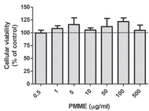
The viability of hippocampal slices incubated in control conditions with increasing concentrations of PMME is shown in Fig. 1. No significant changes on cellular viability were observed at any concentration tested. The results are expressed as the percentage viability with respect to the control group (100%).
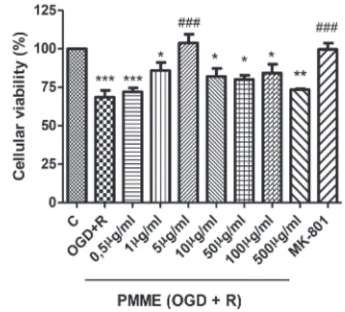
Considering that the extract was not toxic to hippocampal slices, they were pre-incubated in KRB buffer for 15 min with increasing concentrations of PMME (0.5 to 500 µg/ml) and then subjected to OGD and re-oxygenation (OGD+R). Ischemic conditions (OGD+R) significantly decreased cellular viability (p ≤ 0.001) (Fig. 2). Pretreatment with PMME extract (5 µg/ml) significantly inhibited OGD+R induced cell death (p ≤ 0.001) to a similar extent as MK-801 (50 µM), an NMDA receptor antagonist. Based on this result, this PMME concentration was used in all subsequent experiments.
Next, PMME extract was added to slices during 15 min of OGD or during the 2 h re-oxygenation period. PMME treatment significantly reduced cell death in OGD or OGD+R-induced hippocampal slices (Fig. 3). Therefore, not only is PMME
Fig. 3 - Effect of PMME on oxygen/glucose deprivation and re-oxygenation (OGD+R)-induced cell death. PMME (5 µg/ml) was incubated during the 15 min OGD time period or during the 2 h re-oxygenation (R) period. The control group (dashed lines) was considered to be 100%. The results represent the means ± SEM of five experiments carried out in triplicate. * Indicates that the mean is significantly different from all other groups; p ≤ 0.05; F = 7.292 (ANOVA followed by Tukey’s test).
neuroprotective as a pretreatment but, more importantly, it can also reverse cellular injury when it is already been activated. This experiment was performed in a condition that is clinically relevant to the treatment of ischemic injury.
Impairment of glutamate transport and excessive activation of glutamate receptors are involved in ischemia-induced damage in vitro and in vivo (Dal-Cim et al., 2011; Puyal et al., 2013). Because MK-801, an NMDA subtype receptor antagonist, afforded the same level of neuroprotection as PMME extract
Fig. 2 - Effect of preincubation with crescent concentrations of PMME (0.5, 1, 5, 10, 50, 100 and 500 µg/ml) on cellular viability reduction induced by oxygen/glucose deprivation and re-oxygenation (OGD+R). The control group (dashed lines) was considered to be 100%. The results represent the means±SEM of five experiments carried out in triplicate.
*** (p ≤ 0.001), ** (p ≤ 0.01) and * (p ≤ 0.05) indicate that the mean is significantly different from the control (dashed lines), PMME 5 µg/ml or MK-801 groups; ### (p ≤ 0.001; F = 8.809) indicates that the mean is significantly different from the OGD+R group, (ANOVA followed by Tukey’s test).
(Fig. 2), we evaluated the ability of PMME extract to counteract OGD+R-induced glutamate uptake impairment. OGD+R significantly decreased glutamate uptake, and the addition of PMME during the re-oxygenation period counteracted this OGD+R-induced impairment of glutamate uptake (Figure 4). These data suggest that PMME has an inhibitory effect on ODG+R –induced glutamate transport impairment, affording neuroprotection by inhibiting excitotoxicity.
Adenosine levels rise markedly in response to ischemia, hypoxia, excitotoxicity or inflammation, as it is a neuroprotectant under these conditions (Lopes et al., 2011). In hippocampal glutamatergic nerve terminals, A1 and A2A receptors co-localise and functionally interact with a subset of these terminals (Rebola et al., 2005); (Ciruela et al., 2006). In this study, the neuroprotective effect of PMME was abolished in the presence of A1 or A2A adenosine receptor antagonists (Figs. 5 and 6), suggesting that A1 and A2A receptors mediate the neuroprotective effects of PMME extract in hippocampal slices. In contrast, when the antagonists were added to the media during the re-oxygenation period (in the absence of PMME extract), they did not alter cell death, which indicates that blocking adenosine receptors was not detrimental toward hippocampal slices compared to OGD+R per se.
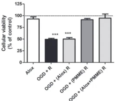
Due to their low density in the brain, the possible involvement of A3 and A2B receptors in brain function and neuroprotection are poorly understood. However, both A3 and A2B are present in neural tissue and can be activated under pathological conditions (Popoli and Pepponi, 2012). As shown in Figs. 7 and 8, the presence of A2B or A3 receptors antagonists (respectively) did not alter the PMME-induced increase in cell viability . Therefore, neither the A2B nor A3 receptors are involved in the neuroprotective effect of PMME against OGD+R in mouse hippocampal slices. Moreover, antagonists added to the media during the re-oxygenation period
Fig. 4 - Effect of PMME on glutamate uptake in hippocampal slices subjected to oxygen/glucose deprivation and re-oxygenation (OGD + R). PMME (5 µg/ml) was added during the re-oxygenation period (R). The values are expressed as nmol L-[3H] glutamate/mg protein/min and represent the
Fig. 6 - Effect of the adenosine A2A receptor antagonist (ZM 241385, 50 nM) on the neuroprotective effect of PMME. Control group (dashed lines) was considered to be 100%. The values are expressed as the percentage of the control and the data represents the mean s± SEM of four experiments carried out in triplicate. *** Indicates that the mean is significantly different from the control, ZM and OGD + (PMME) R groups,
p ≤ 0.001; F = 42.05 (ANOVA followed by Tukey’s test).
Fig. 7 - Effect of the adenosine A2B receptor antagonist (alloxazine 0,1 µM) on the neuroprotective effect of PMME. Control group (dashed lines) was considered to be 100%. The values are expressed as the percentage of the control and the data represent the means ± SEM of seven experiments carried out in triplicate. *** Indicates that the mean is significantly different from the control, alox, OGD + (PMME) R and OGD + (alox + PMME) R groups, p ≤ 0.001; F = 28.67 (ANOVA followed by Tukey’s test).
Fig. 8 - Effect of the adenosine A3 receptor antagonist (VUF 5574, 1 µM) on the neuroprotective effect of PMME. Control group (dashed lines) was considered to be 100%. The values are expressed as the percentage of the control and the data represent the means ± SEM of seven experiments carried out in triplicate. *** indicates that the mean is significantly different from the control, DMSO, VUF, OGD + (PMME)R and OGD+(VUF+PMME)R groups, p ≤ 0.001; F = 10.45 (ANOVA followed by Tukey’s test).
Fig. 5 - Effect of the adenosine A1 receptor antagonist (DPCPX, 100 nM) on the neuroprotective effect of PMME. The control group (dashed lines) was considered to be 100%. The values are expressed as the percentage of the control and the data represent the means ± SEM of five experiments carried out in triplicate. *** indicates that the mean is significantly different from the control, DPCPX and OGD + (PMME) R groups, p ≤ 0.001; F = 17.64 (ANOVA followed by Tukey’s test).
did not alter cell death, which indicates that blocking adenosine receptors was not detrimental toward hippocampal slices compared to OGD+R per se.
Taken together, these results indicate that PMME-induced neuroprotection involves modulation of the adenosine A1 and A2A receptors but not of the A2A or A3 receptors. Presynaptic A2A receptors heterodimerise with A1 receptors and tightly regulate glutamate release (Ciruela et al., 2006). Thus, we hypothesise that PMME extract acts as an agonist of adenosine receptors, or it may increase adenosine release into the synaptic cleft, which in turn can act on the A1 or A2A receptors and provide neuroprotection by decreasing the release of glutamate and counteracting neuronal cell death.
et al., 2000) and isolated polyphenol obtained from Lauraceae species provide neuroprotection against OGD-induced cell death (Lee et al., 2010) by inhibiting glutamate release (Panickar et al., 2012). The Persea genus is rich in polyphenols (Rodriguez-Carpena et al., 2011) and has demonstrated neuroprotective effects against ischemia-induced neuronal death (Eser et al., 2011; Yaman et al., 2007). However, to the best of our knowledge, this is the first evidence that a species from the Lauraceae family exerts neuroprotection against OGD by modulating adenosine receptors.
In conclusion, for the first time, this study demonstrates the neuroprotective effect of PMME and its underlying mechanism of action in hippocampal brain slices. Few studies have isolated and identified the chemical constituents of Persea major. Batista et al., (2010) reported the presence of flavonoids, sesquiterpenes and lignans. Phytochemical studies are currently in progress in our laboratory to elucidate the principles of PMME and its relevant mechanism of action. The medical management of ischemic stroke is still dependent on thrombolytics, anticoagulants and nonspecific neurotonics (Heitsch and Panagos, 2013); thus, the introduction of new natural products that prevent the effects of glutamate during ischemia opens a new chapter for ischemic stroke management that might provide substantial benefits for patients.
Authorship
MLF (undergraduate student) and FKL (PhD student) contributed to the laboratory work, biological studies, data analysis and drafted the paper. SM and CIT designed the study, supervised the laboratory work and critically read the manuscript. All the authors have read the final manuscript and approved its submission.
Acknowledgments
This research was supported by a grant from Fundação de Amparo à Pesquisa e Inovação do Estado de Santa Catarina (FAPESC). The authors thank Reinaldo Ribeiro for his contribution in collecting plant samples and Clarisse Bolfe Poliquesi for assisting with plant identification and herbarium confection.
R E F E R E N C E S
Arias, R.L., Tasse, J.R., Bowlby, M R., 1999. Neuroprotective interaction effects of NMDA and AMPA receptor antagonists in an in vitro model of cerebral ischemia. Brain. Res. 816, 299-308.
Batista, A.N.D., Batista, J.M., Lopez, S.N., Furlan, M., Cavalheiro, A.J., Silva, D.H.S., Bolzani, V.D., Nunomura, S.M., Yoshida, M., 2010. Aromatic compounds from three Brazilian Lauraceae species. Quim. Nova. 33, 321-323.
Brongholi, K., Souza, D.G., Bainy, A.C., Dafre, A.L., Tasca, C.I., 2006. Oxygen-glucose deprivation decreases glutathione levels and glutamate uptake in rat hippocampal slices. Brain. Res. 1083, 211-218.
Brown, P., Dale, N., 2000. Adenosine A1 receptors modulate high voltage-activated Ca2+ currents and motor pattern generation in the xenopus embryo. J. Physiol. 525 Pt 3, 655-667.
Cechinel-Filho, V., Zampirolo, J.A., Stulzer, H.K., Schlemper, V., 2007. Antispasmodic effects of Persea cordata bark fractions on guinea pig ileum. Fitoterapia. 78, 125-128.
Cho, E.Y., Lee, S.J., Nam, K.W., Shin, J., Oh, K.B., Kim, K.H., Mar, W., 2010. Amelioration of oxygen and glucose deprivation-induced neuronal death by chloroform fraction of bay leaves (Laurus nobilis). Biosci. Biotechnol. Biochem. 74, 2029-2035.
Ciruela, F., Casado, V., Rodrigues, R.J., Lujan, R., Burgueno, J., Canals, M., Borycz, J., Rebola, N., Goldberg, S.R., Mallol, J., Cortes, A., Canela, E.I., Lopez-Gimenez, J.F., Milligan, G., Lluis, C., Cunha, R.A., Ferre, S., Franco, R., 2006. Presynaptic control of striatal glutamatergic neurotransmission by adenosine A1-A2A receptor heteromers. J. Neurosci. 26, 2080-2087.
Cosmo, S.A., Mayer, B., Freitas, C.S., Baggio, C.H., Marques, M.C.A., 2007. Gastroprotective effect of hydroalcoholic extract from barks of Persea major Kopp (Lauraceae) in rats. Rev. Bras. Farmacogn. 17, 533-537.
Cunha, R.A., 2001. Adenosine as a neuromodulator and as a homeostatic regulator in the nervous system: different roles, different sources and different receptors. Neurochem. Int. 38, 107-125.
Dal-Cim, T., Martins, W.C., Santos, A.R., Tasca, C.I., 2011. Guanosine is neuroprotective against oxygen/glucose deprivation in hippocampal slices via large conductance Ca(2)+-activated K+ channels, phosphatidilinositol-3 kinase/ protein kinase B pathway activation and glutamate uptake. Neuroscience. 183, 212-220.
Danbolt, N. C., 2001. Glutamate uptake. Prog. Neurobiol. 65, 1-105. Dolphin, A. C., Archer, E. R., 1983. An adenosine agonist inhibits
and a cyclic AMP analogue enhances the release of glutamate but not GABA from slices of rat dentate gyrus. Neurosci. Lett. 43, 49-54.
Eser, O., Songur, A., Yaman, M., Cosar, M., Fidan, H., Sahin, O., Mollaoglu, H., Buyukbas, S., 2011. The protective effect of avocado soybean unsaponifilables on brain ischemia/ reperfusion injury in rat prefrontal cortex. Br. J. Neurosurg. 25, 701-706.
Fredholm, B.B., 2007. Adenosine, an endogenous distress signal, modulates tissue damage and repair. Cell Death Differ. 14, 1315-1323.
Fredholm, B.B., Chen, J.F., Cunha, R.A., Svenningsson, P., Vaugeois, J.M., 2005. Adenosine and brain function. Int. Rev. Neurobiol. 63, 191-270.
Gupta, Y.K., Briyal, S., Gulati, A., 2010. Therapeutic potential of herbal drugs in cerebral ischemia. Indian J. Physiol. Pharmacol. 54, 99-122.
Heitsch, L.E., Panagos, P.D., 2013. Treating the elderly stroke patient: complications, controversies, and best care metrics. Clin. Geriatr. Med. 29, 231-255.
Hofmeijer, J., van Putten, M.J., 2012. Ischemic cerebral damage: an appraisal of synaptic failure. Stroke. 43, 607-615.
Kostandy, B.B., 2012. The role of glutamate in neuronal ischemic injury: the role of spark in fire. Neurol. Sci. 33, 223-237. Lai, T.W., Shyu, W.C., Wang, Y.T., 2010. Stroke intervention
pathways: NMDA receptors and beyond. Trends. Mol. Med. 17, 266-275.
Lee, J., Son, D., Lee, P., Kim, D.K., Shin, M.C., Jang, M.H., Kim, C.J., Kim, Y.S., Kim, S.Y., Kim, H., 2003. Protective effect of methanol extract of Uncaria rhynchophylla against excitotoxicity induced by N-methyl-D-aspartate in rat hippocampus. J. Pharmacol. Sci. 92, 70-73.
Lee, K. Y., Kim, S. H., Jeong, E. J., Park, J. H., Kim, Y. C., Sung, S. H., 2010. New secoisolariciresinol derivatives from Lindera obtusiloba stems and their neuroprotective activities. Planta Med. 76, 294-297.
Lin, B., 2011. Polyphenols and neuroprotection against ischemia and neurodegeneration. Mini. Rev. Med. Chem. 11, 1222-1238. Lopes, L.V., Sebastiao, A.M., Ribeiro, J.A., 2011. Adenosine and
related drugs in brain diseases: present and future in clinical trials. Curr. Top. Med. Chem. 11, 1087-1101.
Martini, L.H., Jung, F., Soares, F.A., Rotta, L.N., Vendite, D.A., Frizzo, M.E., Yunes, R.A., Calixto, J.B., Wofchuk, S., Souza, D.O., 2007. Naturally occurring compounds affect glutamatergic neurotransmission in rat brain. Neurochem. Res. 32, 1950-1956.
Matsuda, T., Hori, T., Nakashima, T., 1992. Thermal and PGE2 sensitivity of the organum vasculosum lamina terminalis region and preoptic area in rat brain slices. J. Physiol. 454, 197-212.
Molz, S., Dal-Cim, T., Tasca, C.I., 2009.
Guanosine-5’-monophosphate induces cell death in rat hippocampal slices via ionotropic glutamate receptors activation and glutamate uptake inhibition. Neurochem. Int. 55, 703-709.
Mosmann, T., 1983. Rapid colorimetric assay for cellular growth and survival: application to proliferation and cytotoxicity assays. J. Immunol. Methods. 65, 55-63.
Oleskovicz, S.P., Martins, W.C., Leal, R.B., Tasca, C.I., 2008. Mechanism of guanosine-induced neuroprotection in rat hippocampal slices submitted to oxygen-glucose deprivation. Neurochem. Int. 52, 411-418.
Oliveira, I.J., Molz, S., Souza, D.O., Tasca, C.I., 2002. Neuroprotective effect of GMP in hippocampal slices submitted to an in vitro model of ischemia. Cell. Mol. Neurobiol. 22, 335-344.
Panickar, K.S., Jang, S., 2013. Dietary and plant polyphenols exert neuroprotective effects and improve cognitive function in cerebral ischemia. Recent. Pat. Food. Nutr. Agric.
Panickar, K.S., Polansky, M.M., Anderson, R.A., 2009. Cinnamon polyphenols attenuate cell swelling and mitochondrial dysfunction following oxygen-glucose deprivation in glial cells. Exp. Neurol. 216, 420-427.
Panickar, K.S., Polansky, M.M., Graves, D.J., Urban, J.F., Jr., Anderson, R.A., 2012. A procyanidin type A trimer from cinnamon extract attenuates glial cell swelling and the reduction in glutamate uptake following ischemia-like injury in vitro. Neuroscience. 202, 87-98.
Pocock, J.M., Nicholls, D.G., 1998. Exocytotic and nonexocytotic modes of glutamate release from cultured cerebellar granule cells during chemical ischaemia. J. Neurochem. 70, 806-813. Popoli, P., Pepponi, R., 2012. Potential therapeutic relevance of
adenosine A2B and A2A receptors in the central nervous system. CNS Neurol. Disord. Drug. Targets. 11, 664-674. Puyal, J., Ginet, V., Clarke, P.G., 2013. Multiple interacting cell
death mechanisms in the mediation of excitotoxicity and ischemic brain damage: A challenge for neuroprotection. Prog. Neurobiol. 105c, 24-48.
Rebola, N., Rodrigues, R.J., Lopes, L.V., Richardson, P.J., Oliveira, C.R., Cunha, R.A., 2005. Adenosine A1 and A2A receptors are co-expressed in pyramidal neurons and co-localized in glutamatergic nerve terminals of the rat hippocampus. Neuroscience. 133, 79-83.
Rodriguez-Carpena, J.G., Morcuende, D., Andrade, M.J., Kylli, P., Estevez, M., 2011. Avocado (Persea americana Mill.) phenolics, in vitro antioxidant and antimicrobial activities, and inhibition of lipid and protein oxidation in porcine patties. J. Agric. Food. Chem. 59, 5625-5635.
Savitz, S.I., Mattle, H.P., 2013. Advances in stroke: emerging therapies. Stroke. 44, 214-315.
Schlemper, S.R., Schlemper, V., da Silva, D., Cordeiro, F., Cruz, A.B., Oliveira, A.E., Cechinel-Filho, V., 2001. Antibacterial activity of Persea cordata stem barks. Fitoterapia. 72, 73-75. Shimada, Y., Goto, H., Kogure, T., Kohta, K., Shintani, T., Itoh, T., Terasawa, K., 2000. Extract prepared from the bark of Cinnamomum cassia Blume prevents glutamate-induced neuronal death in cultured cerebellar granule cells. Phytother. Res. 14, 466-468.
Simonyi, A., Wang, Q., Miller, R.L., Yusof, M., Shelat, P.B., Sun, A.Y., Sun, G.Y., 2005. Polyphenols in cerebral ischemia: novel targets for neuroprotection. Mol. Neurobiol. 31, 135-147. Stella, S.L., Bryson, E.J., Cadetti, L., Thoreson, W.B., 2003.
Endogenous adenosine reduces glutamatergic output from rods through activation of A2-like adenosine receptors. J. Neurophysiol. 90, 165–174.
Strasser, U., Fischer, G., 1995. Protection from neuronal damage induced by combined oxygen and glucose deprivation in organotypic hippocampal cultures by glutamate receptor antagonists. Brain. Res. 687, 167-174.
Xia, W., Han, J., Huang, G., Ying, W., 2010. Inflammation in ischaemic brain injury: current advances and future perspectives. Clin. Exp. Pharmacol. Physiol. 37, 253-258. Yaman, M., Eser, O., Cosar, M., Bas, O., Sahin, O., Mollaoglu, H.,
Fidan, H., Songur, A., 2007. Oral administration of avocado soybean unsaponifiables (ASU) reduces ischemic damage in the rat hippocampus. Arch. Med. Res. 38, 489-494.