* Corresponding author.
E-mail: [email protected] (A. Vijayalakshmi) A R T I C L E I N F O
Article history:
Received 22 August 2013 Accepted 12 September 2013
Keywords Adiantum capillus Propylthiouracil Goitrogenic Antioxidant status
A B S T R A C T
Adiantum capillus-veneris L., Pteridaceae, is a fern reputed to have numerous applications in traditional medicine in the treatment of leprosy, animal bites, thyroid dysfunction and musculoskeletal disorder. The proposed study was aimed to investigate the potency of the fern Adiantum capillus-veneris on the thyroid dysfunction - hypothyroidism in terms of determining thyroid gland weight, thyroid peroxidase activity, estimation of iodine in urine, concentrations of total thyroxine (T4), triiodothyronine (T3) and thyroid stimulating hormone (TSH) in serum. The weight of thyroid gland was decreased, while thyroid peroxidase activity, serum T4 (p < 0.01) and serum T3 levels (p < 0.01) were increased in animals treated with the ethanol plant extract (500 mg/kg); but serum TSH level decreased significantly (p < 0.01) when compared with hypothyroid control animals. The SOD, Catalase and glutathione antioxidant enzyme levels and lipid peroxidation were measured. The results of lipid peroxidation and antioxidant enzyme assay indicated that the level of malondialdehyde was significantly lowered and the levels of antioxidant enzymes were significantly increased when treated with ethanol extract of Adiantum capillus (500 mg/kg). Polyphenolic compounds viz., quercetin and gallic acid were determined by HPTLC analysis. With the outlined results, Adiantum capillus could be used for the regulation of hypothyroidism.
© 2013 Elsevier Editora Ltda. All rights reserved.
Original Article
Evaluation of goitrogenic and antithyroidal effect of the fern
Adiantum capillus-veneris
A. Vijayalakshmi
a,*, Y. Kiran Kumar
ba Department of Pharmacognosy, SRM College of Pharmacy, SRM University, Kattankulathur, Tamil Nadu, India b Department of Pharmacognosy, School of Pharmaceutical Sciences, Vels University, Pallavaram, Tamil Nadu, India
Introduction
The thyroid is one of the sensitive endocrine hormone-producing glands that enhance protein synthesis and oxygen utilization, which in turn, influence the basal metabolic rate (BMR). The level of thyroid hormone production is determined by the level of thyroid stimulating hormone (TSH) released from the pituitary gland; and by the availability of iodine and tyrosine. Thyroid stimulating hormone levels are further regulated by the hypothalamus, which produces a feedback loop to increase TSH production as thyroid
hormones decrease and to decrease TSH productions as thyroid hormones increase. Thyroid function is evaluated by measuring the amount of the thyroid hormones T3 (triiodothyronine) and T4 (thyroxine) in the blood plasma (Joseph Collins, 2007; Yilmaz et al., 2003).
induced by hypothyroidism leads to accelerated production of free radicals such as, superoxide anion, hydrogen peroxide, lipid peroxides and hydroxyl radicals; which consequently leads to oxidative stress (OS) (Pasupathi and Latha, 2008).
The current medical therapies for hypothyroidism are often deemed inadequate because of difficulties in regulating the level of thyroid hormones through use of drugs. Thus, it is worth developing new plant-derived agents that enhance thyroid hormone in hypothyroidism and entail fewer adverse effects. Adiantum capillus-veneris is one such plant, reputed to have numerous applications in traditional medicine. The plant is used as an expectorant, diuretic, antipyretic, and as a tonic to treat leprosy, erysipelas, animal bites, thyroid dysfunction and musculoskeletal disorder (Ansari and Ekhlasi Kazaj, 2012; Pan et al., 2011). Based on the above facts, this study was aimed to investigate the potency of the fern Adiantum capillus-veneris
on the thyroid dysfunction: hypothyroidism.
Materials and methods
Reagents
Propylthiouracil (Sigma Chem. Co.), EDTA (Merck), RIA commercial kits (DPC; Diagnostic Product Corporation USA) were used. For the antioxidant study hydrogen peroxide, nitroblue tetrazolium (NBT) (Sigma, India), thiobarbituric acid (Merck), 5,5-dithiobis (2-nitrobenzoic acid) (DTNB), reduced glutathione (GSH) and glutathione (Sisco, India). Gallic acid and quercetin for HPTLC analysis were purchased from Sigma-Aldrich (Steinheim, Germany).
Plant material
The fern Adiantum capillus-veneris L., Pteridaceae, was collected from the forests of the Tirumala, Andrapradesh, India in September 2010. The plant was identified by its common name Hamsapadi and later authenticated Dr. P. Jayaraman, director of Plant Anatomy Research Center of Tambaram, Chennai, India. A voucher specimen (accession No. 819) was deposited in the herbarium for future reference.
Extraction
About 100 g of a coarsely grinded powder was extracted with 70% ethanol in a Soxhlet apparatus for 18 h, for five cycles. The extract was filtered and concentrated in a rotary evaporator under reduced pressure (rotary vacuum flash evaporator).
Preliminary phytochemical screening
The presence of secondary metabolites in the ethanol extract were screened qualitatively using standard procedures (Tiwari et al., 2011).
High performance thin layer chromatography (HPTLC)
The ethanol extract of Adiantum capillus-veneris was further subjected to HPTLC for the determination of the active
constituents. HPTLC was performed on 10×10 cm aluminum backed plates coated with silica gel 60 F254 (Merck, Mumbai, India). A standard solution of quercetin and the sample solution were applied onto the plates as bands 8.0 mm wide, 30.0 mm apart, and 10.0 mm from the bottom edge of the same chromatographic plate using a Camag Linomat V sample applicator (Muttenz, Switzerland) equipped with a 100 μl Hamilton (USA) syringe. Development was kept at 80 mm of distance at room temperature (28 ± 2 °C) in a Camag glass twin-trough chamber previously saturated with mobile phase vapour for 20 min. Solvent system toluene:ethyl acetate:formic acid (5:4:1 v/v/v), respectively, was used as mobile phase, After development, the plates were dried with a hair dryer and then scanned at 254 nm with a Camag TLC Scanner, operated by WINCAT software, using a deuterium lamp. The same procedure was followed both for gallic acid standard solution and sample solution. The method was validated according to the ICH guidelines (Sasikumar et al., 2011).
Preparation of standard solution (quercetin and gallic acid)
A standard solution was prepared by transferring10 mg of quercetin and 10 mg of gallic acid, into a 100 ml volumetric flask, after dissolving each in 50 ml methanol separately. The solution was then sonicated for 10 min and the final volume of the solutions was made up to 100 ml with methanol to get stock solutions containing 100 μg/ml respectively.
Preparation of sample solution
Sample solution was prepared by transferring 100 mg of the ethanol extract of Adiantum capillus to a 100 ml volumetric flask diluting the extract in 80 ml of methanol. It was then sonicated for 10 min and the contents of the flask were filtered through Whatman No. 1 paper (Merck, Mumbai, India). The final volume of the solution was made up to 100 ml with methanol to get a stock solution containing 1 mg/ml.
Calibration curve of quercetin and gallic acid
A stock quercetin (100 μg/ml) and gallic acid (100 μg/ml) solution was prepared using methanol. Spots of 2, 4, 8, 10 and 16 μl of the stock solution, were placed on to TLC plate to obtaine concentrations of 200, 400, 800, 1000 and 1600 ng/ spot of quercetin and gallic acid, respectively. The calibration curve range (200-1600 ng/spot) for quercetin and gallic acid were found to be linear.
Iodine and inorganic mineral content analysis
The concentration of iodine and other inorganic minerals in the whole plant of Adiantum capillus was determined.
Iodine determination
components were mixed and allowed to dry. The containers were then covered with aluminum foil and placed in a muffle furnace. The samples were heated to for 15 min at 250 , heated further for 15 min at 480 °C, and finally brought to 580 °C. Samples were maintained at this temperature for 3 h, after which they were allowed to cool at room temperature. The resulting ash was extracted with three successive 2 ml portions of a 1.0 mM sodium hydroxide solution dissolved in double-distilled water. The sodium hydroxide was used to keep the iodine in a nonvolatile form, while the potassium nitrate was used to increase the oxidation of the organic matter. The solution was centrifuged at 2500 g for 20 min using polypropylene centrifuge tubes. The supernatant was collected for iodine determination. Iodine was determined by adding 1 ml of sample solution to a cuvette at 35 °C and 1 ml of arsenic reagent was added. The reaction started after the addition of 1 ml of ceric reagent. The initial reaction rate was calculated by the change in absorbance at 420 nm. Iodine concentrations were determined from a standard curve.
Mineral composition
Each sample (2 g) was transferred to a 75 ml digestion tube. A digestion mixture (5 ml) made of nitric acid, perchloric acid and sulfuric acid in (5:2:1 v/v/v) was added to each digestion tube, vortexed and placed in a fume hood. Digestion was allowed for 2 h at 150 °C., Past this time, the tubes were removed from the digester, cooled for 10 min and 3 ml of 6 N HCl were added to each tube. These mixtures were returned and let to digest for another 1.5 h. Afterwards the set up was removed from the digester, cooled and filled up to 50 ml with distilled water. Each tube was vortexed. The resulting digest was used for the determination of copper, zinc, selenium, iron, calcium and magnesium using an atomic absorption spectrophotometer (Ujowundu et al., 2011).
Animals
Eight to ten week-old male rats of the Wistar strain initially weighing 180-200 g and Albino mice weighing between 18-22 g were used in this study. The animals were obtained from Institutional Animal Breeding House, School of Pharmaceutical Sciences, Vels University, Chennai-117. Animals were housed in plastic cages were maintained under standard laboratory conditions (25 ± 2 °C, 45-55% relative humidity and 12-h light-dark cycle) They were fed with balanced rodent pellet diet from Poultry Research Station, Nandanam, Chennai-35 and water was allowed ad libitum. Animals were acclimatized to their environment for at least one week before experimentation.
Toxicity study
The ethanol extract of Adiantum capillus-veneris acute toxicity evaluation was performed using the up-and-down procedure at dose levels of 1000 and 2000 mg/kg body weight. It was orally administered, as pOECD-423 guidelines (OECD, 2002), to two groups of ten mice each and observed for mortality after 24 h. The protocol was approved by the Institutional Animal Ethics Committee (IAEC) (Registration no. 290 CPCSEA dated 22.2.2011).
Propylthiouracil-induced hypothyroidism
Induction of hypothyroidism
Hypothyroidism was induced in animals by propylthiouracil administration, as the compound prevents the transformation of iodides to iodine and in consequence prevents the formation of T3 into T4. Iodine is not able to enter the thyroid gland and hence monoiodothyronine and diiodothyronine do not form, since the residues of thyrosine and iodine do not bound.
Male Wistar rats (200–250 g) were randomly divided into five groups: 1) control, 2) hypothyroid control group, 3) test group I, 4) test group II and 5) standard group. Hypothyroidism was induced in animals by adding 10 mg/kg/day propylthiouracil to drinking water for 20 days. The test I, test II and the standard group were treated with 250, 500 mg/kg, and standard concentrations, respectively, of the ethanol extract of
Adiantum capillus-veneris. The animals were anesthetized after three weeks of treatment. About 5 ml of blood was collected by cardiac puncture, and immediately transferred into tubes containing EDTA. The plasma was separated by centrifuging the blood samples at 1077 x g for 30 min at 4 °C, and the hormone levels in the plasma were determined. Animals were sacrificed, and thyroid glands were taken after removing connective tissues to be weighed and preserved to evaluate thyroid peroxidase activity (TPO) (Blouquit et al., 1990).
Measure of hormone levels
The level of hormones: thyroid stimulating hormone (TSH), triiodothyronine (T3) and thyroxine (T4) present in plasma were measured with a RIA commercial kit (Diagnostic Product Corporation, USA).
Evaluation of iodine in urine
The iodine present in urine was estimated by dry ashing method by drying urine at 600 °C in presence of potassium carbonate. The iodine present in the ash was measured by ceric-arsenite reaction (Ranganathan and Karmarkar, 2006).
Assay of thyroid peroxidase activity (TPO)
The thyroid tissue homogenate (10%) was prepared in phosphate buffer (pH 7.2, 100 mM) and sucrose solution (500 mM) at 4 °C, in a Potter- Elvehjem glass homogenizer at 2000 rpm. The homogenate was centrifuged at 1000 x g for 10 min at 4 °C to obtain the mitochondrial fraction. After centrifugation, the precipitate was solubilized in phosphate buffer. Thyroid peroxidase activity was measured following I3- formation from iodide in presence of H2O2 using a spectrophotometer (UV-1240 Shimadzu, Japan) at 353 nm. The thyroid tissue protein level was determined using bovine serum albumin as standard. The results are expressed as OD/min/mg protein (Lowry et al., 1951).
Antioxidant studies
Fig. 1 - Peak display of marker quercetin (scanned at 254 nm). radicals: superoxide anion, hydrogen peroxide, and hydroxyl radical as well as lipid peroxides, which in consequence lead to oxidative stress (OS). The antioxidant potency of the ethanol extract of Adiantum capillus-veneris was evaluated in terms of lipid peroxidation (LPO), reduced glutathione content (GSH), superoxide dismutase (SOD) and catalase activity (CAT).
Preparation of liver homogenates
Propylthiouracil-induced hypothyroid albino Wistar rat were sacrificed by chloroform inhalation on the 21st day. The liver
was quickly removed and washed several times with ice cold saline solution (0.15 M KCl, pH 7.4). The liver was homogenized in a teflon homogenizer in phosphate buffered saline (pH 7.4), centrifuged at 77 x g for 20 min at 4 °C, afterwards the homogenate (10%) was filtered. The SOD, LPO, catalase and glutathione in supernatant were estimated.
Lipid peroxidation assay
About 2 ml of 28% trichloroacetic acid was added to 2 ml of the supernatant, centrifuged and the supernatant was collected. To the 4 ml of supernatant, 1 ml of 1% thiobarbituric acid was added, heated in boiling water for 60 min and cooled. Lipid peroxidation of supernatant was measured spectrophotometrically at 532 nm. The lipid peroxidation was calculated on the basis of the molar extinction coefficient of malondialdehyde (Czeczot et al., 2010).
Glutathione assay
The amount of glutathione was measured by adding 1.5 ml of phosphate solution and 0.5 ml of DTNB (5,5’dithiobis-(2-nitrobenzoic acid) reagent. The sulphydryl groups present in glutathione form a coloured complex with DTNB, which was measured colorimetrically at 412 nm (Shen et al., 2011).
Superoxide dismutase assay
The SOD was assessed by autoxidation of hydroxylamine at pH 10.2, accompanied by reduction of nitro-blue terazoleum (NBT). Nitrite production in the presence of EDTA was detected
colorimetrically. One enzymatic unit of SOD corresponds to the amount of proteins present in 100 μl of 10% liver homogenate required to inhibit the reduction of 24 mM NBT by 50% and is expressed as units per mg of proteins.
Catalase assay
For 0.5 ml of supernatant, 2 ml of phosphate buffer (pH 7.0) and 1 ml of 1 mM of H2O2 was added. Catalase activity was estimated by determination of decomposition of H2O2 at 240 nm. Catalase activity was calculated using millimolar extinction coefficient of 0.07 and expressed in terms of micromole per minute per milligram of protein (Mathur et al., 2010).
Statistical analysis
Statistical significance was analyzed by one-way analysis of variance (ANOVA), followed by Dunnett’s t- test. p values less than 0.05 (p < 0.05) determined statistical significance.
Result
Preliminary phytochemical screening
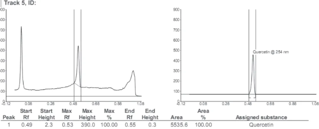
The results of the preliminary phytochemical studies confirmed the presence of carbohydrates, steroids, tannins, glycosides, flavonoids and terpenoids in the ethanol extract of Adiantum capillus-veneris. The polyphenolic compounds quercetin and gallic acid were detected and quantified using HPTLC using the mobile phase toluene:ethyl acetate:formic acid (5:4:1 v/v/v). Flavonoids and tannins were quantified at 254 nm, comparing peak area to a calibration curve derived from the quercetin and gallic acid. It was confirmed that quercetin and gallic acid are active constituents of A. capillus-veneris. The detection of quercetin was observed to be linear over a concentration range of 200 to 1600 ng.l1 with a correlation coefficient of 0.9961.
The detection of gallic acid was observed to be linear over a concentration range of 200 to 1600 ng.l-1 with a correlation
Fig. 2 - Peak display of quercetin in ethanol extract of Adiantum capillus (scanned at 254 nm).
Fig. 3 - Peak display of marker gallic acid (scanned at 254 nm).
Iodine and inorganic mineral content analysis
Iodine concentration of A. capillus-veneris was 0.069 μg/g of the sample, which suggests a moderate iodine levels. The inorganic mineral content of A. capillus-veneris powder showed the absence of metals such as arsenic, mercury, phosphorus, sulphur, vanadium and tin. The results are shown in Table 1.
Thyroid weight
Reversal of the thiouracil-induced increase of thyroid weight (goitre) (21.8 ± 1.45 mg/100 g body weight) in comparison to the reversal produced by test I (17.5 ± 3.68 mg/100 g body weight) and test II (14.2 ± 2.48 mg/100 g body weight) as a measure of thyroxine-like activity. The comparison of thyroid weight between test groups was not significantly different from control animals.
Thyroid peroxidase activity
TPO activity of hypothyroid control rats was 3.04 ± 0.09 ΔOD/ min/mg protein. TPO activity of test I rast was 5.48 ± 3.80 ΔOD/min/mg protein and test II was 5.48 ± 3.80 ΔOD/min/mg protein. Test II showed significant increase in TPO activity compared to hypothyroid controls.
Urinary iodine concentration
There was a significant decrease in urinary excretion of iodine in both the test group I and II, 408.10 ± 4.96 μg/dl, 368.58 ± 3.06 μg/dl respectively when compared to the hypothyroid controls 479.03 ± 2.06 μg/dl. The results are shown in Table 2.
Antioxidant activity
Lipid peroxidation assay
The test group treated with ethanol extract of A. capillus-veneris
(500 mg/kg) showed significant increase of LPO levels (15.89 ± 2.02) when compared with normal control group LPO levels (6.65 ± 0.84); whereas in hypothyroid group, the LPO levels were higher (24.14 ± 2.46). Thus, the marked increase in the oxidative stress found in hypothyroid indicated by increase in LPO levels contrasts with the reduction in oxidative stress of animals treated with Adiantum capillus extract.
Reduced glutathione assay
There was a significant increase in GSH content (12.09 ± 0.21) in the test group treated with ethanol extract of A. capillus-veneris (500 mg/kg) when compared with hypothyroid control group GSH content (8.98 ± 0.06). However the GSH content in the control group was higher than the other two (18.76 ± 0.09). Thus, the marked increase in the oxidative stress was found in hypothyroid groups as indicated by decrease in GSH content, whereas treatment with ethanol extract of Adiantum capillus showed decrease in oxidative stress as indicated by the increased GSH content.
Superoxide dismutase assay
There was a significant increase in SOD content (11.68 ± 3.58) in test group treated with ethanol extract of A. capillus-veneris (500 mg/kg) when compared with hypothyroid control group SOD content (7.50 ± 1.68); whereas in normal group, the SOD content (13.85 ± 0.82). Thus, the marked increase in the oxidative stress was found in hypothyroid groups as indicated by decrease in Metal Concentration/ g sample
Arsenic Nil
Cadmium 0.0012 µg
Calcium 0.07301 µg
Chromium 2.54 µg
Copper 1.094 µg
Iron 1.45 µg
Lead 0.0060 µg
Magnesium 0.1833 µg
Manganese 0.445 µg
Mercury Nil
Nickel 0.0007 µg
Phosphorus Nil
Potassium 2.5158 mg
Selenium 0.002 µg
Sodium 0.1131 mg
Sulphur Nil
Tin Nil
Vanadium Nil
Zinc 0.0062 mg
Table 1
Metal analysis of the whole plant Adiantum capillus.
Acute toxicity
In the acute toxicity study, no mortality was observed up to a dose of 2 g/kg body weight in mice treated with A. capillus-veneris extract. Even at this high dose, there were no gross behavioral changes.
Thyroid hormone enhancing activity in PTU induced hypothyroidism
Serum level of T3, T4 and TSH
GSH content, whereas treatment with ethanolic extract of A. capillus-veneris showed decrease in oxidative stress as indicated by the increased SOD content.
Catalase assay
The test group treated with ethanol extract of Adiantum capillus
(500 mg/kg) showed significant increase in CAT content (125.90 ± 4.48) when compared with hypothyroid control group CAT content (79.04 ± 2.42); whereas in normal group, the CAT content (179.48 ± 1.68). Thus, the marked increase in the oxidative stress was found in hypothyroid groups as indicated by decrease in CAT content. The results are reported in Table 3.
Discussion
Preliminary phytochemical screenings of ethanol extract of
Adiantum capillus-veneris L., Pteridaceae, showed the presence of various secondary metabolites such as flavonoids, tannins, carbohydrates, terpenoids, proteins and steroids. The presence of quercetin and gallic acid was confirmed by HPTLC analysis. The iodine concentration assay showed a moderate iodine concentration, sufficient for optimal thyroid function. The
concentrations of inorganic minerals such as Se, Ca and Zn were not at levels that may cause negative impact on thyroid function. Selenium is a main component of the enzymes deiodinase and thyroperoxidase, vital for the synthesis of thyroid hormones T3 and T4 and suggests that, selenium deficiency contributes to the development of goiter even in the presence of adequate iodine intake. The effects of plant calcium on thyroid function has not been extensively studied (FNB-IM, 2001), however, a high calcium level in drinking water may slow iodine absorption resulting in goiter, particularly if the iodine level is in the limits of meeting body needs (FNB-IM, 2001). The calcium concentration was 0.07301 mg. Studies have shown that low zinc intake exacerbates the effect of low iodine intake. Zinc supplementation has been reported to have favorable effect on thyroid hormone levels, particularly total T3, and resting metabolic rate (Maxwell and Volpe, 2007). The zinc concentrationwas 0.0062 mg.
In PTU induced hypothyroidism, the extract showed significant increase in the levels of T3, T4; a decrease in the level of TSH and reversal of the thiouracil-induced increase in thyroid weight (goitre) suggesting its thyroid hormone enhancing effect and anti-goitrogenic effect. Thyroid peroxidase (TPO) catalyses the biosynthesis of thyroid hormone by oxidization of iodide, iodination of tyrosine residues of
Parameter Control Hypothyroid
control
Test I EAC 250 mg/kg
Test II EAC
500 mg/kg Standard
Thyroid weight (mg/100 g body weight) 7.19 ± 0.18 21.8 ± 1.45 17.5 ± 3.68 14.2 ± 2.48 11.6 ± 5.90
Thyroid stimulating hormone TSH (mIU/ml) 0.40 ± 0.02 2.08 ± 0.06 1.64 ± 0.06** 0. 96 ± 0.02** 0.64 ± 0.06**
Triiodothyronine T3 (ng/dl) 124.4 ± 1.08 88.4 ± 9.02 126.32 ± 4.58 144.1 ± 3.98** 192.36 ± 3.70
Thyroxine T4 (μg/dl) 9.29 ± 0.24 3.15 ± 0.37 7.65 ± 1.36 9.15 ± 0.32** 11.16 ± 0.76
Urinary iodine (μg/dl) 409.35 ± 0.7 479.03 ± 2.06 408.10 ± 4.96** 368.58 ± 3.06** 302.30 ± 6.07**
Thyroid peroxidase activity (ΔOD/min/mg/protein) 4.02 ± 0.03 3.04 ± 0.09 5.40 ± 03.80 7.90 ± 6.08 9.84 ± 1.64
EAC-Ethanolic extract of Adiantum capillus.
Values are mean ± SEM of 6 parallel measurements.
Statistical significant test for comparison was done by ANOVA, followed by Dunnet’s ‘t’ test (n = 6). **p < 0.01, *p < 0.05 when compared against hypothyroid control.
Treatment LPO
(nM of MDA/gHb)
Glutathione (μM/mg of protein)
SOD (U/mg of protein)
Catalase (μM/min/mg of protein)
Normal Control 6.65 ± 0.84 18.76 ± 0.09 13.85 ± 0.82 179.48 ± 1.68
Hypothyroid Control 24.14 ± 2.46 8.98 ± 0.06 7.50 ± 1.68 79.04 ± 2.42
EAC 250 mg/kg 18.48 ± 1.08 10.02 ± 2.67 9.87 ± 2.06 118 ± 3.64
EAC 500 mg/kg 15.89 ± 2.02* 12.09±0.21* 11.68 ± 3.58* 125.90 ± 4.48**
EAC – Ethanol extract of Adiantum capillus
Values are mean ± SEM of 6 parallel measurements.
Statistical significant test for comparison was done by ANOVA, followed by Dunnet’s ‘t’ test (n = 6). **p < 0.01, *p < 0.05 when compared against hypothyroid control.
Table 2
Plasma hormone levels.
Table 3
thyroglobulin and coupling reaction for the synthesis of thyroxine and triiodothyronine attached to thyroglobulin (Chandra et al., 2004). There was a remarkable increase in TPO Test I (250 mg/kg) and Test II (500 mg/kg) treated rats (Table 2) that might be the possible reason for the increased levels of T4 and T3 in serum. The increase in TPO level might be due to increased accumulation of iodide in the thyroid gland, which in turn leads to decreased urinary iodine excretion. There was a significant increase in serum TSH level in Test I (250 mg/kg) and Test II (500 mg/kg) treated rats in comparison to that of the thyroid control group (Table 2). The overall results showed that administration of ethanol extract of A. capillus-veneris has decreased the thyroid gland weight; and an enhanced TPO activity in a relative reversal of hypothyroidism, as evidenced by elevated serum T4, T3 and TSH profiles. With the outlined results, A. capillus-veneris could be used in the regulation of hypothyroidism.
The results of lipid peroxidation and antioxidant enzyme assays indicated that the level of malondialdehyde (MDA) was lowered and the levels of antioxidant enzymes increased when treated with A. capillus-veneris in hypothyroid animals. This suggests that the A. capillus-veneris has marked free radical scavenging activity, probably caused by the increase in the level of endogenous antioxidant enzymes system. This in turn could be useful in the amelioration of disease-induced oxidative stress. The antioxidant potency of A. capillus-veneris
may be attributed to the presence of polyphenolics like flavonoids and tannins. Flavonoids are powerful antioxidants and are described as free-radical scavengers. Flavonoids possess various clinical properties such as antioxidant, anti-inflammatory, antiallergic, antiviral, and anticarcinogenic activities and these can be attributed to their hydrogen donating ability ‘‘H” atoms such that the subsequent radicals produced can be delocalized over the flavonoid structure (Bors and Saran, 1987). Therefore this drug is used for the treatment of stress and strain syndrome, metabolic disorders, hormonal disturbances, chronic illness etc. The overall results showed that consumption of A. capillus-veneris can be stimulatory to thyroid function.
Conclusion
The plant Adiantum capillus-veneris L., Pteridaceae, was found to contain large amounts of polyphenolic compounds: tannins and flavonoids. The plant also possesses moderate iodine content and adequate amount of inorganic minerals, not at levels that may cause negative impact on thyroid function. In PTU induced hypothyroidism, the ethanol extract of the fern A. capillus-veneris increased the level of T3, T4 significantly and reversed the increase in thyroid weight (goitre) induced by thiouracil. This evidence suggests its thyroid hormone enhancing effect and anti-goitrogenic effect. In addition, the level of malondialdehyde (MDA) was lowered and the levels of antioxidant enzymes were increased when treated with A. capillus-veneris in hypothyroid animals. With the outlined results, A. capillus-veneris could be used for hypothyroidism regulation.
Authorship
AV designed the study, interpreted the data and drafted the manuscript. KK collected plant material and participated in all experiments and data analysis.
Acknowledgement
Authors are extremely grateful to the management for the facilities provided to complete this work successfully.
R E F E R E N C E S
Ansari, R., Ekhlasi Kazaj, K., 2012. Adiantum capillus-veneris. L: Phytochemical constituents, traditional uses and pharmacological properties: a review. J. Adv. Sci. Res. 3, 15-20.
Blouquit, M.F., Valens, M., Bagayoko, A., Gripois, D., 1990. Adrenal tyrosine-hydroxylase activation in the developing rat - influence of the thyroid status. J. Dev. Physiol. 14, 325-329. Bors, W., Saran, M., 1987. Radical scavenging by flavonoid
antioxidants. Free Radical Res. Com. 2, 289-294.
Chandra, A.K., Mukhopadhyay, S., Lahari, D., Tripathy, S., 2004. Goitrogenic content of Indian cyanogenic plant foods & their in vitro anti-thyroidal activity. Indian. J. Med. Res. 119, 180-185.
Czeczot, H., Scibior-Bentkowska, D., Skrzycki, M., Podsiad, M., 2010. [Assessment of lipid peroxidation level in serum of patients with gastrointestinal tract tumors]. Wiad. Lek. 63, 180-187.
Fischer, P.W., L’Abbe, M.R., Giroux, A., 1986. Colorimetric determination of total iodine in foods by iodide-catalyzed reduction of Ce+4. J. Assoc. Off. Anal. Chem. 69, 687-689. FNB-IM, F.a.N.B.-I.o.M., 2001. Dietary Reference intakes for
Vitamin A, K, arsenic, boron, chromium, copper iodine, iron, manganese, molybdenum, nickel, silicon, vanadium and zinc, in: Intakes, S.C.o.t.S.E.o.D.R. (Ed.). National Academy Press, Washington DC.
Joseph Collins, N.D., 2007. Phytotherapeutic support of thyroid function. Nutri news. 8, 1-4.
Lowry, O.H., Rosebrough, N.J., Farr, A.L., Randall, R.J., 1951. Protein measurement with the folin phenol reagent. J. Biol. Chem. 193, 265-275.
Mathur, A., Satish, K., Verma, R.P., Santosh, K., Singh, D.M., Prasad, G.B.K.S., Dua, V.K., 2010. Pharmacological investigation of Bacopa monnieri on the basis of antioxidant, antimicrobial and anti-inflammatory properties. J. Chem. Pharm. Res 2, 191-198.
Maxwell, C., Volpe, S.L., 2007. Effect of zinc supplementation on thyroid hormone function. A case study of two college females. Ann. Nutr. Metab. 51, 188-194.
OECD, 2002. Acute oral toxicity. Acute oral toxic class method guideline 423 adopted 23.03.1996, in: Development, T.O.f.E.C.-o.a. (Ed.). OECD, Paris.
Pasupathi, P., Latha, R., 2008. Free radical activity and antioxidant defense mechanisms in patients with hypothyroidism. Thyroid Sci. 3, 1-6.
Ranganathan, S., Karmarkar, M.G., 2006. Estimation of iodine in salt fortified with iodine & iron. Indian J. Med. Res. 123, 531-540.
Sasikumar, J.M., Jinu, U., Shamna, R., 2011. In vitro antioxidant activity and HPTLC analysis of root of Pandanus odoratissimus. Int. J. Pharm. Pharm. Sci. 3, 64-67.
Shen, Q., Shang, N., Li, P., 2011. In vitro and in vivo antioxidant activity of Bifidobacterium animalis 01 isolated from centenarians. Curr. Microbol. 62, 1097-1103.
Tiwari, P., Kumar, B., Kaur, M., Kaur, G., Kaur, H., 2011.
Phytochemical screening and extraction: a review. I. Pharm. Sciencia. 1, 98-106.
Ujowundu, C.O., Kalu, F.N., Nwosunjoku, E.C., Nwaoguikpe, R.N., Okechukwu, R.I., Igwe, K.O., 2011. Iodine and inorganic mineral contents of some vegetables, spices and grains consumed in South Eastern Nigeria. Afr. J. Biochem. Res. 5, 57-64.