Received from Hôpital d’Instruction des Armées (HIA Begin), Saint Mandé, France. 1. MD; MBA; Department of Anesthesia and Intensive Care, HIA Begin 2. MD; Department of Anesthesia and Intensive Care, HIA Begin
3. MD; Anesthesiologist Department of Anesthesia and Intensive Care Centre Hospitalier Universitaire de Caen (CHU Caen)
4. MD; PhD; Professor of Anesthesiology. Department of Anesthesia and Intensive Care, Hôpital Universtitaire Tenon
5. MD; PhD; Professor of Anesthesiology. Department of Anesthesia and Intensive Care Centre Hospitalier Privé (CHP) Saint-Grégoire
Submitted on November 4, 2010. Approved on March 21, 2011. Correspondence to: Alexandre Gnaho, MD, MBA
Department of Anesthesia and Intensive Care Hôpital d’Instruction des Armées
Begin 94160 Saint Mandé, France E-mail: [email protected] SCIENTIFIC ARTICLE
Assessing the Depth of the Subarachnoid Space by
Ultrasound
Alexandre Gnaho
1, Vinh Nguyen
2, Thierry Villevielle
2, Melina Frota
3, Emmanuel Marret
4, Marc E Gentili
5Summary: Gnaho A, Nguyen V, Villevielle T, Frota M, Marret E, Gentili ME – Assessing the Depth of the Subarachnoid Space by Ultrasound.
Background and objectives: To assess the accuracy of the ultrasound (US) to predict the depth to reach lumbar intrathecal and epidural spaces in order to decrease the number of puncture attempts.
Methods: Thirty-one patients (25 males and 6 females), ASA I or II participated in this study. The transversal ultrasound image of the lumbar spine was obtained at the level of the L3-L4 space. An anesthesiologist without prior information performed the spinal anesthesia through the predicted target area. The distance between the skin and the anterior portion of the flavum ligamentum which is supposedly the bottom limit of the intrathe-cal depth or an approximation of the depth of the epidural space (ED-US) was measured by ultrasound and it was compared with the distance between the skin and the anterior portion of the flavum ligamentum on the needle (ED-N).
Results: ED-US and ED-N were respectively 5.15 ± 0.95 cm and 5.14 ± 0.97 cm; these distances were not significantly different (p > 0.0001). A significant correlation r = 0.982 [95% CI 0.963-0.992, p > 0.0001] was observed between the ED-US and ED-N measurements. Bland-Altman analysis showed an accuracy of 0.18 cm; tolerated variations ranged from -0.14 cm to -0.58 cm.
Conclusions: This study supports the idea that the US transversal plane allows the identification of axial anatomical structures and provides physicians with efficient information to perform spinal anesthesia.
Keywords: Ultrasonography, Interventional; Anesthesia, Spinal; Dura mater.
©2012 Elsevier Editora Ltda. All rights reserved.
INTRODUCTION
Ultrasound (US) guidance for regional anesthesia is a subject of major interest worldwide 1. Since a few decades ago
previ-ous papers have been drawing the interest of physicians for the use of US for spinal or epidural anesthesia 2,3,4,5. Recently,
US has been proposed as a preoperative assessment tool predicting the feasibility of neuroaxial blockade 6.
Neverthe-less, physicians currently using US for neuroaxial anesthesia are still only a few, probably because this procedure requires not only a good knowledge of spinal sonoanatomy but also an advanced interventional skills. However, data related to the learning process for this technique are lacking. Spinal US
of-fer valuable information to facilitate neuroaxial blockade. Most previous studies focused on the epidural space 7,8. We
be-lieve that US images could also provide accurate estimation of depth to reach intrathecal space. This pilot study was de-signed to assess the reliability of US to predict lumbar intrath-ecal space depth and help to restrict the number of puncture attempts.
METHODS
Participants
Our Institutional Ethics Committee approved the study. Writ-ten informed consent was obtained from each patient. ASA I or II patients scheduled for lower extremity surgery were en-rolled. Patients with spinal deformities or previous spinal sur-gery were excluded. Data collection was planned as a cohort study.
Measurements and puncture methods
placed by another investigator over the sacrum two or three centimeters to the right of the midline to visualize the hyper-echoic line corresponding to the image of the sacrum.
The probe was then moved cephalad to obtain the clas-sic hyperechoic saw-like image representing the articular pro-cesses, and the interspaces were counted upward till the L3-L4 interspace. At this time, a transverse scan was performed. The midline was identified using the after shadow of the spine, and the L3-L4 interspace was identified by moving the probe slowly cephalad or caudad in order to obtain the absence of after shadow and the view of the complex formed by the ligamentum flavum-dura mater and also to find the vertebral body. However, our equipment does not always allow us to visualize ligamentum flavum and dura mater as two separate structures but often as only one hyperechoic line. At this time the optimum sonogram, depicted according to an agreement between two investigators, was frozen. The outlines of the probe and the target in its center corresponding to the punc-ture point was drawn with a dermographic pen.
Two investigators analyzed the images, and the US visibili-ty of anatomical structures (corpora vertebrae, thecal sac, and ligamentum flavum-dura mater complex) was rated as good (very well defined), moderate (well defined), or none (hardly or not defined). We recorded the time necessary to obtain an op-timum sonogram of the structures (location time), which also included the time elapsed for making the outlines of the probe and the target point. We measured with a built-in caliper the distance from the skin to the anterior part of the ligamentum flavum-dura mater complex (ED), which is supposed to be the bottom limit before reaching intrathecal space or an approxi-mation of the epidural space depth (ED-US). Subsequently, a blinded anesthesiologist to this measure performed spinal anesthesia (25 G pencil spinal needle, B. Braun, Melsungen AG, Germany) through the predicted target point. The mea-surement of the distance from skin to the anterior part of the ligamentum flavum-dura mater complex was obtained after marking the needle with a sterile skin-marking pen (ED-N). The technique: one hand of the anesthesiologist held firmly the needle and the point of measure was pinched between the thumb and the index of the other hand to secure measure-ment. ED-US was compared with ED-N. During puncture, each advancement of needle was considered as puncture attempt. If spinal anesthesia was not possible at L3-L4 interspace, the anesthesiologist punctured a lower vertebral interspace. The number of attempts was recorded.
Statistical methods
Data were summarized as mean ± standard deviation (SD). ED was calculated from ultrasound device (ED-US) and directly during needle punctures (ED-N). Correlation between ED-US and ED-N was determined by simple linear regression analy-sis using the least-squares method. We constructed Bland-Altman plots of differences to averages of the epidural depth measured by the two methods and calculated the bias, preci-sion (1 SD), and 95% limits of agreement (mean bias ± SD).
Excel Microsoft (Chicago, IL) and Medcalc software were used for statistical analysis. Two-sided p value < 0.05 was considered statistically significant.
RESULTS
Thirty one patients underwent spinal anesthesia for lower extremity surgery (25 males and 6 females). The average age was 43 ± 15 yr, weight 79 ± 14 kg, length 174 ± 8 cm, and body mass index 27 ± 3 kg.m-2. The visibility of
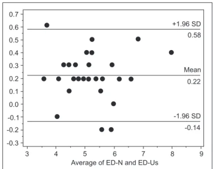
anatomi-cal structures was good in 87% of patients and moderate in 13% of patients (Figures 1A and 1B). The location time was 76 ± 18 seconds. The ED-US and ED-N were 5.15 ± 0.95 cm and 5.14 ± 0.97 cm, respectively. These distances were not significantly different (p > 0.0001). In addition, a significant correlation r = 0.982, [95% CI 0.963-0.992, p > 0.0001] was observed between ED-US and ED-N measurements (Fig-ure 2). Bland-Altman analysis between these two meas(Fig-ures is shown in Figure 3. The mean difference (bias) was 0.22 cm, with a precision of 0.18 cm [95% CI 0.15-0.29]. The limits of agreement were -0.14 cm [95% CI (-0.25; -0.01)] to -0.58 cm [95% CI (-0.46; -0.70)].
Spinal anesthesia was performed at first attempt with 24 patients (78 %), 2 attempts were necessary for 5 patients (16%), and one patient (3%) needed 4 attempts. Dural access was impossible for another patient (3%). Bony surface land-marks were absent before location in 68% of patients. Visibil-ity of anatomical structures was good in 87% of patients and moderate for the remaining participantes.
DISCUSSION
This study supports the condition that US transverse plane is likely to identify axial anatomical structures and may provide ef-ficient information to practitioners to perform spinal anesthesia.
Comparison with previous studies
Previous studies in obese and nonobese patients have dem-onstrated a good correlation between ED-US and ED-N: data are summarized in Table I.
At this time, US guidance was used to improve epidural ap-proach in 76 surgical patients 2,9; in 76 parturients undergoing
caesarean section under epidural anesthesia 10,11; and overall
in 427 parturients receiving epidural analgesia for delivery 13-15.
However most previous studies did not focus on spinal an-esthesia, but epidural anesthesia. The method of indirect US guidance allows identification in a midline sagittal scan of lumbar vertebrae and an accurate measurement of the depth
8.0
ED-US (cm)
7.5
7.0
6.5
6.0
5.5
5.0
4.5
4.0
3.5
3.0
3 4 5 6 7 8 9
ED-N (cm)
r = 0.982 p < 0.001
-0.3 -0.2 -0.1 0.0 0.1 0.2 0.3 0.4 0.5 0.6 0.7
3 4 5 6 7 8 9
Average of ED-N and ED-Us
-1.96 SD
-0.14 0.22 Mean 0.58 +1.96 SD
Figura 2 – Correlation between US Depth (ED-US) in cm and Needle
Depth (ED-N) in cm. Figura 3 – Bland-Altman Analysis: Agreement between Ultrasound Depth (ED-US) and Needle Depth (ED-N).
Table I – Overview on Correlation and Precision of US-assisted Epidural Puncture
Studies Patients Number Depth (mm) Correlation Bland-coefficient Altman*
Cork et al. 2 Ea 36 45.0 0.98 No data
Currie 13 Ea in obstetric 75
41.2 ± 8.1 0.92 5.4
Wallace et al. 10 Ea cesarian section 36
55.1 ± 2.1 0.98 5.4
Bonazzi et al. 9 Herniotome 40
51.0 ± 6.2 0.98 No data
Grau et al. 15 Ea in obstetric 100
53.0 ± 7.0 0.79 6.8
Grau et al. 3 CSE cesarian section 80
51.5 ± 9.3 0.92 5.1
Grau et al. 11 Difficult Ea in obstetric 36
57.5 ± 11.0 0.87 7.7
Grau et al. 4 Ea in obstetric 300
51.2 ± 7.0 0.83 6.9
Arzola et al. 19 Ea in obstetric 61
46.6 ± 6.8 0.88 6.6
Lee et al 12 Ea in obstetric 36
43.8 ± 5.1 No data No data Balki et al. 20 Ea obese in obstetric 46
63.0 ± 8.0 0.85 No data
Tran et al. 18 Ea in obstetric 20
51.0 ± 11.0 0.80 No data
Karmakar et al. 22 Ea 15
57.1 ± 7.1 No data No data
Helavel et al. 7 Ea 60
49.7 ± 5.0 0.66 No data
to reach intrathecal space. This procedure can facilitate the performance for spinal anesthesia and may decrease the rate of complications, most probably in those patients whose ana-tomic landmarks are obscured 9,10,16,17.
US imaging may reduce attempts to lumbar approach, con-sidering that prior to it palpation was the only way to lumbar puncture approach. A significant reduction of number punc-ture has been demonstrated when performing combined spi-nal and epidural anesthesia 3. In addition, the same authors
have shown the value of pre-puncture sonographic informa-tions for lumbar access in obstetric field 4. Grau et al. 5 have
shown changes that occur on spinal and epidural anatomy during pregnancy using US.
Our study reinforces the usefulness of pre-puncture in-formation such as optimum puncture point, depth to reach intrathecal space, and the visibility of neuroaxial anatomical structures. In some patients, performing spinal anesthesia is challenging. However, the presence of these healthy young patients in which bony surface landmarks were not palpable in our study was an unexpected observation. Assessment of palpation upon agreement of two investigators instead of one should have influenced this rate. Nevertheless, this surprising observation did not affect the study results.
In 60 participants (20 parturients and 40 healthy volunteers in general surgery), Grau et al. 5 have shown that longitudinal
paramedian access provides a larger permeable window, and improves the quality of pre-puncture diagnostics for neuroax-ial anesthesia or analgesia 18. Despite this classic superiority
of the longitudinal paramedian access to provide better sono-grams of lumbar spine, we have been able to identify neuroax-ial structures using transverse median plane. Such approach seems to be closer to the reality of our clinical practice. Some recent studies focus on transverse plane providing reliable landmarks for epidural anesthesia 19,20. In some cases such as
impalpable bony landmarks, several unsuccessfuly puncture attempts, patient becoming irritable etc., when things do not go as smoothly like in the textbook, transverse and paramedi-an longitudinal approaches could be used in a complementary way 21. In addition, to our knowledge, there is no randomized
and prospective study comparing longitudinal and transverse approaches for spinal anesthesia. Furthermore, we did not encounter significantly calcifications on scanning area; prob-ably because of our young population. The reflexion involved in these calcifications are often described in transverse scan-ning plane and is supposed to further difficult lumbar access, particularly in elderly patients.
ED-US measurement must be expected to differ slightly from the ED-N measurement. This degree of uncertainty could be erased by a real time monitoring of the lumbar puncture 22.
We used a linear 5-10 MHZ probe, while in most papers the transducer often used was a sector 2-5 MHZ probe for spinal ultrasound imaging, mainly because top managers of our institution were in a context of difficult financial choice to
make. The result was that we could not acquire a complete US device. So the sector 2-5 MHZ probe was not available. In addition, Ferre et al. 23 had been able to identify neuroaxial
structure with a linear array probe with a good delineation of these structures in 93.4% of patients.
Clinical implications
Several reports suggest the use of US to perform neuroxial blockade in difficult anatomical situations, such as obesity, scoliosis, or edema. Nevertheless, training is probably needed in easier clinical presentations as to acquire proficiency in or-der to perform later a successfully spinal anesthesia that could be difficult or impossible without sonographic landmarks.
In a randomized trial evaluating epidural access in 72 pa-tients with difficult history of epidural anesthesia and/or sub-stantial alterations of lumbar spine (such as scoliosis, kypho-sis, hyperlordosis and BMI > 33 kg.m-2), Grau et al. 5 have
demonstrated in the US group (36 patients) a higher satisfac-tion, an improved visual analogical scale of pain score, and fewer puncture attempts 11.The advantages included a target
definition facilitating lumbar puncture, direct visualization of all neuroaxial structures with fewer bone contacts and thus reducing complications and side effects. Lumbar spine ultra-sound could be a useful clinical tool to facilitate needle inser-tion. Lee et al have shown that ultrasonography makes it pos-sible to detect abnormal sonoanatomy. 12 We can decrease
complications such as unintentional dural punctures.
Pediatric anesthesia is the other field in which spinal or epi-dural blockade is challenging. The exclusive technique of loss of resistance usually performed with a child under general an-esthesia has been associated with complications and adverse outcomes, including more significantly neurological deficit as a result of unintentional spinal cord trauma 24,25. In a
prospec-tive randomized study of 64 children evaluating the feasibility, number of bone contacts, and duration for the performance of epidural anesthesia using US guidance compared with the loss of resistance method, Willschke et al. 26 have shown that
direct visualization of intra-epidural spread of local anesthetic was a reliable way to verify position of epidural catheter. In addition, they have demonstrated that US guidance in experi-enced hands reduces both the risk of bone contact as well as duration for catheter placement.
Potential limitations
anat-omy knowledge and training are strongly necessary to use this technology. Interpretation of images may be also difficult some times. In addition, the acoustic windows for US spine are very limited 15. This is part of the explanation why US
im-aging for neuroaxial anesthesia had been such a poor source of interest for decades.
9. Bonazzi M, Bianchi De Grazia L, Di Gennaro S et al. – Ultrasonogra-phy-guided identification of the lumbar epidural space. Minerva Anes-thesiol, 1995;61:201-205.
10. Wallace DH, Currie JM, Gilstrap LC et al. – Indirect sonographic gui-dance for epidural anesthesia in obese pregnant patients. Reg Anes-th, 1992;17:233-236.
11. Grau T, Leipold W, Conrandi R et al. – Ultrasound control for presumed difficult epidural puncture. Acta Anaesthesiol Scand, 2001;45:766-771.
12. Lee Y, Tanaka M, Carvalho JCA. Sonoanatomy of the lumbar spine in patients with previous unintentional dural punctures during labor epidurals. 2008; 33: 266-270
13. Currie JM – Measurement of the depth to the extradural space using ultrasound. Br J Anaesth, 1984;56:345-347.
14. Grau T, Leipold RW, Conrandi R et al. – Paramedian access to the epidural space: the optimum window for ultrasound imaging. J Clin Anesth, 2001;13:213-217.
15. Grau T, Leipold R, Conradi R et al. – Ultrasonography and peridural anesthesia. Technical possibilities and limitations of ultrasonic exami-nation of the epidural space. Anaesthesist, 2001;50:94-101. 16. O’Donnell D, Prasad A, Perlas A – Ultrasound-assisted spinal
anes-thesia in obese patients. Can J Anaesth, 2009;56:982-983.
17. Prasad GA, Tumber PS, Lupu CM – Ultrasound guided spinal anes-thesia. Can J Anaesth, 2008;55:716-717.
18. Tran D, Kamani AA, Lessoway VA et al. – Preinsertion parame-dian ultrasound guidance for epidural anesthesia. Anesth Analg, 2009;109:661-667.
19. Arzola C, Davies S, Rofaeel A et al. – Ultrasound using transverse approach to the lumbar spine provides reliable landmarks for labor epidurals. Anesth Analg, 2007;104:1188-1192.
20. Balki M, Lee Y, Halpern S et al. – Ultrasound imaging of the lumbar spine in the transverse plane: the correlation between estimated and actual depth to the epidural space in obese parturients. Anesth Analg, 2009;108:1876-1881.
21. O’Donnell D, Prasad A, Perlas A – Ultrasound assisted spinal anes-thesia in obese patient. Can J Anesth, 2009;56:982-983.
22. Karmakar MK, Li X, Ho AMH et al. – Real time ultrasound-guided paramedian access evaluation of a novel in plane technique. Br J An-aesth, 2009;120:845-854.
23. Ferre RM, Sweeney TW – Emergency physicians can easily obtain ultrasound images of anatomical landmarks relevant to lumbar punc-ture. J Emerg Med, 2007;25:291-296.
24. Flandin -Bletry C, Barrier G – Accidents following extradural analge-sia in children. A result of a retrospective study. Paediatr Anaesth, 1995;5:41-46.
25. Rose JB – Spinal cord injury in a child after single shot epidural anes-REFERENCES
thesia. Anesth Analg, 2003;96:3-6.
26. Willschke H, Marhofer P, Bösenberg A et al. – Epidural catheter pla-1. Gray AT – Ultrasound-guided regional anesthesia: current state of the cement in children: comparing a novel approach using ultrasound
art. Anesthesiology, 2006;104:368-373. guidance and standard loss of resistance technique. Br J Anaesth, 2. Cork RC, Kryc JJ, Vaughan RW – Ultrasonic localization of the lumbar 2006;97:200-207.
epidural space. Anesthesiology, 1980;52:513-516.
3. Grau T, Leipold RW, Conradi R, Martin E, Motsch J. Ultrasound faci-litates localization of the epidural space during combined spinal and epidural anesthesia. Reg Anesth Pain Med. 2001; 26:64-67
4. Grau T, Leipold RW, Conradi R, Martin E, Motsch J.Efficacy of ultra-sound imaging in obstetric epidural anesthesia. J Clin Anesth 2002; 14:169-75
5. Grau T, Leipold RW, Horter J et al. –The lumbar epidural space in preg-nancy: visualization by ultrasonography. Br J Anaesth, 2001;86:798-804.
6. Chin KJ, Chan V – Ultrasonography as a preoperative assessment tool: predicting the feasibility of central neuraxial blockade. Anesth Analg, 2010;110:252-3.
7. Helavel PE, Conceiçao DB, Swarovsky C et al. – Evaluating the depth of the epidural space with the use of ultrasound. Rev Bras Anestesiol, 2010;60:376-82.