GRAZIANO L ETAL.
618 REV ASSOC MED BRAS 2016; 62(7):618-621
IMAGE IN MEDICINE
Metaplastic squamous cell carcinoma of the breast: A case report
and literature review
LUCIANA GRAZIANO1, PASCHOAL GRAZIANO FILHO1, ALMIR GALVÃO VIEIRA BITENCOURT2*, DANIEL BERNAL SOTO1, ALEXANDRE HIRO1,
CÍNTIA CAMILLO NUNES3
1MD – Radiologist at Gimi Medicina Diagnóstica, São Paulo, SP, Brazil 2PhD – Radiologist at AC Camargo Cancer Center, São Paulo, SP, Brazil
3MD – Pathologist at CDAP – Clínica de Diagnóstico Anátomo-Patológico, São Paulo, SP, Brazil
S
UMMARYStudy conducted at Gimi Medicina Diagnóstica, São Paulo, SP, Brazil Article received: 7/5/2015 Accepted for publication: 7/6/2015
*Correspondence: Address: Rua Isabel, 131 São Paulo, SP – Brazil Postal code: 03647-020 [email protected]
http://dx.doi.org/10.1590/1806-9282.62.07.618
Metaplastic tumors are rare and represent a heterogeneous group of neoplasms showing dominant areas of non-glandular differentiation. Etiology and patho-genesis of this type of lesion in the breast is uncertain. The most common sources of metastatic squamous cell carcinoma of the breast are lung, esophagus, cervix, and urinary bladder. Squamous cell carcinomas may present clinically with inlammation and average size greater than breast adenocarcinoma. As for imaging studies, mammography shows no typical indings and ultrasound can show a complicated cyst or an inlammatory process, among the differential diagnoses. Therefore, knowing this pathological entity, its clinical course and imaging indings is important to safely treat such a rare and aggressive disease. We herein report a case of metaplastic carcinoma, squamous subtype, diagnosed by core needle biopsy.
Keywords: breast, breast neoplasms, needle biopsy, carcinoma.
I
NTRODUCTIONBreast cancer is a heterogeneous entity regarding clinical and imaging presentation and biological behavior. The ductal type is the most common, followed by lobular carcinoma. Together they account for over 70% of carci-nomas. Metaplastic tumors, in turn, are rare and represent a heterogeneous group of neoplasms showing dominant areas of non-glandular (spindle cell, squamous, and/or mesenchymal) differentiation.
Etiology and pathogenesis of this type of lesion in the breast is uncertain.1 It is believed that it arises directly from
the epithelium of the mammary ducts, while another the-ory is that the tumor grows from foci of squamous meta-plasia within a pre-existing breast adenocarcinoma.2
An-other theory defended by Stevenson et al. is that the lesion is a disease with varying degrees of squamous metaplasia, representing an extreme form of squamous metaplasia inside the adenocarcinoma.3
Squamous cell carcinoma of the breast is an extremely rare type, representing less than 1% of all invasive breast carcinomas with a mean age for onset among women of 54 years.2 When found in the breast, other sites of extra
mammary lesions should be sought, since there is a pos-sibility that this is a metastatic tumor and not a primary lesion. The most common sources of metastatic squamous cell carcinoma of the breast are lung, esophagus, cervix, and urinary bladder. There are no speciic clinical and radio-logical signs for squamous cell carcinoma. For this reason, the nature or origin of the lesion needs to be determined.3
The objective of our study is to report a case of meta-plastic carcinoma, squamous subtype, diagnosed by core needle biopsy.
C
ASE REPORTFemale patient, 59 years old, with a palpable mass in the lateral quadrant of the left breast. She denies having any systemic diseases or any family history of breast or ovary cancer. Physical examination revealed large breasts, with a palpable lesion in the upper-outer quadrant of the left breast, mobile and hard.
METAPLASTICSQUAMOUSCELLCARCINOMAOFTHEBREAST: A CASEREPORTANDLITERATUREREVIEW
REV ASSOC MED BRAS 2016; 62(7):618-621 619
We performed an ultrasound-guided core needle bi-opsy (Figure 3), which led to the diagnosis of invasive ductal carcinoma, not otherwise speciied (NOS), subse-quently treated with neoadjuvant chemotherapy. During treatment, due to reduction in size of the lesion, a new core needle biopsy was requested. The new biopsy revealed a metaplastic squamous cell carcinoma, which was con-irmed after surgical resection.
D
ISCUSSIONSquamous cell carcinomas may present clinically with inlammation and average size greater than breast adeno-carcinoma.4 As for imaging studies, mammography shows
no typical indings and ultrasound can show a
compli-cated cyst or an inlammatory process, among the dif-ferential diagnoses.1
At diagnosis, lesions are large (greater than 4 cm) and predominantly cystic in over 50% of cases.5 They present
lymphatic dissemination less frequently than adenocar-cinomas, in which the range is from 40 to 60%. In 10 to 30% of cases there is iniltration of lymph nodes at surgery and about 30 to 33% of patients develop distant metas-tases.1,3,5 Positron emission tomography (PET-scan) is an
imaging method that can be used to search for distant metastases or a primary site of tumor cells.1
The pathological criteria for establishing a secure diagnosis of squamous cell carcinoma of the breast in-clude: (a) origin of the tumor does not depend on the
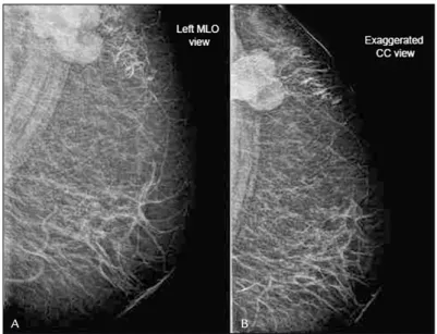
FIGURE 1 Left mammogram shows oval mass without microcalcifications and with circumscribed margins.
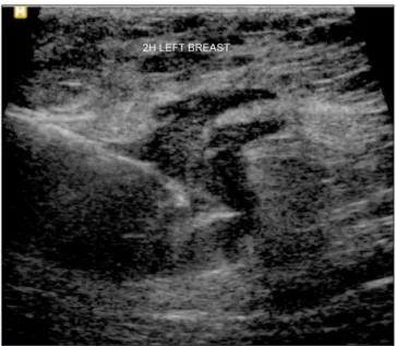
FIGURE 2 Ultrasound revealed a complex cystic and solid mass with internal vascularity on color Doppler, located in the upper-outer quadrant of the left breast.
A
A
B
GRAZIANO L ETAL.
620 REV ASSOC MED BRAS 2016; 62(7):618-621
skin, nipple or adnexal elements of the skin; (b) more than 90% of the tumor must be squamous; (c) there can be no other neoplastic invasive elements, ductal or mes-enchymal, on the entire sample, and other sites of pri-mary tumor must be ruled out.6 To determine whether
the tumor is a metaplastic carcinoma with squamous differentiation or a squamous cell carcinoma, a large sample of the lesion is required. However, it should be noted that the distinction between a primary tumor and a metastatic squamous cell carcinoma is unlikely to be solved by needle biopsy.3
In the case presented, the imaging indings were not classic invasive ductal carcinoma. The core needle biopsy represented only a small sample of the lesion and the representative elements of the irst procedure indicated an invasive ductal tumor. However, keep in mind that the metaplastic elements can progress from one type to an-other after induction cheman-otherapy.4
Squamous cell carcinoma of the breast is usually a high-grade and hormone receptor-negative tumor. This means that the hormone-based therapy may not be efica-cious in these tumors. Additionally, HER2 is usually not overexpressed or ampliied in this disease. The high fre-quency of EGFR positivity is interesting and can be ex-ploited in the development of future treatments.5
Treat-ment of this type of tumor does not differ from other histologic types common in the breast and may involve surgery, chemotherapy, hormonal therapy, and radio-therapy. The therapeutic regimen most suitable for this rare disease is still unclear.5
Therefore, knowing this pathological entity, its clin-ical course and imaging indings is important to safely treat such a rare and aggressive disease.
R
ESUMOCarcinoma metaplásico espinocelular da mama: relato de caso e revisão da literatura
Os tumores metaplásicos são raros e representam um grupo heterogêneo de neoplasias que mostram áreas dominantes de diferenciação não glandular. A etiologia e patogenia desse tipo de lesão na mama é incerta. As causas mais comuns de carcinoma metastático de células escamosas na mama são o pulmão, o esôfago, o colo uterino e a bexiga urinária. Os carcinomas espinocelula-res podem apespinocelula-resentar-se clinicamente com inlamação e tamanho médio maior do que o do adenocarcinoma da mama. A mamograia não apresenta achados típicos, e a ultrassonograia pode mostrar um cisto complicado ou um processo inlamatório, entre os diagnósticos di-ferenciais. Conhecer essa entidade patológica, seu curso clínico e os achados de imagem é importante para um manejo seguro, pois trata-se de entidade rara e agressiva. Este trabalho relata um caso de carcinoma metaplásico, subtipo espinocelular, diagnosticado por biópsia com agulha grossa.
Palavras-chave: mama, neoplasias da mama, biópsia por agulha, carcinoma.
METAPLASTICSQUAMOUSCELLCARCINOMAOFTHEBREAST: A CASEREPORTANDLITERATUREREVIEW
REV ASSOC MED BRAS 2016; 62(7):618-621 621
R
EFERENCES1. Flikweert ER, Hofstee M, Liem MSL. Squamous cell carcinoma of the breast: a case report. World J Surg Oncol. 2008; 6:135.
2. Hennessy BT, Krishnamurthy S, Giordano S, Buchholz TA, Kau SW, Duan Z, et al. Squamous cell carcinoma of the breast. J Clin Oncol. 2005; 23(31):7827-35. 3. Guerriero G, Zagami MG, Montesano M, Primavera A, Carino R, Battista
C, et al. Squamous cell carcinoma of the breast diagnosis by vacuum-assisted core biopsy. Tumori. 2005; 91(5):418-20.
4. Liu J, Yu Y, Sun J, He S, Wang X, Yin J, et al. Clinicopathologic characteristics and prognosis of primary squamous cell carcinoma of the breast. Breast Cancer Res Treat. 2015; 149(1):133-40.
5. Murialdo R, Boy D, Musizzano Y, Tixi L, Murelli F, Ballestrero A. Squamous cell carcinoma of the breast: a case report. Cases J. 2009; 2:7336. 6. Behranwala KA, Nasiri N, Abdullah N, Trott PA, Gui GPH. Squamous cell