Adult T-cell leukemia/lymphoma
PEDRO DANTAS OLIVEIRA1*, LOURDES FARRE2, ACHILÉA LISBOA BITTENCOURT3
1PhD, MD – Professor of Dermatology, Universidade Federal de Sergipe, Aracaju, SE, Brazil 2PhD – Researcher, Centro de Pesquisa Gonçalo Moniz – Fiocruz Bahia, Salvador, BA, Brazil 3PhD, MD – Pathologist and Researcher, Universidade Federal da Bahia, Salvador, BA, Brazil
SUMMARY
Study conducted at Complexo Hospitalar Universitário Professor Edgard Santos (Hupes), Universidade Federal da Bahia (UFBA), Salvador, BA, Brazil
Article received: 7/14/2015 Accepted for publication: 9/15/2015
*Correspondence: Address: Av. Augusto Viana, s/n, Canela Salvador, BA – Brazil Postal code: 40110-060 [email protected]
http://dx.doi.org/10.1590/1806-9282.62.07.691
Adult T-cell leukemia/lymphoma (ATL) is a malignancy of mature CD4+ T-cells caused by human T-cell lymphotropic virus type 1 (HTLV-1). Twenty million people are believed to be infected throughout the world, mostly in Japan, Africa, the Caribbean, and South America, particularly in Brazil and Peru. ATL affects about 5% of infected individuals and is classiied in the following clinical forms: acute, lymphoma, primary cutaneous tumoral, chronic (favorable and unfavor-able), and smoldering (leukemic and non-leukemic). Although it is considered an aggressive disease, there are cases with a long progression. We emphasize the importance of clinical classiication as an indispensable element for evaluating prognosis and appropriate therapeutic approach. Since several cases have been published in Brazil and this disease is still poorly known, we decided to make a review paper for dissemination of clinical, hematological and pathological aspects, diagnosis, and therapy. The best way to reduce the occurrence of ATL would be halting the transmission of the virus through breastfeeding.
Keywords: human T-cell lymphotropic virus 1, adult T-cell leukemia/lymphoma, T-cell lymphoma, peripheral T-cell lymphoma, mycosis fungoides, cutaneous T-cell lymphoma.
I
NTRODUCTIONAdult T-cell leukemia/lymphoma (ATL) is a distinct neo-plasia of peripheral T-lymphocytes caused by human T-cell lymphotropic virus type 1 (HTLV-1). It was de-scribed by Uchiyama et al. (1977),1 in southwest Japan, when HTLV-1 had not yet been discovered, through ob-servation of many patients with a different pattern of T-cell neoplasia.1 These authors suspected a possible viral etiology.
HTLV-1 was discovered in 1980 after being isolated from cells derived from a cutaneous lymphoma, probably a mycosis fungoides (MF) lesion. Soon after, it was cor-related to ATL.2 In 1986 and in 1990, it was correlated to two other serious diseases, HTLV-1-associated myelopathy/ tropical spastic paraparesis (HAM/TSP)3 and infective dermatitis associated with HTLV-1 (IDH), respectively.4 Although most infected patients remain asymptomatic, it is believed that in up to 10% of them the disease pro-gresses during their lifetime.5
Several other inlammatory and autoimmune condi-tions, such as polymyositis, arthropathy, Sjögren’s
syn-drome, and facial nerve paralysis have been associated with this virus.6 Furthermore, infected individuals are more predisposed to developing infectious and parasitic diseases, and may also develop ophthalmic diseases, such as HTLV-1 uveitis.
HAM/TSP affects the central nervous system (CNS) and is characterized by progressive spastic paraplegia, sensory disorders of the lower limbs, neurogenic bladder, and bowel rhythm changes.3 In Bahia, it occurs associ-ated with ATL in 14% of cases.7 IDH almost exclusively affects child/adolescent age ranges and is characterized by infected, intense, and recurrent eczema that mainly affects the scalp, face, and skin folds.8 It has been noted that 37.5% of cases of ATL with cutaneous involvement described in Bahia have a history compatible with IDH. Furthermore, there are some well-documented cases of IDH associated with ATL.9-13
E
PIDEMIOLOGYthere are around 5 to 10 million infected individuals worldwide. It is most highly prevalent in Japan, Africa, the Caribbean Islands, and Central and South America, particularly Peru and Brazil.14
In Brazil, several regions are endemic for HTLV-1. A seroprevalence study of blood donors in the capitals showed a high prevalence of infection in São Luís (10.0/1,000), Salvador (9.4/1,000), Belém (9.1/1,000), and Recife (7.5/1,000). In Salvador, a study of a population sample identiied the rate of carriers of the virus as 1.8%.15
The risk of carriers of the virus developing ATL during their lifetime is 6 to 7% in men and 2 to 3% in women, usu-ally after a long latency period (20 to 30 years).16 ATL cor-responds to around 33% of the cases of cutaneous T-cell lymphoma at a reference service in Bahia,17 and occurs predominantly in those of African descent.7
Although this disease is considered aggressive, cas-es with very long progrcas-ession have been recorded.18 ATL has been observed in children and adolescents, but not frequently.18,19
It is believed that the route of transmission respon-sible for the development of ATL is vertical, through breastfeeding,20 although HTLV-1 may also be transmit-ted by blood transfusion, sharing of needles and unpro-tected sex. In Brazil, until November 1993 there were no mandatory serological tests on blood and organ donors, and to this day there is no standardization for prenatal HTLV-1 tests.21,22
P
ATHOGENESISATL pathogenesis is not yet completely understood. The virus multiplies in the carrier through virological synapse and mitotic division. Through the synapses, various components of the virus, including its RNA, are trans-ferred from the infected cell to an uninfected one. Inside the newly infected cell, the viral RNA is transcribed into DNA, becoming part of the human nuclear DNA, and giving rise to a newly infected clone. Using the second mechanism, the virus induces mitotic division of the infected cell, producing other identical infected cells with the proviral DNA inserted in the same site in the human genome, thereby increasing the number of cells of the infected clone. The expression of viral genes such as tax and HBZ stimulates the proliferation of infected lymphocytes and inhibits apoptosis. However, expression of the tax gene is not detected in infected cells originat-ing from patients with ATL. In about 5% of HTLV-1 carriers, continuous and prolonged stimulation induc-es the accumulation of genetic and/or epigenetic
al-terations in infected cells, which acquire greater prolif-erative capacity, becoming established as the major clone and leading to ATL.23,24
Changes in the pattern of the cytotoxic immune re-sponse by both CD8 T-cells and natural killer (NK) cells from the innate immune response may lead to the develop-ment of the disease and may be conditioned by genetic factors. In this context, speciic haplotypes of the human leukocyte antigen (HLA) and killer immunoglobulin-like receptors (KIR) genes may be associated with an abnormal immune response that could contribute to or slow down the progression of ATL, as already observed in HAM/TSP.25 There is marked evidence that MHC class I genotyping inluences the course of infection with HTLV-1.26 For ex-ample, in a population from southern Japan, class I HLA-A2 and HLA-Cw8 alleles were considered as protective factors for the development of HAM/TSP, and were associated with a lower proviral load in asymptomatic carriers.27,28
C
LINICAL CHARACTERISTICSThe natural history, clinical characteristics, and prog-nosis of ATL vary greatly, serving as the basis for the classiication of the disease into ive clinical types: smol-dering, chronic, primary cutaneous tumoral (PCT), lym-phoma, and acute. The smoldering type is subdivided into leukemic and non-leukemic, and the chronic type into favorable and unfavorable.7,29,30 The acute, lymphoma, unfavorable chronic, and PCT types are considered ag-gressive, while the favorable chronic and non-leukemic smoldering types have a better prognosis.7,29 There is still no data in the literature to assess the prognosis of the leukemic smoldering type.
In our case series, the median survival time (MST) of ATL is 4 months in the acute form, 9 in the lymphoma form, 21 in the PCT form, 18 in the chronic form, and 58 months in the smoldering form.7
The non-leukemic smoldering form without pulmo-nary involvement and the PCT are considered as primary cutaneous ATL.31
Less aggressive types may develop into more serious forms in up to 25% of cases, and this may be associated with speciic changes in the gene expression proile.32
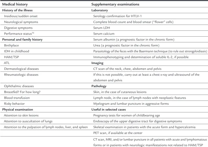
Table 1 presents the suggested conduct at the irst consultation of a patient with suspected ATL and pro-poses in the medical history and physical examination the points that require more attention.
TABLE 1 Conduct at the first consultation of a patient with suspected ATL.
Medical history Supplementary examinations
History of the illness Laboratory
Insidious/sudden onset Serology confirmation for HTLV-1
Neurological symptoms Complete blood count and blood smear (“flower” cells)
Digestive symptoms Serum LDH
Performance status70 Serum calcium
Personal and family history Serum albumin (a prognostic factor in the chronic form) Birthplace Urea (a prognostic factor in the chronic form)
IDH in childhood Parasitology of the feces with the Baermann technique (to rule out strongyloidiasis) HAM/TSP Immunophenotyping and determination of soluble IL-2, if possible
ATL Imaging
Dermatological diseases CT scan of the neck, chest, abdomen and pelvis
Rheumatologic diseases If this is not possible, carry out at least a chest x-ray and ultrasound of the abdomen and pelvis
Ophthalmic diseases Pathology
Breastfed? For how long? Skin, in the case of cutaneous lesions
Blood transfusion Lymph node, in the case of lymph nodes with neoplastic features Risky behavior Myelogram and lumbar puncture in aggressive forms
Physical examination Useful in selected cases
Attention to skin lesions Pregnancy tests for women of childbearing age
Attention to auscultation of lungs Endoscopy of the upper digestive tract for digestive symptoms Attention to the palpation of lymph nodes, liver, and spleen Skeletal examination in patients with the acute form and hypercalcemia
PET scan, if available at the center
CT scan, MRI, and/or lumbar puncture in all patients with acute and lymphomatous forms or in patients with neurologic manifestations not related to HAM/TSP
ATL: adult T-cell leukemia/lymphoma; IDH: infective dermatitis associated with HTLV-1; HAM/TSP: HTLV-1-associated myelopathy/tropical spastic paraparesis; LDH: lactic dehydrogenase; PET: positron emission tomography; CT: computed tomography; MRI: magnetic resonance imaging.
TABLE 2 Clinical classification of ATL (adapted from Shimoyama’s classification).7,29
Clinical form Lymphocytosis (> 4 x 10⁹/L)
Atypical lymphocytes
LDH level Hypercalcemia Organs involved
Smoldering* – < 5% or ≥ 5% ≤ 1.5 x N – Skin and/or lungs only¥
PCT – ... ≤ 1.5 x N – Cutaneous nodule/tumor lesions, mandatorily Chronic ** + ≥ 5% < 2 x N or ≥ 2 x N – Any organ except bone, GIT, and CNS Lymphoma – ≤ 1% Variable -/+ Lymph node, mandatorily, and/or any other organ Acute Usually + Usually ≥ 5% Usually ≥ 2 x N +/- Any organ and pleural effusions
*This form is divided into non-leukemic (< 5% atypical lymphocytes) and leukemic (≥ 5% atypical lymphocytes); **This form is divided into favorable and unfavorable, the latter being characterized by increased LDH (≥ 2 x N) and/or increased urea and/or decreased serum albumin; PCT: primary cutaneous tumoral; ... : not determined; LDH: serum lactic dehydrogenase; N: upper limit of the reference value; ¥skin and/or lung involvement may be lacking in the leukemic form; GIT: gastrointestinal tract; CNS: central nervous system.
Below is a list of the main characteristics of the various forms of ATL:
• Smoldering form: There is only involvement of the
skin and/or lungs; however, involvement of these or-gans may be absent in the leukemic form. Lymphocy-tosis (≥ 4,000 cells/mL) and hypercalcemia are absent,
• Primary cutaneous tumoral (PCT): The only diffe-rences in relation to the non-leukemic smoldering form are the presence of nodules or tumors on the skin and a worse prognosis.7 In many studies, this type is inclu-ded in the smoldering form.35-37
• Chronic: This is marked by lymphocytosis that may
remain stable for months or years, an increase in LDH over 1.5 times the normal value, absence of hypercal-cemia, with possible moderate lymphadenomegaly. There is an unfavorable subtype that is deined by low levels of serum albumin and high levels of serum LDH and/or urea, having a prognosis similar to the aggres-sive forms.38 In the chronic form there is no involve-ment of the CNS, bone, gastrointestinal tract (GIT) or pleural effusions. There are often skin lesions, mainly in the form of disseminated papules.
• Lymphoma: This is characterized by marked
lympha-denopathy without lymphocytosis and ≤ 1% abnormal lymphocytes in the peripheral blood. There may be creased serum LDH and serum calcium as well as in-volvement of the CNS, GIT and bones.7,29 Histological proof of iniltration of T-cell lymphoma in the lymph nodes is required, associated with extranodal involve-ment or otherwise.
• Acute: This form displays high levels of lymphocyto-sis and atypical cells, including “lower” cells in the peripheral blood smear.1,30 Any organ may be involved, including the CNS, GIT, and bone. Pleural effusions occur frequently.39 Lytic bone lesions are frequent and may include up to 80% of cases.40 A sharp increa-se in levels of increa-serum LDH can also be noted. Lympha-denomegaly and cutaneous involvement are frequent. It should be taken into consideration that this form may present different aspects including, less com-monly, the absence of lymphocytosis and hypercal-cemia. In the absence of lymphocytosis, differential diagnosis against the lymphoma form depends on the presence of a high percentage of atypical lym-phocytes in the peripheral blood.
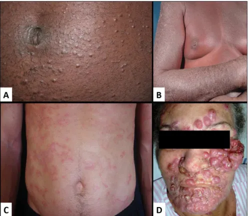
ATL involves the skin in around 60% of cases and in all clinical forms, and is most frequent in the smoldering and chronic forms.7 The lesions are multiple and generalized in around 50% of cases (Figure 1). Erythroderma, inil-trated plaques, papules, nodules, and tumors can be ob-served. Macular lesions are seen less frequently. Nodules and tumors are present in the aggressive forms (PCT, lym-phoma, and acute forms). Erythroderma has been observed
in all clinical forms, with the exception of PCT, mimicking Sézary syndrome.31 Although rare, vesicular lesions41 and purpuric lesions42 may also appear in ATL, similarly to that seen in MF. According to Sawada et al. (2011)43 the skin lesions that correspond to cases with a worse prog-nosis are the erythroderma and nodular/tumor lesions. In their case series, all cases of erythroderma occurred in the acute form.
D
IAGNOSISClinically, ATL diagnosis should be based on seropositiv-ity for HTLV-1 associated with hematological and/or histopathological diagnoses of peripheral T-cell leukemia and/or lymphoma.30
Conirmation of infection with HTLV-1 is generally performed by enzyme-linked immunosorbent assay (ELISA), and should always be conirmed by Western blot and/or polymerase chain reaction (PCR).
Distinctive “lower” cells can be seen in peripheral blood smears, that is, medium and/or large lymphocytes with multi-lobed nuclei, densiication of chromatin, and absent or small nucleoli. These are seen mostly in the acute and chronic forms (Figure 2A). These cells are considered pathognomonic of ATL and enable diagnosis alone.44 Other atypical cells may have the following morphologies: chronic lymphocytic leukemia, lymphoblastic type, and pleomorphic with granular or vacuolar cytoplasm.44
Flow cytometry is an important test for the diagnosis of ATL. Most patients display a phenotype of mature CD4 cells. The following markers should be used: CD2, CD3, CD4, CD5, CD7, CD8, CD25, CD29, CD26, CD45RO, αβ
T-cell, and HLA-DR receptors. Many cases of ATL do not express CD7 and CD26 and show decreased expression of CD3. The minimum markers required for this examination should include: CD3, CD4, CD7, CD8, CD25, and Ki-67.30,31 This examination can also be performed on cerebrospinal luid and pleural effusions.45
Patients with tissue iniltration should undergo a biopsy and pathological examination. Whenever possible, the ideal action is to investigate the type of viral integra-tion in the peripheral blood mononuclear cells (PBMC) and/or fresh neoplastic tissue, which conirms the diag-nosis of ATL if monoclonal.30 The techniques used are the reverse and long-range PCR46 and Southern blot.47 Southern blot is mainly used when there is a greater amount of DNA (Figure 2B). These techniques are per-formed by few laboratories, and thus are not generally accessible. However, they are not essential to the diagno-sis in most cases. Their importance is greater as scien-tiic proof in cases with atypical aspects, such as those
presenting very long progression. On the other hand, it is known that the occurrence of T-cell leukemia/lym-phoma not associated with HTLV-1 is rare in patients infected with the virus.30
There are several differential diagnoses of ATL, includ-ing mature T-cell neoplasms such as MF, peripheral T-cell lymphoma not otherwise speciied (PTCL-NOS), anaplastic large cell lymphoma (ALCL), angioimmunoblastic T-cell lymphoma, and even Hodgkin’s lymphoma.7,48,49
A
NATOMICAL-
PATHOLOGICAL ASPECTSIn 120 cases observed in Bahia, the organs most affected by ATL were: the skin (66.7%), lymph nodes (56.7%), pe-ripheral blood (53.3%), spleen (32.5%), bone marrow (27.5%), and liver (25%), although several other organs may be involved (data not published).
The histopathological patterns of ATL vary and mimic different types of T-cell lymphomas not associ-ated with HTLV-1. However, in the World Health Orga-nization’s classiication of cutaneous lymphomas,50 all cases of leukemia/lymphoma associated with HTLV-1 are classiied as ATL, regardless of the histological pat-tern and without taking into account that the patholo-gists can only diagnose this disease if they are aware of HTLV-1 infection. Without this information, the pa-thologist classiies these cases as PTCL-NOS, MF or, less often, ALCL.51
In cases with MF morphology, iniltration is by small and irregular cells, usually associated with epidermotro-pism, obliteration of the basal layer by atypical lympho-cytes (Figure 3A) and Pautrier’s abscesses. PTCL-NOS is characterized by moderate to marked pleomorphism (Figure 3B), and may also present epidermotropism of lymphocytes and Pautrier’s abscesses. In the ALCL pattern, large, cohesive cells with abundant cytoplasm and ana-plastic nuclei are noted.31
FIGURE 3 A. Skin biopsy of a patient with the chronic form and with a pattern of mycosis fungoides. Infiltration of small and medium lymphocytes in the superficial dermis, with pagetoid infiltration of the epidermis (HE, 100x). B. Skin biopsy of a patient with the primary tumor of skin form and with a pattern of peripheral T-cell lymphoma not otherwise specified. Note the accentuated cellular pleomorphism (HE, 500x). C. Biopsy of the same patient in figure B, showing a high proliferative index (Ki-67, 400x). D. Lymph node of chronic patient with a Hodgkin’s lymphoma pattern. Reed-Sternberg and Hodgkin type cells seen amid a background of medium sized lymphocytes, with a T-cell phenotype (HE, 400x). A Reed-Sternberg cell highlighted in the lower right corner (HE, 560x).
MST.7 In 60 to 70% of cases, the tumor cells express FoxP3 on the surface, which is a marker of regulatory T-cells.52
Conirming the stated above, a comparative study showed no signiicant difference between the histopath-ological aspects of PTCL-NOS and MF in individuals with and without infection with HTLV-1.53
Besides the aspects of PTCL-NOS and ALCL, a Hodg-kin’s type pattern may be observed in the lymph nodes, al-though infrequent, with a background of small and medium--sized T phenotype cells, with sparsely scattered Hodgkin and Reed-Sternberg type cells (Figure 3D). These cells are CD30, CD20, and/or CD15.48,54 In our case series we found one case of Hodgkin type ATL among the 120 individuals studied. Rarely, ATL may be present in the lymph node presenting a pattern similar to angioimmunoblastic T-cell lymphoma.48 It is important that pathologists consider Hodgkin-type ATL in the diagnosis of Hodgkin’s disease.
As with cutaneous lymphomas in general, it is of par-amount importance to differentiate between primary and secondary cutaneous ATL, as there is a statistically sig-niicant difference between them with respect to MST (48 months vs. 7 months).31 In Bahia, among the cases of pri-mary cutaneous T-cell lymphoma, 26.4% correspond to primary cutaneous ATL, while secondary ATL corresponds to 66.7%.17 This data shows that ATL is frequent in Brazil.
T
REATMENTThe treatment of ATL is based on the clinical type. Patients with aggressive forms, such as the acute, lymphoma or unfavorable chronic types, often receive chemotherapy. Recently, the Brazilian Ministry of Health published a guideline for ATL treatment including zidovudine (AZT) and interferon-α (IFN-α) as the irst-line treatment for all clinical types, and associated chemotherapy only for lym-phoma form.55 In the United States and Europe, the as-sociation of AZT and IFN-α is the standard treatment for the leukemic forms. In Europe, chemotherapy alone is the irst line treatment only for the lymphoma form of ATL, because survival with antiviral treatment alone is shorter.56
Traditionally, patients with the smoldering and favor-able chronic forms are not submitted to speciic treatments. In these forms, NB-UVB phototherapy is used for more supericial lesions and PUVA for more iniltrated lesions, with good results.57,58 A recent study of patients with the smoldering form of ATL and cutaneous involvement dem-onstrated better survival in those treated with photother-apy combined with etoposide (25 to 75 mg/day for 2 to 4 weeks with a one-week interval or on alternate weeks).35
As such, the favorable chronic and smoldering types of ATL are considered less aggressive and should be kept
under observation until possible progression of the disease, similar to management of chronic lymphocytic leukemia and smoldering myeloma. The treatment of the smolder-ing form with chemotherapy worsens the prognosis, which is similar to the unfavorable chronic form.59
Antiviral therapy using AZT and IFN was described in 199560 as an alternative treatment for ATL and has been used ever since. A meta-analysis with 254 patients recruited from four Western countries has been published, where all of the patients with the chronic and smoldering forms that were initially treated with AZT/IFN survived for more than 5 years. In acute patients treated initially with antivirals who had a complete response, survival at 5 years was 82%.56 A summary of the recommendations of the 16th International Conference on HTLV-1 held in Montreal in June 2013 deined the combination of AZT and IFN as effective in the leukemic forms of ATL, which should be considered as the standard procedure and irst-line therapy in this situation. In these cases, chemother-apy should only be started when a response to antivirals is not obtained.61
In relation to chemotherapy treatment, various combi-nations have been evaluated in Japan among ATL patients. However, MST ranged between 6 and 8.5 months.62 The Japanese Clinical Oncology Group (JCOG) has conducted various clinical trials with several chemotherapeutic regimens. The best results for aggressive clinical forms (acute, lym-phoma, and unfavorable chronic) were obtained with the VCAP-AMP-VECP regimen (vincristine, cyclophosphamide, doxorubicin, prednisone-doxorubicin, ranimustine, and prednisone-vindesine, etoposide, carboplatin, prednisone), which obtained a complete response rate of 40 vs. 25%, and an MST of 13 vs. 11 months, respectively, compared with the biweekly CHOP (cyclophosphamide, doxorubicin, vincristine, prednisone) regimen. However, due to the high toxicity of this regimen, especially in patients over 70 years old, CHOP regimens are preferred.63 As some of these drugs are not available in the United States, hyper-CVAD (cyclophospha-mide, vincristine, doxorubicin, and dexamethasone – meth-otrexate, cytarabine) is acceptable as an alternative regimen.64 Due to frequent CNS impairment in aggressive forms (from 10 to 25%) intrathecal prophylaxis is recommended.65
Autologous and allogeneic bone marrow transplanta-tion (BMT) has been attempted in ATL in order to improve the outcome of these patients. Autologous BMT does not seem to have many beneits due to frequent relapses and the occurrence of infections.67 Several researchers refer to an improvement in survival with allogeneic BMT, espe-cially using myeloablative regimens. However, a high mortality rate has limited its use.68 Studies using a reduced intensity conditioning regimen show an interesting and even curative option in approximately 15% of cases, prob-ably due to a graft versus tumor effect.68 In a retrospective analysis of 386 patients in Japan treated with allogeneic BMT under any induction regimen, survival at 3 years was 33%. Four factors were associated with having a poor prognosis: being older than 50 years, being male, disease without complete remission at the time of BMT, and having an unrelated donor.69
Many new agents for ATL are under study with promising results in the treatment of ATL, for example, anti-CCR4 monoclonal antibody (mogamulizumabe), IL-2 fusion inhibitor (denileukin diftitox), histone deacetylase inhibitors (HDAC), purine nucleoside phos-phorylase inhibitor (forodesine), proteasome inhibitor (bortezomib), etc.68
P
ROGNOSISA study of 854 patients using a multivariate analysis determined the indicators of a poor prognosis as being: high performance status,70 high levels of LDH, being aged > 40 years, more than three areas involved and hy-percalcemia.71 Most of these indicators are present in the acute form, which has the worst prognosis.30 In relation to the chronic form of the disease, as mentioned above, patients who have high levels of LDH and urea and low levels of albumin have the worst prognosis.38 A recent multicenter retrospective study with 807 patients newly diagnosed with the acute and lymphoma forms of ATL identiied Ann Arbor clinical staging, performance sta-tus and three continuous variables (age, serum albumin, and dosage of the soluble IL-2 receptor) as independent prognostic factors.72
In a study in Bahia that included 70 cases of ATL as-sessed using a univariate analysis, the factors related to poor prognosis were: the acute, lymphoma and PCT clin-ical forms, a proliferative index higher than 18%, presence of large cells in the histology, and the absence of cutaneous lesions. However, cutaneous involvement predominated in the forms with a better prognosis, and was present in all cases of the smoldering form and in 90% of cases of the chronic form.7
P
REVENTIONFor the prevention of ATL it is also important to halt vertical transmission of the HTLV-1, with infected moth-ers recommended not to breastfeed and being provided with formula and suitable pediatric assistance to children, as is already the case with HIV-infected mothers.22,73
Given that strongyloidiasis predisposes the develop-ment of ATL due to clonal expansion of lymphocytes, and considering that this form of parasitosis may be asymp-tomatic, frequent investigations for such in asymptomatic carriers of HTLV-1 are important, having in mind that proper treatment of this parasitosis can reverse clonal ex-pansion.74 Atypical cells, including “lower” cells, can be found in 10 to 43% of asymptomatic carriers of HTLV-1, and thus they are considered as being at high risk of devel-oping ATL.75 These patients should be monitored at regu-lar intervals in order to detect the early development of ATL.
C
ONCLUSION1. Clinical classiication of ATL is fundamental to de-termining the prognosis and therapeutic conduct. 2. ATL can simulate other T-cell lymphomas not
clini-cally and histologiclini-cally associated with the virus, such as MF, PTCL-NOS, and ALCL.
3. Serology for HTLV-1 should be performed in all patients with a diagnosis of mature T-cells leukemia/lympho-ma, so that cases of ATL receive adequate orientation. 4. Although new therapeutic options are gradually
im-proving the prognosis of ATL patients, treatment continues to be a major challenge. New studies and measures will be necessary in order to optimize the-rapeutic combinations.
5. It is important for the Brazilian Ministry of Health to consider the inclusion of HTLV-1 serology in pre-natal programs to decrease the incidence of ATL.
R
ESUMOLeucemia/linfoma de células T do adulto
e não leucêmica). Embora seja considerada uma doença agressiva, há casos com longa evolução. Salientamos a importância da classiicação clínica como elemento im-prescindível para avaliação do prognóstico e conduta terapêutica adequada. Como já foram publicados vários casos no Brasil e essa doença ainda é pouco conhecida, decidimos fazer um trabalho de revisão para divulgar os seus aspectos clínicos, hematológicos, anatomopatológi-cos, diagnósticos e terapêuticos. O melhor meio de redu-zir a ocorrência de LLcTA seria sustando a transmissão vertical do vírus pela amamentação.
Palavras-chave: vírus 1 linfotrópico T humano, leucemia--linfoma de células T do adulto, linfoma de células T, lin-foma de células T periférico, micose fungoide, linlin-foma cutâneo de células T.
R
EFERENCES1. Uchiyama T, Yodoi J, Sagawa K, Takatsuki K, Uchino H. Adult T-cell leukemia: clinical and hematologic features of 16 cases. Blood. 1977; 50(3):481-92. 2. Poiesz BJ, Ruscetti FW, Gazdar AF, Bunn PA, Minna JD, Gallo RC. Detection
and isolation of type C retrovirus particles from fresh and cultured lymphocytes of a patient with cutaneous T-cell lymphoma. Proc Natl Acad Sci USA. 1980; 77(12):7415-9.
3. Osame M, Usuku K, Izumo S, Ijichi N, Amitani H, Igata A, et al. HTLV-I associated myelopathy, a new clinical entity. Lancet. 1986; 1(8488):1031-2. 4. LaGrenade L, Hanchard B, Fletcher V, Cranston B, Blattner W. Infective dermatitis of Jamaican children: a marker for HTLV-I infection. Lancet. 1990; 336(8727):1345-7.
5. Verdonck K, González E, Van Dooren S, Vandamme AM, Vanham G, Gotuzzo E. Human T-lymphotropic virus 1: recent knowledge about an ancient infection. Lancet Infect Dis. 2007; 7(4):266-81.
6. Manns A, Hisada M, La Grenade L. Human T-lymphotropic virus type I infection. Lancet. 1999; 353(9168):1951-8.
7. Bittencourt AL, da Graças Vieira M, Brites CR, Farre L, Barbosa HS. Adult T-cell leukemia/lymphoma in Bahia, Brazil: analysis of prognostic factors in a group of 70 patients. Am J Clin Pathol. 2007; 128(5):875-82. 8. de Oliveira Mde F, Fatal PL, Primo JR, da Silva JL, Batista Eda S, Farré L, et al.
Infective dermatitis associated with human T-cell lymphotropic virus type 1: evaluation of 42 cases observed in Bahia, Brazil. Clin Infect Dis. 2012; 54(12):1714-9.
9. Farre L, de Oliveira Mde F, Primo J, Vandamme AM, Van Weyenbergh J, Bittencourt AL. Early sequential development of infective dermatitis, human T cell lymphotropic virus type 1-associated myelopathy, and adult T cell leukemia/lymphoma. Clin Infect Dis. 2008; 46(3):440-2.
10. Gonçalves DU, Guedes AC, Carneiro-Proietti AB, Lambertucci JR. HTLV-I associated infective dermatitis may be an indolent HTLV-I associated lymphoma. Braz J Infect Dis. 2000; 4(2):100-2.
11. Hanchard B, LaGrenade L, Carberry C, Fletcher V, Williams E, Cranston B, et al. Childhood infective dermatitis evolving into adult T-cell leukaemia after 17 years. Lancet. 1991; 338(8782-8783):1593-4.
12. Oliveira PD, Magalhaes M, Argolo JM, Bittencourt AL, Farre L. Double integration band of HTLV-1 in a young patient with infective dermatitis who developed an acute form of adult T-cell leukemia/lymphoma. J Clin Virol. 2013; 56(2):163-6. 13. Bittencourt A, Brites C, Pereira Filho C, Dias N, Vieira M. Linfoma/leucemia de células T associado ao HTLV-I (ATL) em criança e adolescente. An Bras Dermatol. 2001; 76(Suppl 2):88.
14. Gessain A, Cassar O. Epidemiological aspects and world distribution of HTLV-1 infection. Front Microbiol. 2012; 3:388.
15. Dourado I, Alcantara LC, Barreto ML, da Gloria Teixeira M, Galvão-Castro B. HTLV-I in the general population of Salvador, Brazil: a city with African ethnic and sociodemographic characteristics. J Acquir Immune Deic Syndr. 2003; 34(5):527-31.
16. Iwanaga M, Watanabe T, Yamaguchi K. Adult T-cell leukemia: a review of epidemiological evidence. Front Microbiol. 2012; 3:322.
17. Bittencourt AL, Oliveira PD, Andrade AC, Santos TC, Oliveira RF, Farré L, et al. Analysis of cutaneous lymphomas in a medical center in Bahia, Brazil. Am J Clin Pathol. 2013; 140(3):348-54.
18. Bittencourt AL, Barbosa HS, Pimenta A, Farre L. A case of adult T-cell leukemia/lymphoma (ATL) with a survival of more than 13 years. Acta Oncol. 2008; 47(5):981-3.
19. do Valle AC, Galhardo MC, Leite AC, Araujo AQ, Cuzzi-Maya T, Maceira JP, et al. Adult T-cell leukemia/lymphoma associated with HTLV-1 infection in a Brazilian adolescent. Rev Inst Med Trop São Paulo. 2001; 43(5):283-6. 20. Takahashi K, Takezaki T, Oki T, Kawakami K, Yashiki S, Fujiyoshi T, et al.
Inhibitory effect of maternal antibody on mother-to-child transmission of human T-lymphotropic virus type I. The Mother-to-Child Transmission Study Group. Int J Cancer. 1991; 49(5):673-7.
21. de Oliveira Mdo S, Hamerschlak N, Chiattone C, Loureiro P. HTLV-I infection and adult T-cell leukemia in Brazil: an overview. São Paulo Med J. 1996; 114(3):1177-85.
22. Ministério da Saúde. Atenção ao pré-natal de baixo risco. Brasília: Editora do MS; 2013.
23. Brand H, Alves JGB, Pedrosa F, Lucena-Silva N. Leucemia de células T do adulto. Rev Bras Hematol Hemoter. 2009; 31(5):375-83.
24. Matsuoka M. Human T-cell leukemia virus type I (HTLV-I) infection and the onset of adult T-cell leukemia (ATL). Retrovirology. 2005; 2:27. 25. Talledo M, López G, Huyghe JR, Verdonck K, González E, Clark D, et al.
Role of killer cell immunoglobulin-like receptor gene content and human leukocyte antigen-C group in susceptibility to human T-lymphotropic virus 1-associated myelopathy/tropical spastic paraparesis in Peru. Hum Immunol. 2010; 71(8):804-8.
26. Bangham CR, Osame M. Cellular immune response to HTLV-1. Oncogene. 2005; 24(39):6035-46.
27. Jeffery KJ, Siddiqui AA, Bunce M, Lloyd AL, Vine AM, Witkover AD, et al. The inluence of HLA class I alleles and heterozygosity on the outcome of human T cell lymphotropic virus type I infection. J Immunol. 2000; 165(12):7278-84.
28. Jeffery KJ, Usuku K, Hall SE, Matsumoto W, Taylor GP, Procter J, et al. HLA alleles determine human T-lymphotropic virus-I (HTLV-I) proviral load and the risk of HTLV-I-associated myelopathy. Proc Natl Acad Sci USA. 1999; 96(7):3848-53.
29. Shimoyama M. Diagnostic criteria and classiication of clinical subtypes of adult T-cell leukaemia-lymphoma. A report from the Lymphoma Study Group (1984-87). Br J Haematol. 1991; 79(3):428-37.
30. Tsukasaki K, Hermine O, Bazarbachi A, Ratner L, Ramos JC, Harrington W Jr, et al. Deinition, prognostic factors, treatment, and response criteria of adult T-cell leukemia-lymphoma: a proposal from an international consensus meeting. J Clin Oncol. 2009; 27(3):453-9.
31. Bittencourt AL, Barbosa HS, Vieira MD, Farré L. Adult T-cell leukemia/ lymphoma (ATL) presenting in the skin: clinical, histological and immunohistochemical features of 52 cases. Acta Oncol. 2009; 48(4):598-604. 32. Tsukasaki K, Tanosaki S, DeVos S, Hofmann WK, Wachsman W, Gombart AF,
et al. Identifying progression-associated genes in adult T-cell leukemia/lymphoma by using oligonucleotide microarrays. Int J Cancer. 2004; 109(6):875-81. 33. Matutes E. Adult T-cell leukaemia/lymphoma. J Clin Pathol. 2007;
60(12):1373-7.
34. Ratner L. Human T cell lymphotropic virus-associated leukemia/lymphoma. Curr Opin Oncol. 2005; 17(5):469-73.
35. Sawada Y, Shimauchi T, Yamaguchi T, Okura R, Hama-Yamamoto K, Fueki-Yoshioka H, et al. Combination of skin-directed therapy and oral etoposide for smoldering adult T-cell leukemia/lymphoma with skin involvement. Leuk Lymphoma. 2013; 54(3):520-7.
36. Setoyama M, Katahira Y, Kanzaki T. Clinicopathologic analysis of 124 cases of adult T-cell leukemia/lymphoma with cutaneous manifestations: the smouldering type with skin manifestations has a poorer prognosis than previously thought. J Dermatol. 1999; 26(12):785-90.
37. Germain M, Williams J, Skelton HG, Smith KJ. Smoldering HTLV-1-induced T-cell lymphoma localized within the skin; a radiation-resistant tumor. Int J Dermatol. 2000; 39(11):815-21.
38. Takatsuki K (ed.). Adult T-cell Leukemia. New York: Oxford University Press; 1994.
40. Kiyokawa T, Yamaguchi K, Takeya M, Takahashi K, Watanabe T, Matsumoto T, et al. Hypercalcemia and osteoclast proliferation in adult T-cell leukemia. Cancer. 1987; 59(6):1187-91.
41. Bittencourt AL, Mota K, Oliveira RF, Farré L. A dyshidrosis-like variant of adult T-cell leukemia/lymphoma with clinicopathological aspects of mycosis fungoides. A case report. Am J Dermatopathol. 2009; 31(8):834-7. 42. Oliveira PD, Torres IS, Oliveira RF, Bittencourt AL. Acute adult T-cell
leukemia/lymphoma (ATL) presenting with cutaneous purpuric lesions: a rare presentation. Acta Oncol. 2010; 50(4):595-7.
43. Sawada Y, Hino R, Hama K, Ohmori S, Fueki H, Yamada S, et al. Type of skin eruption is an independent prognostic indicator for adult T-cell leukemia/lymphoma. Blood. 2011; 117(15):3961-7.
44. Tsukasaki K, Imaizumi Y, Tawara M, Fujimoto T, Fukushima T, Hata T, et al. Diversity of leukaemic cell morphology in ATL correlates with prognostic factors, aberrant immunophenotype and defective HTLV-1 genotype. Br J Haematol. 1999; 105(2):369-75.
45. Dahmoush L, Hijazi Y, Barnes E, Stetler-Stevenson M, Abati A. Adult T-cell leukemia/lymphoma: a cytopathologic, immunocytochemical, and low cytometric study. Cancer. 2002; 96(2):110-6.
46. Etoh K, Tamiya S, Yamaguchi K, Okayama A, Tsubouchi H, Ideta T, et al. Persistent clonal proliferation of human T-lymphotropic virus type I-infected cells in vivo. Cancer Res. 1997; 57(21):4862-7.
47. Kamihira S, Sugahara K, Tsuruda K, Minami S, Uemura A, Akamatsu N, et al. Proviral status of HTLV-1 integrated into the host genomic DNA of adult T-cell leukemia cells. Clin Lab Haematol. 2005; 27(4):235-41.
48. Karube K, Suzumiya J, Okamoto M, Takeshita M, Maeda K, Sakaguchi M, et al. Adult T-cell lymphoma/leukemia with angioimmunoblastic T-cell lymphomalike features: report of 11 cases. Am J Surg Pathol. 2007; 31(2):216-23.
49. Huang CT, Lee YH, Chow KC, Yang CF, Chen PC, Hsiao LT, et al. Adult T-cell leukaemia/lymphoma can mimic other lymphomas in a non-endemic area: dilemmas in diagnosis and treatment. Intern Med J. 2014; 44(4):374-83. 50. Willemze R, Jaffe ES, Burg G, Cerroni L, Berti E, Swerdlow SH, et al.
WHO-EORTC classiication for cutaneous lymphomas. Blood. 2005; 105(10): 3768-85.
51. Bittencourt AL, de Oliveira Mde F. Cutaneous manifestations associated with HTLV-1 infection. Int J Dermatol. 2010; 49(10):1099-110.
52. Roncador G, Garcia JF, Garcia JF, Maestre L, Lucas E, Menarguez J, et al. FOXP3, a selective marker for a subset of adult T-cell leukaemia/lymphoma. Leukemia. 2005; 19(12):2247-53.
53. Bittencourt AL, Barbosa HS, Brites C, Ferraz N, Freitas V, Sampaio Jr C, et al. Clinicopathological aspects of HTLV- I positive and negative cutaneous T-cell lymphoma: a comparative study. Eur J Dermatol. 2000; 7(4):283-9. 54. Ohshima K, Niino D, Karube K. Microenvironment of adult T-cell leukemia/
lymphoma-associated nodal lesions. Int J Hematol. 2014; 99(3):240-8. 55. Ministério da Saúde – Secretaria de Vigilância da Saúde. Portaria n. 54 de
18/07/2016 – Aprova o Protocolo de Uso da Zidovudina para Tratamento do Adulto com Leucemia/Linfoma Associação ao Vírus HTLV-1. Diário Oicial da União. 2016. Available in: http://bvsms.saude.gov.br/bvs/saudelegis/ svs/2016/prt0054_18_07_2016.html.
56. Bazarbachi A, Plumelle Y, Carlos Ramos J, Tortevoye P, Otrock Z, Taylor G, et al. Meta-analysis on the use of zidovudine and interferon-alfa in adult T-cell leukemia/lymphoma showing improved survival in the leukemic subtypes. J Clin Oncol. 2010; 28(27):4177-83.
57. Kudo H, Fukushima S, Masuguchi S, Sakai K, Jinnin M, Ihn H. Cutaneous type adult T-cell leukaemia/lymphoma successfully treated with narrowband ultraviolet B phototherapy. Clin Exp Dermatol. 2012; 37(2):183-4.
58. Takemori N, Hirai K, Onodera R, Saito N, Yokota K, Kinouchi M, et al. Satisfactory remission achieved by PUVA therapy in a case of crisis-type adult T-cell leukaemia/lymphoma with generalized cutaneous leukaemic cell iniltration. Br J Dermatol. 1995; 133(6):955-60.
59. Takasaki Y, Iwanaga M, Imaizumi Y, Tawara M, Joh T, Kohno T, et al. Long-term study of indolent adult T-cell leukemia-lymphoma. Blood. 2010; 115(22):4337-43.
60. Gill PS, Harrington W, Jr., Kaplan MH, Ribeiro RC, Bennett JM, Liebman HA, et al. Treatment of adult T-cell leukemia-lymphoma with a combination of interferon alfa and zidovudine. N Engl J Med. 1995; 332(26):1744-8. 61. Barbeau B, Hiscott J, Bazarbachi A, Carvalho E, Jones K, Martin F, et al.
Conference highlights of the 16th International Conference on Human Retrovirology: HTLV and related retroviruses, 26-30 June 2013, Montreal, Canada. Retrovirology. 2014; 11(1):19.
62. Uozumi K. Treatment of adult T-cell leukemia. J Clin Exp Hematopathol. 2010; 50(1):9-25.
63. Tsukasaki K, Utsunomiya A, Fukuda H, Shibata T, Fukushima T, Takatsuka Y, et al.; Japan Clinical Oncology Group Study JCOG9801. VCAP-AMP-VECP compared with biweekly CHOP for adult T-cell leukemia-lymphoma: Japan Clinical Oncology Group Study JCOG9801. J Clin Oncol. 2007; 25(34):5458-64. 64. Di Venuti G, Nawgiri R, Foss F. Denileukin diftitox and hyper-CVAD in the treatment of human T-cell lymphotropic virus 1-associated acute T-cell leukemia/lymphoma. Clin Lymphoma. 2003; 4(3):176-8.
65. Teshima T, Akashi K, Shibuya T, Taniguchi S, Okamura T, Harada M, et al. Central nervous system involvement in adult T-cell leukemia/lymphoma. Cancer. 1990; 65(2):327-32.
66. Hande KR, Garrow GC. Acute tumor lysis syndrome in patients with high-grade non-Hodgkin’s lymphoma. Am J Med. 1993; 94(2):133-9. 67. Tsukasaki K, Maeda T, Arimura K, Taguchi J, Fukushima T, Miyazaki Y, et
al. Poor outcome of autologous stem cell transplantation for adult T cell leukemia/lymphoma: a case report and review of the literature. Bone Marrow Transplant. 1999; 23(1):87-9.
68. Utsunomiya A, Choi I, Chihara D, Seto M. Recent advances in the treatment of adult T-cell leukemia-lymphomas. Cancer Sci. 2015; 106(4):344-51. 69. Hishizawa M, Kanda J, Utsunomiya A, Taniguchi S, Eto T, Moriuchi Y, et
al. Transplantation of allogeneic hematopoietic stem cells for adult T-cell leukemia: a nationwide retrospective study. Blood. 2010; 116(8):1369-76. 70. Oken MM, Creech RH, Tormey DC, Horton J, Davis TE, McFadden ET, et
al. Toxicity and response criteria of the Eastern Cooperative Oncology Group. Am J Clin Oncol. 1982; 5(6):649-55.
71. Lymphoma Study Group (1984-1987). Major prognostic factors of patients with adult T-cell leukemia-lymphoma: a cooperative study. Leukemia Res. 1991; 15(2-3):81-90.
72. Katsuya H, Yamanaka T, Ishitsuka K, Utsunomiya A, Sasaki H, Hanada S, et al. Prognostic index for acute- and lymphoma-type adult T-cell leukemia/ lymphoma. J Clin Oncol. 2012; 30(14):1635-40.
73. Ribeiro MA, Martins ML, Teixeira C, Ladeira R, Oliveira Mde F, Januário JN, et al. Blocking vertical transmission of human T cell lymphotropic virus type 1 and 2 through breastfeeding interruption. Pediatr Infect Dis J. 2012; 31(11):1139-43. 74. Gabet AS, Mortreux F, Talarmin A, Plumelle Y, Leclercq I, Leroy A, et al. High circulating proviral load with oligoclonal expansion of HTLV-1 bearing T cells in HTLV-1 carriers with strongyloidiasis. Oncogene. 2000; 19(43):4954-60.