Correlation between maximum voluntary contraction and
endurance measured by digital palpation and manometry: An
observational study
FÁTIMA FANÍ FITZ1*, LILIANA STÜPP2, THAÍS FONSECA COSTA3, MARAIR GRACIO FERREIRA SARTORI4, MANOEL JOÃO BATISTA CASTELLO GIRÃO4,
RODRIGO AQUINO CASTRO4
1PT, MSc, Department of Gynecology, Universidade Federal de São Paulo (Unifesp), São Paulo, SP, Brazil 2PT, PhD, Department of Gynecology, Unifesp, São Paulo, SP, Brazil
3PT, Department of Gynecology, Unifesp, São Paulo, SP, Brazil 4MD, PhD, Department of Gynecology, Unifesp, São Paulo, SP, Brazil
S
UMMARYStudy conducted at Departamento de
Ginecologia, Universidade Federal de São Paulo (Unifesp), São Paulo, SP, Brazil
Article received: 6/29/2015 Accepted for publication: 7/6/2015
*Correspondence: Departamento de Ginecologia
Address: Rua Napoleão de Barros, 608 São Paulo, SP – Brazil
Postal code: 04024-002 [email protected]
http://dx.doi.org/10.1590/1806-9282.62.07.635
Financial support: This study was funded by the National Council for Scientiic and Technological Development (CNPq)
research foundation, grant nº 140190/2013-9
Introduction: Digital palpation and manometry are methods that can provide information regarding maximum voluntary contraction (MVC) and endurance of the pelvic loor muscles (PFM), and a strong correlation between these variables can be expected.
Objective: To investigate the correlation between MVC and endurance, measured by digital palpation and manometry.
Method: Forty-two women, with mean age of 58.1 years (±10.2), and predominant symptoms of stress urinary incontinence (SUI), were included. Examination was irstly conducted by digital palpation and subsequently using a Peritron ma-nometer. MVC was measured using a 0-5 score, based on the Oxford Grading Scale. Endurance was assessed based on the PERFECT scheme.
Results: We found a signiicant positive correlation between the MVC measured by digital palpation and the peak manometric pressure (r=0.579, p<0.001), and between the measurements of the endurance by Peritron manometer and the PERFECT assessment scheme (r=0.559, p<0.001).
Conclusion: Our results revealed a positive and signiicant correlation between the capacity and maintenance of PFM contraction using digital and manometer evaluations in women with predominant symptoms of SUI.
Keywords: pelvic loor, stress urinary incontinence, palpation/methods, vaginal squeeze pressure, manometry.
I
NTRODUCTIONAccording to the International Continence Society (ICS), pelvic loor muscle (PFM) function is deined by the abil-ity to perform a normal or strong voluntary contraction, with the presence of an involuntary contraction, resulting in a “circular closing of the vagina, the urethra, and the anus” and in a “cranioventral movement of the perineum
and upward movement of the pelvic organs.”1
PFM training should be recommended as a irst-line conservative management in the treatment of urinary incontinence,2,3 as demonstrated by numerous randomized
controlled trials.4-7 The success of treatment with
exer-cises is dependent on the achievement of strength and
endurance, which consequently leads to improvement of the PFM function. Evaluation of PFM function is a dif-icult task, as there is no consensus regarding the best method to evaluate or control the effects of PFM training. There are various methods to verify and quantify PFM function supported by the ICS, which include visual in-spection, intravaginal palpation, electromyography, pres-sure meapres-surements, and imaging methods, such as ultra-sound, magnetic resonance imaging (MRI), and video urodynamics. Visual inspection and digital palpation are
the most common methods used by physiotherapists.1,8
The vaginal palpation was irst described by Kegel,9
muscles, classifying muscle contraction subjectively as correct or incorrect. Currently, digital palpation is still considered an essential part of the PFM examination, and has become widespread due to its low cost, and also be-cause it is well accepted by the patients. The evaluation of muscle strength and endurance provides information about the severity of muscle weakness and forms the basis for patient-speciic exercise programs.10
In recent years, different methods have been developed to evaluate PFM function quantitatively.11,12 The
measure-ment of vaginal pressure has been considered a reproduc-ible method.13,14 However, practitioners should be aware
that increased intra-abdominal pressure might occur during the evaluation and inluence the results. Thus, this method should not be used alone.13
Considering that both digital and manometric meth-ods are able to provide information with respect to max-imal voluntary contraction (MVC) and endurance, a strong correlation between these variables can be expected. Thus, the aim of this study was to investigate this correlation, as measured by digital palpation and manometry.
M
ETHODStudy design
We present an observational and correlational study as-sessing the correlation between MVC and endurance measured by digital palpation and manometry.
Women admitted with untreated mixed stress urinary incontinence (SUI) and more than 2 g of leakage, as proven by a pad test with a standardized bladder volume,15
were enrolled in this trial at the Division of Urogynecol-ogy and Reconstructive Pelvic Surgery of the Universidade Federal de São Paulo (Unifesp), Brazil. This study was approved by the Review Board Committee of this institu-tion (CEP 1981/10). Each participant provided a written informed consent.
Patients with less than 2 g of urinary leakage (by pad test) and/or inability to contract the PFM were not in-cluded. Potential subjects were excluded if they had chronic degenerative diseases affecting the muscular and nerve tissues, diabetes, cerebrovascular diseases or overt neurological conditions, or autoimmune connective tis-sue disorders; if they were pregnant; or if they had previ-ously undergone pelvic loor re-education programs and/ or pelvic loor surgery.
To ascertain adequate PFM contraction, each volun-teer was assessed by inspection and digital vaginal palpa-tion to observe a lift of the pelvic loor in a superior, an-terior direction and a constriction around the urethra,
vagina, and rectum while in supine position.16 The patients
were requested to ‘‘lift and squeeze the PFM as hard as possible.’’ The co-contraction of the gluteal, hip adductor and rectus abdominal muscles was discouraged.
Once enrolled by a physiotherapist investigator, each subject completed a questionnaire designed to collect demographic characteristics such as age, body mass index (BMI), parity, and hormonal status.
Procedure
The assessments of the MVC and muscle endurance by digital palpation and vaginal squeeze pressure measurement were conducted by a physiotherapist specialized in PFM rehabilitation. Digital and vaginal pressure evaluations were carried out randomly, on the same day, with a 1-hour interval between measurements. The sequence of measure-ments was MVC followed by endurance. Three consecutive muscle contractions were recorded, with a 10-second in-terval between efforts,17 and the best of three was registered.18
One researcher (T.F.) was responsible for evaluating all patients and did not have knowledge about the analysis of correlation between the measurements. This researcher was instructed to use the same verbal command in all measurements. These results are part of a larger study involving pre- and post-physical therapy treatment. Sub-sequently, the main investigator (F.F.) performed the analysis of data. Both researchers are physiotherapists specialized in pelvic loor dysfunctions.
Digital palpation
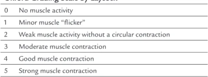
Digital palpation was used to assess PFM strength and endurance. To quantify muscle strength, a score from 0-5 was given based on the previously validated Oxford Grading Scale (Table 1).19 Endurance was recorded via
the PERFECT assessment scheme.20 Endurance was
ex-pressed as the length of time, up to 10 seconds, that an MVC could be sustained. Thus, the contraction was reg-istered until the muscle began to fatigue.
TABLE 1 Assessment of PFM activity according to the Oxford Grading Scale modified by Laycock.
Oxford Grading Scale by Laycock 0 No muscle activity
1 Minor muscle “flicker”
2 Weak muscle activity without a circular contraction 3 Moderate muscle contraction
Vaginal squeeze pressure measurement
The vaginal squeeze pressure measurement was performed using a Peritron manometer (Cardio Design™, Victoria, Australia). This equipment has a conical vaginal catheter, with diameter and length of 26 mm and 108 mm, respec-tively. The vaginal catheter was connected to a handheld microprocessor with latex tubing, allowing the transmis-sion of pressure (cmH2O) when the insert is compressed
by external pressure. The catheter was covered with a sterile latex sleeve for each patient. The vaginal catheter was inserted into the vaginal canal until the full extent of the compressible portion of the device was above the level of the hymenal ring. The baseline pressure reading was recorded after the catheter was inlated to 100 cmH2O,
and then the device was reset.
Statistical analysis
SPSS (Statistical Package for Social Sciences, IBM Com-pany, Chicago, USA) version 21.0 was chosen for the sta-tistical analyses. Spearman’s correlation test was used to correlate the values obtained using Peritron manometer, the modiied Oxford Grading Scale and the PERFECT assessment scheme. P-values were set to <0.05 to indicate statistical signiicance. The power of the relationship be-tween the variables was classiied as high reliability (0.80 to 1.00), moderate reliability (0.60 to 0.80), and question-able reliability (<0.59), according to Richman et al.21
R
ESULTSRecruitment, retention, and compliance
Forty-six (46) women diagnosed with mixed and SUI in the period from March 2011 to October 2013 were in-cluded in the study. Four women were exin-cluded from the study because they were unable to perform a proper PFM contraction. The remaining 42 participants underwent digital assessment and vaginal pressure measurement. None of the women declined to participate in this study.
Baseline characteristics
The mean age was 58.1 years (±10.2 years), BMI was 29.3 kg/m2 (±5.8 kg/m2), and the mean parity was 3.3 (±2.6).
Thirty-one (73.8%) women were menopaused. The mean of urinary leakage registered in pad test was 18.1 g (±24.8 g).
Digital and vaginal pressure measurements
MVC was classiied based on the Oxford Grading Scale system as licker (n=2), weak (n=20), moderate (n=13), good (n=3), and strong (n=4). The vaginal pressure mea-surements revealed an average score of 22.0 cmH2O (±15.0
cmH2O), and the Oxford Grading Scale revealed an
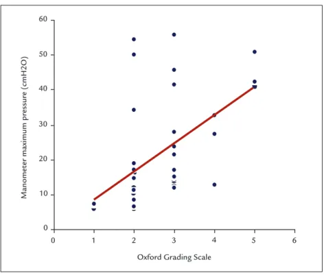
aver-age score of 2.6 (±1.0). There was a signiicant positive correlation between MVC according to the Oxford Grad-ing Scale score and the peak pressure of manometry (r=0.579, p<0.001) (Figure 1).
Measurements of endurance by Peritron manometer and the PERFECT assessment scheme yielded an average score of 3.8 seconds (±1.6 seconds) and 3.0 seconds (±1.4 seconds), respectively. There was a signiicant positive correla-tion between these variables (r=0.559, p<0.001) (Figure 2).
D
ISCUSSIONAbility to contract the PFM has been addressed by many studies. Instruction is mandatory and should be performed by verbal commands, followed by digital palpation and/ or manometry.5,6,22 Digital palpation is not considered a
reproducible or valid method for measuring the PFM strength,17 and peak pressure of manometry should not
be used alone.16 Therefore, it is noteworthy for clinical
practice that the combined use of both methods has a good correlation.
A recent prospective cohort study was conducted to verify the correlation between PFM function as determined by the Oxford Grading Scale and perineometry in pregnant and postpartum women. The authors found a positive correlation, indicating that both vaginal palpation and perineometry are valid and reliable methods for measure the PFM function.12 Accordingly, Ferreira et al. reported
good inter-observer reliability for the modiied Oxford Grading Scale and moderate reliability for manometry.23
Frawley et al. investigated the intra-observer reliabil-ity of bidigital evaluation and vaginal manometry, as well as resistance in different positions. The authors stated that both methods are reliable for quantifying MVC in standing and supine positions. Additionally, manometry is more reliable than vaginal palpation.24
Two studies investigated the inter-rater reliability of other palpation scoring systems, with squeeze pressures ranging from moderate to high (r=0.60 to r=0.90).25,26 Our
indings suggest that the correlation coeficient is ques-tionable with respect to MVC (r=0.57) and muscle endur-ance (r=0.55). Likewise, previous studies have shown weak inter-rater reliability for the Oxford Grading Scale using Cohen’s Kappa (0.37)17 and moderate inter-rater
reliabil-ity for the Peritron manometer.23 Da Roza et al. have also
found a moderate correlation between digital evaluation and manometry (r=0.65) in nulliparous athlete students.27
Ultrasound imaging is considered a responsive and reliable method to assess the PFM movement during contraction.28 Dietz et al. correlated the cranioventral
FIGURE 1 Maximum voluntary contraction measured by the Oxford Grading Scale.
FIGURE 2 PERFECT assessment scheme vs. Peritron manometer – endurance measurements.
Peritron manometer measurements
PERFECT assessment scheme
0 0 1 2 3 4 5 6 7 8
2 4 6 8 10
Oxford Grading Scale 0
60
50
40
30
20
10
0
1 2 3 4 5 6
Manomet
er maximum pr
essur
perineometry and found a highly signiicant correlation (r=0.62 and r=0.52, respectively).29 Another study found
a moderate association between ultrasound and perine-ometry in women with pelvic organ prolapse.30 However,
the perineal ultrasound does not offer the possibility of quantifying PFM contraction.31
Dietz et al. performed a comparative study of bidigital palpation and 4D ultrasound to evaluate trauma in the levator ani muscle. The authors found poor agreement between the two methods and concluded that imaging has a higher reliability than vaginal palpation, even when per-formed by a trained and experienced physiotherapist.32
Further studies using perineometry to evaluate the PFM are required to avoid capturing the action of the other muscle groups that form the wall of the abdomi-nopelvic cavity,33 because an increase in abdominal
pres-sure will affect the urethral, vaginal and rectal prespres-sures.34
However, Perschers et al. assessed the effect of contraction of the abdominal muscles concomitant with the pelvic loor and reported no signiicant increase in readings during digital palpation, perineometry, electromyography or ultrasound.31 In the present study, all women who were
able to contract the PFM correctly were included, and only contractions with a simultaneous inward movement of the catheter or perineum were considered valid.13
Measurements of vaginal squeeze pressure depend on the vaginal probe that is used. Differences may arise due to the length and diameter of the probes, straining, a learn-ing effect or different placement of the devices inside the vagina.17 Bo et al. found mean values of maximum squeeze
pressure of 19.7 cmH2O and 36.5 cmH2O after evaluating
with different types of manometers (p<0.01).35 Some
fac-tors, such as age, BMI, size of genital hiatus and parity, must be taken into consideration to assess the reliability of the evaluation of PFM by manometry.36 Nevertheless,
Hundley et al. reported that none of these variables inlu-ence the examination.33 Brækken et al. reported that
thicker muscles and a smaller levator hiatus were associ-ated with greater strength and muscular endurance; ad-ditionally, a smaller levator hiatus was associated with higher vaginal resting pressure.30
The strength of the present study was the evaluation of muscle endurance, which is recognized but not com-monly reported in the literature. Endurance reveals the severity of muscle weakness and is recommended to be included in all PFM training prescriptions.36 The weakness
of this study was the limited sample size.
In our study, the correlation found could be considered questionable because these methods are grounded on different principles. Vaginal pressure detects the
compres-sion of the PFM, while the Oxford Scale analyzes the com-pression and elevation of these muscles. In our opinion, the evaluation of compression and elevation performed separately should be considered and investigated.
We have demonstrated the importance of pelvic loor bidigital evaluation and manometry in providing various data that can enrich existing clinical and scientiic knowl-edge. These methods have limitations, and their reliabil-ity in the academic ield is still questioned. The training and experience of the evaluator are of extreme importance, as these metrics determine how reliable and realistic the results are. Our indings suggest there is still a gap in the existing information regarding the relationships among these variables, particularly pelvic muscle endurance. We recommend further studies with strong methodological design should be performed.
C
ONCLUSIONOur results revealed a positive and signiicant correlation between the capacity and maintenance of PFM contrac-tion using digital and manometer evaluacontrac-tions in women with predominant symptoms of SUI. However, this cor-relation was classiied as questionable.
A
CKNOWLEDGMENTSThis study was funded by the National Council for Sci-entiic and Technological Development (CNPq) research foundation, grant n. 140190/2013-9.
R
ESUMOCorrelação entre contração voluntária máxima e endurance
avaliados por palpação digital e manometria: um estudo observacional
Introdução: a palpação digital e a manometria são mé-todos capazes de fornecer informações sobre contração voluntária máxima (CVM) e endurance da musculatura do
assoalho pélvico (MAP), e pode-se esperar uma forte cor-relação entre essas variáveis.
Objetivo: investigar a correlação entre CVM e endurance,
avaliados por palpação digital e manometria.
mano-métrica de pico (r=0,579; p<0,001), e entre as medições do endurance avaliado pelo Peritron e o esquema PERFECT
(r=0,559; p<0,001).
Conclusão: os resultados revelaram correlação positiva e signiicativa entre a capacidade e a manutenção de con-tração dos MAP por meio das avaliações digital e mano-métrica em mulheres com IUE.
Palavras-chave: assoalho pélvico, incontinência urinária de esforço, palpação/métodos, pressão de contração va-ginal, manometria.
R
EFERENCES1. Messelink B, Benson T, Berghmans B, Bo K, Corcos J, Fowler C, et al. Standardization of terminology of pelvic floor muscle function and dysfunction: report from the pelvic loor clinical assessment group of the international continence society. Neurourol Urodyn. 2005; 24(4):374-80. 2. Fitz FF, Resende AP, Stüpp L, Sartori MG, Girão MJ, Castro RA. Biofeedback
for the treatment of female pelvic loor muscle dysfunction: a systematic review and meta-analysis. Int Urogynecol J. 2012; 23(11):1495-516. 3. Hay-Smith E, Bø K, Berghmans LC, Hendriks HJ, de Bie RA, van Waalwijk
van Doorn ES. WITHDRAWN: Pelvic loor muscle training for urinary incontinence in women. Cochrane Database Syst Rev. 2013; (1):CD001407. 4. Berghmans LC, Frederiks CM, de Bie RA, Weil EH, Smeets LW, van Waalwijk van Doorn ES, et al. Eficacy of biofeedback, when included with pelvic loor muscle exercise treatment, for genuine stress incontinence. Neurourol Urodyn. 1996; 15(1):37-52.
5. Mørkved S, Bø K, Fjørtoft T. Effect of adding biofeedback to pelvic loor muscle training to treat urodynamic stress incontinence. Obstet Gynecol. 2002; 100(4):730-9.
6. Bø K, Talseth T, Holme I. Single blind, randomised controlled trial of pelvic loor exercises, electrical stimulation, vaginal cones, and no treatment in management of genuine stress incontinence in women. BMJ. 1999; 318(7182): 487-93.
7. Castro RA, Arruda RM, Zanetti MRD, Santos PD, Sartori MG, Girão MJ. Single-blind, randomized, controlled trial of pelvic loor muscle training, electrical stimulation, vaginal cones, and no active treatment in the management of stress urinary incontinence. Clinics (São Paulo). 2008; 63(4):465-72.
8. Talasz H, Gosch M, Enzelsberger H, Rhomberg HP. [Female geriatric patients with urinary incontinence symptoms and their control over pelvic loor muscles]. Z Gerontol Geriatr. 2005; 38(6):424-30.
9. Kegel AH. Progressive resistance exercise in the functional restoration of the perineal muscles. Am J Obstet Gynecol. 1948; 56(2):238-49. 10. Bø K, Scherburn M. Evaluation of female pelvic loor muscle function and
strength. Phys Ther. 2005; 85(3):269-82.
11. Barbosa PB, Franco MM, Souza F de O, Antônio FI, Montezuma T, Ferreira CH. Comparison between measurements obtained with three different perineometers. Clinics (São Paulo). 2009; 64(6):527-33.
12. Riesco ML, Caroci AS, de Oliveira SM, Lopes MH. Perineal muscle strength during pregnancy and postpartum: the correlation between perineome-try and digital vaginal palpation. Rev Lat Am Enfermagem. 2010; 18(6):1138-44.
13. Bø K, Kvarstein B, Hagen R, Larsen S. Pelvic loor muscle exercise for the treatment of female stress urinary incontinence: I. Reliability of vaginal pressure measurements of pelvic loor muscle strength. Nerourol Urodyn. 1990; 9(5):471-7.
14. Dougherty MC, Abrams R, McKey PL. An instrument to assess the dynamic characteristics of the circumvaginal musculature. Nurs Res. 1986; 35(4):202-6.
15. Lose G, Rosenkilde P, Gammelgaard J, Schroeder T. Pad-weighing test performed with standardized bladder volume. Urology. 1988; 32(1):78-80.
16. Bø K, Kvarstein B, Hagen R, Larsen S. Pelvic loor muscle exercise for the treatment of female stress urinary incontinence: II. Validity of vaginal pressure measurements of pelvic loor muscle strength and the necessity of supplementary methods for control of correct contraction. Neurourol Urodyn. 1990; 9(5):479-87.
17. Bø K, Finckenhagen HB. Vaginal palpation of pelvic loor muscle strength: inter-test reproducibility and comparison between palpation and vaginal squeeze pressure. Acta Obstet Gynecol Scand. 2001; 80(10):883-7. 18. Grape HH, Dedering A, Jonasson AF. Retest reliability of surface
electromyography on the pelvic loor muscles. Neurourol Urodyn. 2009; 28(5):395-9.
19. Laycock J. Clinical evaluation of the pelvic loor. In: Schussler B, Laycock J, Norton P, Stanton S, editors. Pelvic loor re-education principles and practice. London: Springer; 2002. p. 42-8.
20. Laycock J, Jerwood D. Pelvic loor muscle assessment: the PERFECT scheme. Physiotherapy. 2001; 87(12):631-42.
21. Richman J, Mackrides L, Prince B. Research methodology and statistics. Part 3: measurement procedures in research. Physiother Can. 1980; 32:253-7. 22. Rett MT, Simões JA, Herrmann V, Pinto CL, Marques AA, Morais SS.
Management of stress urinary incontinence with surface electromyography– assisted biofeedback in women of reproductive age. Phys Ther. 2007; 87(2):136-42.
23. Ferreira CH, Barbosa PB, de Oliveira Souza F, Antônio FI, Franco MM, Bø K. Inter-rater reliability study of the modiied Oxford Grading Scale and the Peritron manometer. Physiotherapy. 2011; 97(2):132-8.
24. Frawley HC, Galea MP, Phillips BA, Sherburn M, Bø K. Reliability of pelvic floor muscle strength assessment using different test positions and tools. Neurourol Urodyn. 2006; 25(3):236-42.
25. Brink CA, Wells TJ, Sampselle CM, Taillie ER, Mayer R. A digital test for pelvic muscle strength in women with urinary incontinence. Nurs Res. 1994; 43(6):352-6.
26. Hove MCPS, Pool-Goudzwaard AL, Eijkemans MJC, Steegers-Theunissen RP, Burger CW, Vierhout ME. Face validity and reliability of the irst digital assessment scheme of pelvic floor muscle function conform the new standardization terminology of the International Continence Society. Neurourol Urodyn. 2009; 28(4):295-300.
27. Da Roza T, Mascarenhas T, Araujo M, Trindade V, Jorge RN. Oxford Grading Scale vs manometer for assessment of pelvic loor strength in nulliparous sports students. Physiotherapy. 2013; 99(3):207-11.
28. Dietz HP. Ultrasound in the assessment of pelvic loor muscle and pelvic organ descent. In: Bø K, Berghmans B, Morkved S, Van Kampen M. Evidence based physical therapy for the pelvic loor. Amsterdam: Elsevier; 2007. p. 81-92. 29. Dietz HP, Jarvis SK, Vancaillie TG. The assessment of levator muscle strength:
a validation of three ultrasound techniques. Int Urogynecol J Pelvic Floor Dysfunct. 2002; 13(3):156-9.
30. Brækken IH, Majida M, Engh ME, Bø K. Are pelvic loor muscle thickness and size of levator hiatus associated with pelvic loor muscle strength, endurance and vaginal resting pressure in women with pelvic organ prolapse stages I–III? A Cross Sectional 3D Ultrasound Study. Neurourol Urodyn. 2014; 33(1):115-20.
31. Peschers UM, Gingelmaier A, Jundt K, Leib B, Dimpl T. Evaluation of pelvic loor muscle strength using four different techniques. Int Urogynecol J Pelvic Floor Dysfunct. 2001; 12(1):27-30.
32. Dietz HP, Hyland G, Hay-Smith J. The assessment of levator trauma: a comparison between palpation and 4D pelvic loor ultrasound. Neurourol Urodyn. 2006; 25(5):424-7.
33. Hundley AF, Wu JM, Visco AG. A comparison of perineometer to brink score for assessment of pelvic loor muscle strength. Am J Obstet Gynecol. 2005; 192(5):1583-91.
34. Bump RC, Mattiasson A, Bø K, Brubaker LP, DeLancey JO, Klarskov P, et al. The standardization of terminology of female pelvic organ prolapse and pelvic loor dysfunction. Am J Obstet Gynecol. 1996; 175(1):10-7. 35. Bø K, Raastad R, Finckenhagen HB. Does the size of the vaginal probe affect
measurement of pelvic loor muscle? Acta Obstet Gynecol Scand. 2005; 84(2):129:33.