Comparison and efects of two diferent airway
occlusion times during measurement of maximal
inspiratory pressure in adult intensive care unit
neurological patients
Comparação e efeitos de dois diferentes tempos de oclusão da via aérea
durante a mensuração da pressão inspiratória máxima em pacientes
neurológicos na unidade de terapia intensiva de pacientes adultos
INTRODUCTION
Maximal inspiratory pressure (IPmax) is a simple and reproducible test, used to measure the inspiratory muscle strength reflecting the combi-nation of force capacity generated by inspiratory muscle in a short almost-static contraction.(1,2)
Although IPmax continues to be used in severely ill patients as an over-all respiratory muscles function indicator, it highly depends on several variables, which may be specifically difficult to control in the intensive care unit (ICU) settings.(3) Recent findings suggest that 40% of mechani-cally ventilated patients have a reduced IPmax during their artificially ventilated time.(4)
Gilvan Reis Pinheiro Filho1, Helena
França Correia dos Reis2, Mônica
Lajana de Almeida3, Wandalvo
de Souza Andrade4, Rodolfo Leal
Sampaio Rocha5, Petrônio Andrade
Leite6
1. Preceptor for the ICU Physiotherapy Course of Universidade Federal da Bahia – UFBA – Salvador (BA), Brazil. 2. MSc, Professor of Escola Bahiana de Medicina e Saúde Pública; Physiotherapist of Hospital Geral do Estado – Salvador (BA), Brazil. 3. Physiotherapist of Hospital Geral do Estado; Professor of Faculdade Social da Bahia and União Metropolitana de Educação e Cultura – UNIME – Salvador (BA), Brazil.
4. Physiotherapist of Hospital Geral do Estado – Salvador (BA), Brazil. 5. Preceptor for the ICU Physiotherapy Course of Universidade Federal da Bahia – UFBA – Salvador (BA), Brazil. 6. Physiotherapist of Hospital Geral do Estado; Professor of Faculdade Adventista de Fisioterapia - FAFIS – Salvador (BA), Brazil.
ABSTRACT
Objective: To verify if the maximal inspiratory pressure values with 40 sec-onds occlusion time are greater than with the 20 seconds occlusion time, and the impacts on the following patient’s physiological variables: respiratory rate, pulse oxygen saturation, heart rate and blood pressure, before and after the mea-surements.
Methods: his was a transversal pro-spective randomized study. Fifty-one patients underwent maximal inspiratory pressure measurement, measured by one single investigator. he manometer was calibrated before each measurement, and then connected to the adapter and this to the unidirectional valve inspira-tory branch for 20 or 40 seconds.
Results: he values with 40 seconds occlusion (57.6 ± 23.4 cmH2O) were signiicantly higher than the measure-ments taken with 20 seconds occlusion (40.5 ± 23.4 cmH2O; p=0.0001). he variables changes between the before and
after measurement respiratory and hemo-dynamic parameters monitoring showed: heart rate variation for the 20 seconds occlusion 5.13 ± 8.56 beats per minute and after 40 seconds occlusion 7.94 ± 12.05 beats per minute (p = 0.053), ver-sus baseline. he mean blood pressure change for 20 seconds occlusion was 9.29 ± 13.35 mmHg and for 40 seconds occlu-sion 15.52 ± 2.91 mmHg (p=0.021). he oxygen saturation change for 20 seconds occlusion was 1.66 ± 12.66%, and for 40 seconds 4.21 ± 5.53% (p=0.0001). he respiratory rate change for 20 seconds oc-clusion was 6.68 ± 12.66 movements per minute and for 40 seconds 6.94 ± 6.01 (p=0.883).
Conclusions: he measurement of maximal inspiratory pressure using a longer occlusion (40 seconds) produced higher values, without triggering clini-cally signiicant stress according to the selected variables.
Keywords: Ventilator weaning;
Respiratory muscles; Intensive care units
Received from Hospital Geral do Estado - Salvador (BA), Brazil.
Submitted on August 14, 2009 Accepted on February 12, 2010
Author for correspondence:
Gilvan Reis Pinheiro Filho Rua Altino Seberto de Barros, 119 - Apto. 902 - Ed. Itaigara Parque Residence - Itaigara
CEP: 41840-020 – Salvador (BA), Brazil.
IPmax measuring has been used through an uni-directional valve in order to prevent results changes due to consciousness level, sedation or lack of moti-vation, all common features in ICU.(4) Although its reproducibility and accuracy are questioned, by the unidirectional valve method it was possible establish-ing a more reproducible form to reach higher IPmax values than with simple end-expiratory occlusion.(5-8)
The number of respiratory movements and occlu-sion time to observe are controversial. The recommen-dations range from one single respiratory movement to 20 seconds as minimum occlusion time.(8-12)
IPmax relevance for monitoring respiratory muscle training as well as predicting mechanic ventilated pa-tients weaning failure has been well accepted, as it has low equipment costs and is an easy to perform test.(12)
A reliable IPmax evaluation, in conjunction with other findings, may predict mechanic ventilation (MV) weaning failure and consequently the need of respiratory muscle training. However, there is no uni-formity on bed side methodology for measuring the inspiratory muscle strength in artificial airway non-cooperative patients.
Thus, this study aimed to verify if maximal inspi-ratory pressure values with 40 seconds occlusion time are greater that with 20 seconds occlusion and the im-pact on the patient’s physiologic variables respiratory rate (RR), pulse oxygen saturation (SpO2), heart rate (HR) and mean blood pressure (MBP) before and af-ter the measurements.
METHODS
This study was conducted in male and female pa-tients above 18 years-old, staying in a general ICU, all under ventilatory weaning, intubated, tracheosto-mized, or under spontaneous ventilation and trache-ostomy disconnected for less than 48 hours from MV. Patients with IPmax measurement contraindica-tions, such as cranial hypertension, chest wall instabil-ity, bronchial-pleural or tracheal-esophageal fistulae, hemodynamic instability with mean blood pressure (MBP) < 70 mmHg even after volume resuscitation, alveolar hemorrhage, known coronary artery disease and upper airway leakage even after cuff hyperinsufla-tion, were excluded.
This was a transversal prospective randomized tri-al, in a sample of polytraumatized, clinical and surgi-cal neurologic predominantly chronic patients, with stable no vasoactive drugs hemodynamics, no
seda-tion, not cooperative, with Glasgow Coma Scale Score (GCSS) below 15, with an artificial airway, under MV weaning with support pressure ventilation (SPV) mode, 5 cmH2O Positive End Expiratory Pressure (PEEP), inspired oxygen fraction (FiO2) ≤ 40%, or spontaneous ventilation (SV) with a T piece discon-nected from MV for less than 48 hours.(13)
The measurements were always performed in the morning.(12) The patients were positioned with 45º head of bed elevation, underwent tracheal aspiration 10 minutes before the measurement, the cuff pressure was adjusted accordingly for no air leakage on lung auscultation. Next the patients were disconnected from MV to SV for at least 10 seconds, and the RR, SpO2, HR and MBP parameters were collected before and after measurements, divided in respiratory and hemodynamic variables. The measurement was per-formed by one single investigator, with the manom-eter calibrated before each measurement keeping the pointer on zero, and next connected to the adapter and this to the unidirectional valve inspiratory branch.(14)
The randomization was made by simple lottery, ac-cording to which was decided the initial airway occlu-sion time used for the subjects entering the trial. This random order of the initial times had a 15 minutes interval.
One single measurement was performed according to the randomization with each occlusion time in the ifty one non-cooperative neurological patients with GCSS < 15, as this is an unidirectional valve method used for patients who would not beneit from repeating the ma-neuver to obtain increased IPmax values, as patients with this proile would not beneit from learning.(13,15-17)
The data were collected with an analogical model MV-120 Instrumentation Industries manometer, with a 0 to 120 cmH2O range, silicone pressure line, inspi-ratory and expiinspi-ratory force adapter, adapter/reducer, a chronometer and an unidirectional valve. As maneu-ver interruption criteria were used the association of two or more of the following criteria: SpO2 ≤ 90%; RR ≥ 40 mpm, HR ≥ 140 bpm, MBP ≥ 120 mmHg.
used, and the significance level adopted was 5%. Ac-cording to the Law 196/96, all evaluations were only conducted after consent of the immediate responsible for the subjects, as manifested by signing the Informed Consent Form clarifying the entire process and assur-ing data confidentiality. This project was approved by the Faculdade Adventista de Fisioterapia’s Ethics Committee, approval opinion nr. 092/2008.
RESULTS
Fifty one patients were screened. The demograph-ics are described on Table 1 and Chart 1. The 40 sec-onds occlusion IPmax values (IP40) were significantly higher than with the 20 seconds occlusion (IP20) as shown in Figure 1. The mean IPmax value with 20 seconds was 40.6 ± 23.4 cmH2O versus 57.6 ± 23.4 cmH2O for 40 seconds occlusion (p=0.0001).
Among the respiratory variables, the post-IP20 measurement RR was 29.2 ± 12.5 mpm versus 29.2 ± 7.6 mpm following IP40 (p=0.001). Mean post-IP20 SpO2 was 95.4 ±3# versus 93 ± 6.04% following IP40 (p=0.001).
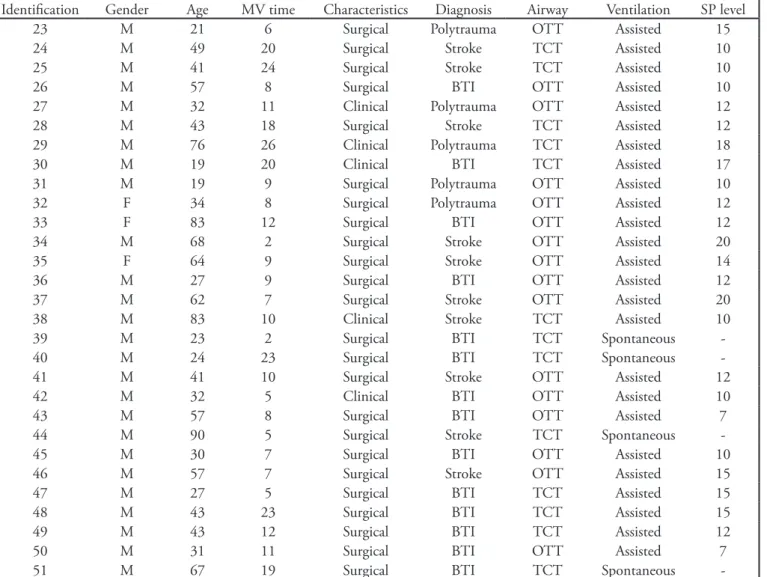
Chart 1 - Individual sample demographics
Identiication Gender Age MV time Characteristics Diagnosis Airway Ventilation SP level
1 F 18 1 Surgical Polytrauma OTT Assisted 7
2 M 52 13 Surgical BTI TCT Assisted 15
3 F 55 21 Surgical Stroke OTT Assisted 12
4 M 23 21 Clinical BTI OTT Assisted 10
5 F 47 7 Surgical BTI OTT Assisted 14
6 F 22 5 Surgical BTI OTT Assisted 7
7 M 45 7 Surgical BTI OTT Assisted 18
8 M 30 5 Surgical BTI OTT Assisted 7
9 F 25 10 Surgical BTI OTT Assisted 12
10 F 62 10 Surgical Polytrauma OTT Assisted 15
11 M 41 19 Surgical Polytrauma TCT Assisted 12
12 M 35 14 Surgical BTI TCT Assisted 10
13 M 31 8 Clinical BTI OTT Assisted 10
14 M 50 8 Surgical Polytrauma OTT Assisted 12
15 M 30 7 Surgical BTI OTT Assisted 15
16 M 80 8 Surgical Polytrauma OTT Assisted 7
17 F 30 5 Clinical Stroke OTT Assisted 12
18 M 28 17 Surgical BTI TCT Assisted 15
19 M 63 9 Surgical BTI OTT Assisted 10
20 M 24 18 Clinical BTI TCT Assisted 20
21 M 49 9 Surgical BTI OTT Assisted 10
22 F 35 12 Clinical Stroke OTT Assisted 12
Continued...
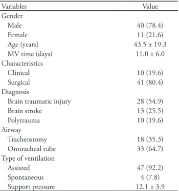
Table 1 - Overall samples characteristics
Variables Value
Gender
Male 40 (78.4)
Female 11 (21.6)
Age (years) 43.5 ± 19.3
MV time (days) 11.0 ± 6.0
Characteristics
Clinical 10 (19.6)
Surgical 41 (80.4)
Diagnosis
Brain traumatic injury 28 (54.9)
Brain stroke 13 (25.5)
Polytrauma 10 (19.6)
Airway
Tracheostomy 18 (35.3)
Orotracheal tube 33 (64.7)
Type of ventilation
Assisted 47 (92.2)
Spontaneous 4 (7.8)
Support pressure 12.1 ± 3.9
Chart 1 - Continuation
Identiication Gender Age MV time Characteristics Diagnosis Airway Ventilation SP level
23 M 21 6 Surgical Polytrauma OTT Assisted 15
24 M 49 20 Surgical Stroke TCT Assisted 10
25 M 41 24 Surgical Stroke TCT Assisted 10
26 M 57 8 Surgical BTI OTT Assisted 10
27 M 32 11 Clinical Polytrauma OTT Assisted 12
28 M 43 18 Surgical Stroke TCT Assisted 12
29 M 76 26 Clinical Polytrauma TCT Assisted 18
30 M 19 20 Clinical BTI TCT Assisted 17
31 M 19 9 Surgical Polytrauma OTT Assisted 10
32 F 34 8 Surgical Polytrauma OTT Assisted 12
33 F 83 12 Surgical BTI OTT Assisted 12
34 M 68 2 Surgical Stroke OTT Assisted 20
35 F 64 9 Surgical Stroke OTT Assisted 14
36 M 27 9 Surgical BTI OTT Assisted 12
37 M 62 7 Surgical Stroke OTT Assisted 20
38 M 83 10 Clinical Stroke TCT Assisted 10
39 M 23 2 Surgical BTI TCT Spontaneous
-40 M 24 23 Surgical BTI TCT Spontaneous
-41 M 41 10 Surgical Stroke OTT Assisted 12
42 M 32 5 Clinical BTI OTT Assisted 10
43 M 57 8 Surgical BTI OTT Assisted 7
44 M 90 5 Surgical Stroke TCT Spontaneous
-45 M 30 7 Surgical BTI OTT Assisted 10
46 M 57 7 Surgical Stroke OTT Assisted 15
47 M 27 5 Surgical BTI TCT Assisted 15
48 M 43 23 Surgical BTI TCT Assisted 15
49 M 43 12 Surgical BTI TCT Assisted 12
50 M 31 11 Surgical BTI OTT Assisted 7
51 M 67 19 Surgical BTI TCT Spontaneous
-MV – mechanic ventilation; SP – support pressure; F – female; M – male; BTI – brain traumatic injury; OTT – orotracheal tube; TCT – trache-ostomy.
Figure 1 – IP20 and IP40 results.
Regarding hemodynamic variables, mean post-IP20 HR was 98.4 ± 18.5 bpm (p=0.001) and post-IP40 101.2 ± 19.7 (p=0.001). Post-IP20 MBP was 114.9 ± 15.4 (p=0.001), while post-IP40 MBP was 121.7 ± 21.2 (p=0.001). Table 2 shows the respiratory and hemody-namic impacts of the diferent IPmax measurements.
DISCUSSION
Increased artificial airway occlusion time lead to increased IPmax values when the two different types of measurement in the studied subjects are compared. The IP40 measurement found a mean 57.6 ± 23.4 cmH2O value, versus 40.6 ± 23.4 cmH2O mean for IP20 measurement (p=0.0001).
Airway occlusion methods
A recent trial investigated the IPmax evaluation in thirty non-cooperative and under MV weaning patients using two occlusion times, 20 and 40 sec-onds, showing significant differences for the IP40 versus IP20 comparison (56.6 ± 23.3 versus 43.4 ± 24 cmH2O; p<0.001), agreeing with this trial results, reporting as the best IPmax evaluation for non-coop-erative patients a 40 seconds occlusion.(14)
Another trial evaluated two occlusion methods in a heterogeneous 28 subjects population, either with or without consciousness level changes, under ventila-tory weaning process, concluding that IP20 looks to be insufficient to measure the real IPmax in GCSS < 15 patients.(3) However, the measurement should have been performed in a more homogeneous patients population, such as in traumatic and non-traumatic, clinical and surgical neurological patients.
This study’s sample was limited to traumatic brain injury (TBI), stroke and polytrama patients, also using the GCSS, differently from other authors who classi-fied the patients as either alert or non-alert, rendering the real subject’s cooperation level subjective.(8) It was assumed, based on previous studies, that a GCSS < 15 is characteristic of a non-cooperative patient.(3)
In a recent twenty three patients trial, IPmax val-ues were compared with four different occlusion times (IPmax 20, 30, 45 and 60 seconds). The maneuver
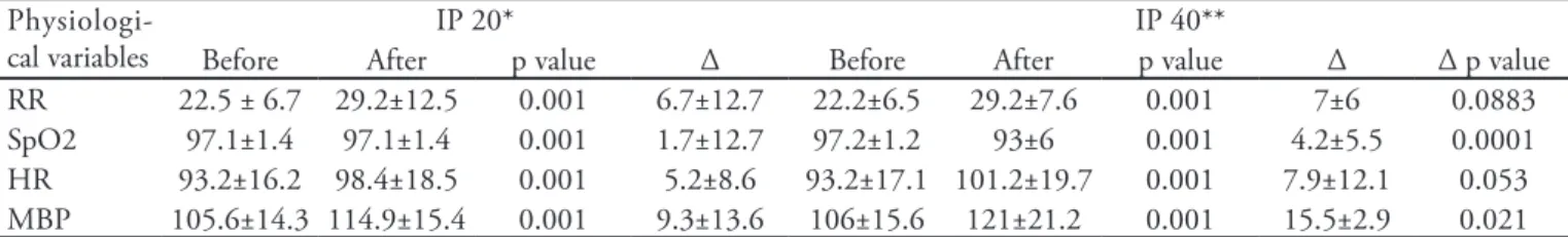
Table 2 – Maximal inspiratory pressure measurement respiratory and hemodynamic changes with range (∆)
Physiologi-cal variables
IP 20* IP 40**
Before After p value Δ Before After p value Δ Δ p value
RR 22.5 ± 6.7 29.2±12.5 0.001 6.7±12.7 22.2±6.5 29.2±7.6 0.001 7±6 0.0883 SpO2 97.1±1.4 97.1±1.4 0.001 1.7±12.7 97.2±1.2 93±6 0.001 4.2±5.5 0.0001 HR 93.2±16.2 98.4±18.5 0.001 5.2±8.6 93.2±17.1 101.2±19.7 0.001 7.9±12.1 0.053 MBP 105.6±14.3 114.9±15.4 0.001 9.3±13.6 106±15.6 121±21.2 0.001 15.5±2.9 0.021
IP – inspiratory pressure; Δ - range; RR – respiratory rate; SpO2 – pulse oxygen saturation; HR – heart rate; MBP – mean blood pressure. Results expressed as mean ± standard deviation. *Maximal inspiratory pressure measured with 20 seconds occlusion time. **Maximal inspiratory pressure measured with 40 seconds occlusion time.
was repeated thrice, with five to ten minutes between measurements intervals.(9,10,15,16,18) IPmax differences were seen in the studied times (p=0.001) IP20 (29 ± 9.2); IP30 (34.4 ± 10); IP45 (41.9 ± 13.2); IP60 (46.8 ± 14.9); no statistically significant differences were found for the three IP60 measurements. Thus, the greatest IPmax value was found for a 60 seconds occlusion time, with no additional maneuvers needed for the maximal inspiratory pressure.
IPmax measurement effects
In this trial 40 seconds was selected as longest mea-surement time because this occlusion method impacts were not known so far, thus being necessary to know about eventual additional IPmax measurement stress for longer than 40 seconds occlusion.
A recent trial mentioned that patients were moni-tored regarding HR and SpO2, however pre- and post-measurement values were not recorded, render-ing impossible to evaluate prolonged maneuver im-pacts, particularly at 40 seconds.(14)
The risks for this technique use include increased RR, HR and BP, and decreased SpO2, however, among the benefits can be mentioned being able to predict possible MV discontinuation failure and obtaining a parameter for muscle strength loss and gain in respira-tory training subjects.
The role of hyperoxygenation
IPmax values improvement.(17) However, this option was not used in this trial, as it was considered to pos-sibly influence the IPmax results.
Clinical and statistical changes analysis
This trial proposed to evaluate the impact of pro-longed maneuver on respiratory and hemodynamic functions. When the HR, MBP, RR and SpO2 vari-ables were compared after both occlusion times, no statistically significant post-measurement difference was found for HR and RR (p=0.053 and p=0.883, re-spectively), and a statistically significant impact was found for MBP and SpO2 (p=0.021 and p=0.0001, respectively).
As the absence of RR and HR variables impact, the SpO2 and MBP variables impact was not neces-sarily translated into clinical changes. We believe that tie IPmax measurement is safe, and can be per-formed using a longer occlusion time, however it is important to define validated clinical boundaries, if the maneuver needs to be withheld. Consonant with the literature data, considering as clinically significant 20% increase/decrease MBP values, or 10% increase of resting heart rate, and SpO2 drop to values below 90%,(18-21) this study found a mean change of IP40 HR of 7.84%, MBP 12.76% and SpO2 drop to 93%.
Thus, during IP40 measurement, no clinically sig-nificant impact was detected for the respiratory and hemodynamic variables, being their variations consid-ered within the safety interval, not related with addi-tional stress to the studied population. Nevertheless, we do not recommend airway occlusion longer than 40 seconds for IPmax measurement, and much less performing the maneuver without appropriate patient monitoring.
Study limitations
One limitation for this trial was not capturing the selected patients’ severity score, and that they weren’t necessarily under invasive MBP monitoring, which would provide more accurate information. Neverthe-less, this trial has shown considerable and homoge-neous sample when compared to other studies using smaller samples and evaluating diverse types of respi-ratory failure.
Perspective for mechanic ventilation weaning
Although IPmax measurement still has no clear correlation with mechanic ventilation weaning success or critical patients clinical outcome, it is believed that
higher IPmax values may be associated with improved lung ventilation, airways clearance and outcomes in these subjects.
This trial didn’t aim to evaluate neurological pa-tients ventilatory weaning success or failure. This study has no sufficient power to detect superiority between the occlusion times, as no follow-up was per-formed regarding mechanic ventilation weaning out-come.
CONCLUSION
IPmax measurement with 40 seconds occlusion time has shown values greater than the traditional 20 seconds method, and, although the statistical signifi-cance found for some variables analyzed with both air-way occlusion times, no clinically significant impact was found during IP40 measurement for these neuro-logical patients; however, we highlight the importance of monitoring the hemodynamic and respiratory vari-ables evaluated in this trial.
Further studies are necessary to confirm the feasi-bility of increased airway occlusion times for IPmax measurements, and how this evaluation is associated with ICU mechanic ventilated patients outcome.
RESUMO
Objetivo: Verificar se os valores de pressão inspiratória máxima com o tempo de oclusão de 40 (PI 40) segundos são maiores do que no tempo de oclusão de 20s (PI 20) e as repercussões apresentadas pelo paciente através das variáveis fisiológicas freqüência respiratória, saturação de pulso de oxigênio, freqüência cardíaca e pressão arterial antes e após as medidas.
Métodos: Estudo transversal prospectivo randomizado.
Foram selecionados cinqüenta e um pacientes para realiza-ção da medida de pressão inspiratória máxima, mensurada por um único investigador, com calibração do manova-cuômetro antes de cada aferição, conectado em seguida ao adaptador e este ao ramo inspiratório da válvula unidirecio-nal durante 20 e 40 segundos.
Resultados:Os valores da PI 40 (57,6 ± 23,4 cmH2O)
foram signiicativamente superiores às medidas realizadas durante um período de 20 segundos (40,5 ± 23,4cmH2O;
arterial média PI 40 foi de 15,52 ± 2,91 mmHg (p = 0,021). O Δ saturação de oxigênio para PI 20 1,66 ± 12,66%, e Δ saturação de oxigênio para PI 40 4,21 ± 5,53% (p = 0,0001). O Δ freqüência respiratória para PI 20 6,68 ± 12,66 ipm, e Δ freqüência respiratória PI 40 foi 6,94 ± 6,01 ipm (p= 0,883).
Conclusões:A mensuração da pressão inspiratória
má-xima utilizando uma oclusão superior (40s) produziu
maio-res valomaio-res quanto à pmaio-ressão inspiratória máxima encontra-da, sem desencadear estresse clinicamente significativos nas variáveis selecionadas.
Descritores: Desmame da ventilação mecânica;
Múscu-los respiratórios; Unidades de terapia intensiva
REFERENCES
1. Voliatinis S, McConnell AK, Jones DA. Assessment of maximum inspiratory pressure. Assessment of maximum inspiratory pressure. Prior submaximal respiratory muscle activity (‘warm-up’) enhances maximum inspiratory activi-ty and attenuates the learning efect of repeated measure-ment. Respiration. 2001;68(1): 22-7.
2. Black LF, Hyatt RE. Maximal respiratory pressures: nor-mal values and relationship to age and sex. Am Rev Respir Dis. 1969;99(5):696-702.
3. Monteiro LS, Veloso CA, Araújo S, Terzi RG. Compara-ção de dois métodos de mensuraCompara-ção da pressão inspirató-ria máxima em pacientes com e sem alterações do nível de consciência. Rev Bras Ter Intensiva. 2006;18(3):256-62. 4. Caruso P, Carnieli DS, Kagoharac KH, Anciães A, Segarra
JS, Deheinzelin D. Trend of maximal inspiratory pressu-re in mechanically ventilated patients: ppressu-redictors. Clinics (São Paulo). 2008;63(1):33-8.
5. Vitacca M, Paneroni M, Bianchi L, Clini E, Vianello A, Ceriana P, et al. Maximal inspiratory and expiratory pres-sure meapres-surement in tracheotomised patients. Eur Respir J. 2006;27(2):343-9.
6. Multz AS, Aldrich TK, Prezant DJ, Karpel JP, Hendler JM. Maximal inspiratory pressure is not a reliable test of inspi-ratory muscle strength in mechanically ventilated patients. Am Rev Respir Dis. 1990;142(3):529-32.
7. Polese G, Serra A, Rossi A. Respiratory mechanics in the intensive care unit. Eur Respir Mon. 2005;31(1):195-206. 8. Marini JJ, Smith TC, Lamb V. Estimation of inspiratory
muscle strength in mechanically ventilated patients: the measurement of maximal inspiratory pressure. J Crit Care. 1986;1(1):32-8.
9. Godfrey S, Campbell EJ. he control of breath holding. Respir Physiol. 1968;5(3):385-400.
10. Sassoon CS, Te TT, Mahutte CK, Light RW. Airway oc-clusion pressure. An im portant indicator for successful weaning in patients with chronic obstructive pulmonary disease. Am Rev Respir Dis. 1987;135(1):107-13.
11. Monteiro LS, Veloso CA, Araújo S, Figueiredo LC, Ter-zi RGG. Comparação de dois métodos de mensuração da pressão inspiratória máxima com o uso de uma válvula unidirecional. Rev Bras Ter Intensiva. 2004;16(2):74-7. 12. Caruso P, Friedrich C, Denari SD, Ruiz SA, Deheinzelin
D. he unidirectional valve is the best method to determi-ne maximal inspiratory pressure during weaning. Chest. 1999;115(4):1096-101.
13. Knaus WA, Draper EA, Wagner DP, Zimmerman JE. APACHE II: a severity of disease classiication system. Crit Care Med. 1985;13(10):818-29.
14. Guimarães FS, Alves FF, Constantino SS, Dias Menezes SLS. Avaliação da pressão inspiratória máxima em pacintes críticos não cooperativos: comparação entre dois métodos. Rev Bras Fisioter. 2007;11(3):233-8.
15. Yamagutti WPS, Alves LA, Kauss IAM, Galvan CCR, Bru-netto AF. Comparação entre a pressão inspiratória máxima medida pelo método da válvula unidirecional e pelo mé-todo convencional em pacientes submetidos ao processo de desmame da ventilação mecânica invasiva. Rev Bras Ter Intensiva. 2004;16(3);142-5.
16. Machado AF, Reis HF, Almeida ML, et al. Mensuração da pressão inspiratória máxima com diferentes tempos de oclu-são em pacientes submetidos ao desmame da ventilação me-cânica. Rev Bras Ter Intensiva. 2008;20(3Suppl):A0-122. 17. Correia Junior MAV, Ramos FF, Souza VV, et al - Efeito da
hiperoxigenação sob a medida da pressão inspiratória má-xima (PImáx) em pacientes em desmame da ventilação me-cânica. Rev Bras Ter Intensiva. 2008;20(3Suppl):A0-121. 18. Clanton TL, Diaz PT. Clinical assessment of the
respira-tory muscles. Phys her. 1995;75 (11):983-95.
19. Stiller K, Phillips A. Safety aspects of mobilising acutely ill in patients. Physiother heory Pract. 2003;19(4):239-57. 20. Stiller K, Philips AC, Lambert P. he safety of
mobilisa-tion and its efect on haemodynamic and respiratory sta-tus of intensive care patients. Physiother heory Pract. 2004;20(3):175-85.