Arquivos Brasileiros de Cardiologia - Volume 83, Nº 1, Julho 2004
8 7
were normal (potassium, creatinine, glycemia, total cholesterol, triglycerides, type I urine, and electrocardiography) at that time. In 1999, the patient was using hydrochlorothiazide and me-thyldopa, but, as blood pressure control deteriorated, the pres-cription was changed to indapamide (2.5 mg/day), perindopril (4 mg/day), and nadolol (80 mg/day). However, no satisfactory blood pressure control was achieved, and the patient began to complain of intense weakness characterized by the incapacity to perform her normal activities, such as sweeping the floor, washing the clothes, and even answering the phone.
The patient reported the following personal antecedents: arterial hypertension for 31 years, gastritis for 8 years, and hypertriglyce-ridemia for 4 years. The patient denied having diabetes mellitus, Chagas’ disease, stroke, acute myocardial infarction, or any other disease.
In regard to familial antecedents, the patient reported 4 hy-pertensive paternal uncles, 2 with deep venous thrombosis, 1 with myocardial infarction, and another with stroke. Her mother was hypertensive.
On physical examination, the patient was in regular general condition, eupneic, afebrile, acyanotic, hydrated, and with healthy coloring. Her pulse was 76 bpm; her BP in the sitting position (right upper limb) was 170/110 mmHg; her BP in the supine position (right upper limb) was 190/120 mmHg; her BP in the supine position (left upper limb) was 180/120 mmHg; and her BP in the standing position (right upper limb) was 170/110 mmHg. On auscultation, her lungs were clear with no rales. Her heart auscultation showed regular cardiac rhythm with cardiac sounds of normal intensity and no murmurs. Her heart rate was 76 bpm. Her abdomen showed hydro-aerial noise, no visceromegaly, and no abdominal murmur. Her lower limbs showed symmetric palpable pulses and no edema.
Differently from the normal results of the various measurements of serum potassium in 1999, potassium levels were extremely
altered (K+= 1.5 mEq/L; normal = 3.5 – 5.3), and the patient
was using indapamide, perindopril, and nadolol. All other routine laboratory tests were within the normal range (glycemia, creatinine, total cholesterol, and urinary sediment). Chest X-rays showed a normal cardiac area with no alterations in the pulmonary paren-chyma. The electrocardiogram showed sinus rhythm and an alte-ration in ventricular repolarization of the anterolateral wall. The echocardiogram showed left ventricular hypertrophy (IVS = 13;
LVPW = 13; LV mass = 292.8 g; LV mass index = 160.9 g/m2).
The laboratory finding of important hypokalemia drew attention to the following 2 situations: hypokalemia secondary to the use of
Case Report
Conn’s Adenoma. A Cause of Hypertension
and Hypokalemia
José Fernando Vilela Martin, Adriano Roberto Tarifa Vicente, Patrícia Maluf Cury,
Jorge Adas Dib, José Paulo Cipullo
São José do Rio Preto, SP - Brazil
Faculdade de Medicina de São José do Rio Preto/SP (FAMERP) Discipline of Internal Medicine and Pathological Anatomy Mailing address: José Fernando Vilela Martin - Av. Anísio Haddad, 7800 - Casa 129 - São José do Rio Preto, SP, Brazil
Cep 15093-000 – E-mail: [email protected] Received: 3/26/03
Accepted: 6/5/03
English version by Stela Maris Costalonga
Secondary hypertension accounts for approximately 5 to 10% of the causes of arterial hypertension, among which primary hyperaldosteronism has an incidence ranging from 0.05 to 2% in hypertensive individuals with characteristic findings of hypo-kalemia, increased production of aldosterone, a reduction or suppression in renin, an increased aldosterone/renin ratio, and metabolic alkalosis. We report the case of a patient with con-trolled primary arterial hypertension, who evolved with adrenal adenoma and worsening of blood pressure levels.
The aldosterone-producing adenoma (aldosteronoma) is the most important cause of hyperaldosteronism and represents one of the few curable causes of secondary arterial hypertension. The patients may be asymptomatic or oligosymptomatic with symptoms resulting from hypertension itself or from the complications gene-rated by hypokalemia (polyuria, nocturia, muscle cramps, excessive muscle weakness, paresthesias, tetany, and even muscle paralysis). The aldosterone-producing adenoma is characterized by arterial hypertension, hypokalemia, excessive urinary excretion of potas-sium, and metabolic alkalosis. We report the case of a patient with evolving primary hypertension for 31 years, who had satis-factory control over blood pressure and developed secondary hy-pertension with accentuated worsening of blood pressure levels.
Case report
Arquivos Brasileiros de Cardiologia - Volume 83, Nº 1, Julho 2004
8 8
Conn’s Adenoma. A Cause of Hypertension and Hypokalemia
as shown on the ABPM performed in 1992 (fig. 1), which favors the initial diagnosis of controlled primary hypertension, despite its beginning at an age suggesting secondary hypertension. That was not investigated, because the results of the basic laboratory tests (potassium, glycemia, total cholesterol, triglycerides, type I urine, and electrocardiography) were within the normal range. In addition, the patient had a favorable response to the treatment used, with normalization of blood pressure levels, and these criteria rendered the hypothesis of secondary hypertension very remote at that time. The lack of blood pressure control with elevated levels of arterial blood pressure detected in 1999 together with intense weakness, adynamia, and significant hypokalemia led us to consider a cause of hypertension secondary in origin superimposed on primary hypertension. It is worth noting that the patient did not have hypokalemia during the initial follow-up at our service, another fact contrary to the presence of hypertension due to primary or secondary hyperaldosteronism, on that occasion. On the other diuretic or hypokalemia due to primary hyperaldosteronism
ag-gravated by the use of the diuretic. Based on this, a diagnostic investigation was carried out.
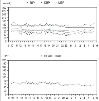
The measurements of sodium and potassium in the 24-hour urine were 192 mEq/L (normal = 50 to 250 mEq/L) and 59 mEq/L (normal = 25 to 125 mEq/L), respectively. With the suspension of indapamide, the clinical findings of the patient and her serum potas-sium levels significantly improved with no replacement (K+= 3.2 mEq/L). The measurements of Na+ and K+ in the 24-hour urine after 10 days of no diuretic use were 135 mEq/L and 24 mEq/L, respectively. The plasma renin level was 0.6 ng/mL/h and aldosterone level was 56.2 ng/100 mL (normal = 1-16 ng/100 mL), the aldos-terone/renin ratio being 93.6. Another ABPM was performed (fig. 2). The arterial blood gas analysis was as follows: pH = 7.50; pO2 = 90; pCO2 = 35; HCO3 = 30 mEq/L; BE = + 6.0; O2 Sat = 98%. After using 100 mg/day of spironolactone + 5mg/day of am-lodipine + 80 mg/day of nadolol, the BP normalized (BP = 130/ 90 mmHg), the asthenia and weakness significantly improved,
and the serum levels of Na+ and K+ were 147 mEq/L and 4.8
mEq/L, respectively.
The patient underwent abdominal ultrasonography and tomogra-phy, which showed a nodular image in the right adrenal topography measuring approximately 1cm in diameter (fig. 3). The patient was referred to the endocrinology surgery service and underwent exeresis of the right adrenal gland and the gallbladder. The adrenal gland weighed 7.2 g and measured 4.7 x 4.5 x 1.5 cm. On sectioning, a single, well-delimitated, orangish nodule measuring 1.5 cm in diameter was revealed (fig. 4). Histologically, the adrenal gland showed an encapsulated epithelial neoplasia with cells of abundant clear cyto-plasm and occasional bizarre nuclei; however, no mitoses were found (fig. 5). The anatomicopathological diagnosis was adenoma of the adrenal gland.
Approximately 3 years after surgery, the patient remains clini-cally stable using 1.5 mg/day of indapamide and nadolol and has adequate control over blood pressure levels observed on ABPM, with a mean blood pressure level of 138/71 mmHg during 24 hours (fig. 6).
Discussion
Our patient had a history of long-term arterial hypertension with satisfactory control over blood pressure levels during 24 hours
Heart Rate (BPM)
260 240 220 200 180 160 140 120 100 80 60 40 200 180 160 140 120 100 80 60 40 20
10 12 14 16 18 20 22 0 2 4 6 8
Blood Pressure (mmHg)
Time (hour)
Fig. 1 - Ambulatory blood pressure monitoring performed in 1992 showing 24-hour blood pressure control with a mean blood pressure level of 113/67 mmHg, presence of nocturnal decrease, and 5% systolic and diastolic blood pressure load during wakefulness.
Fig. 3 - Tomographic section of the abdomen, showing a nodular image in the right adrenal topography, enlarging the adrenal gland (arrows).
SBP
200 180 160 140 120 100 80 60 40 20 0
11 12 13 14 15 16 17 18 19 20 21 2223 0 1 2 3 4 5 67 8 9 10
DBP MBP
mmHg
HEART RATE
bpm 300 270 240 210 180 150 120 90 60 30 0
11 12 13 14 15 16 17 18 19 20 21 22 ,23 0 1 2 3 4 5 6 ,7 8 9 10
Arquivos Brasileiros de Cardiologia - Volume 83, Nº 1, Julho 2004
8 9
Conn’s Adenoma. A Cause of Hypertension and Hypokalemiahand, the severe diuretic-induced hypokalemia, more discrete after suspension of the diuretic, was suggestive of hyperaldostero-nism. The following factors pointed to the diagnosis of primary hyperaldosteronism, which was confirmed on abdominal tomo-graphy: use of spironolactone with good control of arterial blood pressure and normalization of potassium levels; urinary potassium level > 30 mEq/L/24 hours; metabolic alkalosis due to an increase in the serum level of bicarbonate resulting from the urinary loss of potassium and hydrogen ions; and elevated plasma aldosterone with hyporeninemia.
The aldosterone-producing adenoma (aldosteronoma), the most important cause of hyperaldosteronism (60% of the cases), was first reported by Conn in 1955 1,2. Currently, it is one of the few potentially curable causes of arterial hypertension. These tumors are usually small (less than 2cm in diameter) and benign, have a yellowish capsule, and different adrenal cell types visible on mi-croscopy 3,4. After Conn’s report, several other causes of hyperal-dosteronism were reported, such as idiopathic hyperalhyperal-dosteronism, and bilateral (20 to 40% of the cases) 1,3,5 or unilateral (less frequently) adrenal hyperplasia 6,7. The occurrence of adrenocortical carcinoma is rare, and only a few patients with ectopic aldostero-ne-producing tumors have been reported 8. Genetic forms of hy-peraldosteronism responsive to glycocorticoids occur as a dominant autosomalinheritance 3.
The incidence of primary aldosteronism ranges from 0.05 to
2% of the hypertensive population 4,9-16. Some patients are com-pletely asymptomatic or have minimum symptoms resulting from hypertension (ex: headache) and hypokalemia (polyuria, nocturia, muscle cramps). Occasionally, excessive muscle weakness, pa-resthesias, tetany, and even muscle paralysis may occur 1,3,4. Usually, primary aldosteronism is characterized by hypertension, hypokalemia, excessive urinary excretion of potassium, hyperna-tremia, and metabolic alkalosis 4.
The following are the 10 major situations in which primary hy-peraldosteronism should be suspected 13,14: 1) spontaneous hypoka-lemia (K< 3.5 mEq/L); 2) severe hypokahypoka-lemia (K< 3.0 mEq/L) or diuretic-induced hypokalemia; 3) difficulty in maintaining normal potas-sium levels despite the concomitant use of supplements or potaspotas-sium- potassium-sparing diuretics; 4) potassium levels that do not normalize 4 weeks after suspending the diuretic; 5) refractory hypertension; 6) satisfactory therapeutic response to spironolactone regarding blood pressure levels and serum and urinary potassium levels; 7) elevated plasma aldosterone levels (> 20 ng/mL); 8) inadequate kaliuresis (urinary potassium > 30 mEq/L); 9) renin < 1 ng/mL/h; and 10) presence of a tumor image in the adrenal gland on tomography.
Although the presence of spontaneous hypokalemia in a hy-pertensive patient is a strong indicator of aldosteronism, less than 20% of the patients with primary hyperaldosteronism have potas-sium levels below normal, while other hypertensive patients have hypokalemia without primary aldosteronism, a fact that may be explained by the use of diuretics or by the presence of secondary hyperaldosteronism 3.
The activity of plasma renin is suppressed in most patients with untreated primary hyperaldosteronism and in some patients with primary hypertension 15, while in secondary hyperaldostero-nism, renin plasma levels are elevated. Currently, measurement of serum potassium and plasma renin is recommended as the most reliable method for the initial investigation of primary hype-raldosteronism 3,16.
Fig. 5 - Microscopic appearance of the adrenal gland. Note the encapsulated epithelial neoplasia with cells of abundant clear cytoplasm and occasional bizarre nuclei. The anatomicopathological diagnosis was adenoma of the adrenal gland. HE, 400X.
Fig. 4 - Gross appearance of the adrenal gland and gallbladder. Adrenal gland measuring 4.7 x 4.5 x 1.5 cm with a single nodule of 1.5 cm in diameter. The gallbladder showed no alterations.
SBP
200 180 160 140 120 100 80 60 40 20 0
DBP MBP
mmHg
HEART RATE
bpm 300 270 240 210 180 150 120 90 60 30 0
9 10 11 12 13 14 15 16 17 18 19 20 21 2223 0 1 2 3 4 5 6
Fig. 6 - Ambulatory blood pressure monitoring performed after exeresis of the adrenal gland. Note the more satisfactory 24-hour control of blood pressure (mean = 138/71 mmHg), attenuation of the nocturnal decrease, and systolic blood pressure load during sleep (83%).
Arquivos Brasileiros de Cardiologia - Volume 83, Nº 1, Julho 2004
9 0
Conn’s Adenoma. A Cause of Hypertension and Hypokalemia
1. Weinberger MH. Primary aldosteronism: diagnosis and differentiation of subtypes. Ann Intern Med 1984; 100: 300-7.
2. Conn JW. Primary aldosteronism: a new clinical syndrome. J Lab Clin Med 1955; 45: 3-17.
3. Ganguly A. Primary aldosteronism. N Eng J Med 1998; 339: 1829-34. 4. Steward PM. Mineralocorticoid hypertension. Lancet 1999; 353: 1341-7. 5. Vallotton MB. Primary aldosteronism. Differential diagnosis of
hyperaldostero-nism and pseudoaldosterohyperaldostero-nism. Clin Endocrinol 1996; 45: 53-60.
6. Irony I, Kater CE, Biglieri EG, Shackleton CHL. Correctable subsets of primary al-dosteronism: primary adrenal hyperplasia and renin responsive adenoma. Am J Hy-pertens 1990; 3: 576-82.
7. Ganguly A, Zager PG, Luetscher JA. Primary aldosteronism due to unilateral adre-nal hyperplasia. J Clin Endocrinol Metab 1980; 51: 1190-4.
8. Farge D, Chatellier G, Pagny J-Y et al. Isolated clinical syndrome of primary aldoste-ronism in four patients with adrenocortical carcinoma. Am J Med 1987; 83: 635-40. 9. Ganguly A, Donohue JP. Primary aldosteronism: pathophysiology, diagnosis and
treatment. J Urol 1983; 129: 241-7.
References
10. Puccini M, Iacconi P, Bernini G, Miccoli P, Lynn JA. Conn Syndrome: 14 year’s ex-perience from two European centres. Eur J Surg 1998; 164: 811-7.
11. Hiramatsu K, Yamada T, Yukimura Y et al. A screening test to identify aldosterone-producing adenoma by measuring plasma renin activity: results in hypertensive pa-tients. Arch Intern Med 1981; 141: 1589-93.
12. Streeten DHP, Tomycz N, Anderson GH. Reliability of screening methods for the diagnosis of primary aldosteronism. Am J Med 1979; 67: 403-13.
13. Cohen DL, Townsend RR. Secondary Hypertension: Diagnosis and Management of an Adrenal Adenoma. JCOM 2002; 09: 525-31.
14. Ganguly A, Weinberg MH. Low renin hypertension: a current review of definitions and controversies. Am Heart J 1979; 98: 642-52.
15. Carey RM. Screening for surgically correctable hypertension caused by primary al-dosteronism. Arch Intern Med 1981; 141: 1594.
16. Young WF Jr, Hogan MJ, Klee GG, Grant CS, Van Heerden JA. Primary aldostero-nism: diagnosis and treatment. Mayo Clin Proc 1990; 65: 96-110.
17. Blumenfeld JD, Scaley JE, Schlussel Y et al. Diagnosis and treatment of primary hy-peraldosteronism. Ann Intern Med 1994; 121: 877-85.
The relation between plasma aldosterone and plasma renin activity in untreated hypertensive individuals remains the most accepted screening test for distinguishing patients with primary hypertension from those with primary aldosteronism; the cut off point remains around 30 or 50 in most cases with primary aldos-teronism 11,12,16,17.
Recent reviews 3,13,16 recommend that all spontaneous or diu-retic-induced hypokalemia should be investigated. Patients with difficult to control hypertension and low serum potassium levels
(≤ 3.5 mEq/L) should be assessed, and a plasma aldosterone/
plasma renin ratio greater than 30 indicates that a more detailed evaluation should be undertaken 3.
The definitive biochemical diagnosis of aldosteronism may also be established with both the inhibition and stimulation of the secretion of renin and aldosterone using physiological maneuvers of sodium overload and depletion, respectively. In hypertensive patients (with no treatment or 2 weeks after suspension of medi-cations), a high aldosterone urinary excretion rate with a sodium-rich diet (2 to 3 g of salt in each meal for 2 or 3 days) or a high aldosterone level after intravenous infusion of saline solution, to-gether with a low renin activity in conditions of low sodium con-sumption or use of a diuretic, are highly suggestive of primary hyperaldosteronism 3,5.
In conclusion, primary aldosteronism is the generic term for a series of disorders associated with a chronic excess of aldosterone. The most common cause is a solitary aldosterone-producing ade-noma. Hypertensive patients complaining of weakness and malaise are not rarely labeled as having a somatoform disorder, being referred to mental health care professionals. Our patient had a
history of 31 years of hypertension, which was adequately con-trolled until 1999. The diuretic-induced hypokalemia, which in our case was indapamide at the dosage of 2.5 mg/day (the only available dose on the market at the time), led us to consider the possibility of secondary hypertension (primary hyperaldosteronism) and to proceed with the investigation for its diagnosis. The thera-peutic test with the use of spironolactone normalized blood pres-sure, which per se is an important presumptive fact of excessive aldosterone levels.