1 1
Angiographically visible dissections occur in 30% of cases after balloon coronary angioplasty 1-3 and in 5 to 10% of patients when the procedure involves stent implantation 4. These dissections may be extensive and complicated, with contrast retention rapidly evol-ving to occlusion of the treated vessel, or they can be limited, without contrast retention and flow compromise 5-8. Cutlip et al 9 demonstrated that dissections, even the uncomplicated ones, are one of the main predictors of subacute thrombosis following stent implantation. These residual dissections are generally treated with a second stent, but the total length of the implanted stent is also associated with a greater risk of adverse events 9 and is consistently reported as one of the factors most strongly associated with res-tenosis 10-12.
The studies 13-15 that analyzed the influence of uncomplicated residual dissections after coronary angioplasty in long-term follow-up do not suggest that a greater risk of restenosis exists in these patients. On the other hand, late follow-up of patients with un-complicated coronary dissections after stent implantation is rarely studied 16,17. Thus, the objective of this study was to assess the influence of uncomplicated residual dissections after coronary stent implantation in the incidence of major cardiovascular events after 1 year.
Methods
The patients in this study had symptomatic ischemic heart disease and were treated with coronary stent implantation, from June 1996 to December 2000, at the Instituto de Cardiologia do Rio Grande do Sul/Fundação Universitária de Cardiologia, Porto Alegre, RS. Of 1149 patients treated with 1221 stents, 145 patients with coil stents were excluded. The other exclusion criteria were patients who had at least one of the following features: residual stenosis ≥ 30% (31), postprocedure vessel flow TIMI 0 or 1 (14), acute vessel occlusion immediately after the procedure or within the first 24 hours (33), procedure failure due to stent loss (1), procedure failure due to stent positioning outside the lesion (2), impossibility to cross the lesion with the stent (4), acute myocardial infarction related to the procedure (9), emergency surgery (4), and death within the first 24 hours of the procedure (12), amounting to 87 of the 998 patients experiencing angiogra-phic failure, clinical failure, or acute ischemic complications within the first 24 hours of the procedure.
Patients included in the dissection group had clinically suc-cessful stent implantation without greater cardiovascular
compli-Original Article
Clinical Meaning of Uncomplicated Coronary
Dissections After Stent Implantation
Alexandre Schaan de Quadros, Carlos A. M. Gottschall, Rogério Sarmento-Leite, Lenise Valler,
André Bussmann
Porto Alegre, RS - Brazil
Instituto de Cardiologia do Rio Grande do Sul/Fundação Universitária de Cardiologia
Mailing address: Alexandre Schaan de Quadros
Unidade de Pesquisa do IC/FUC - Av. Princesa Isabel, 395 Cep 90620-001 - Porto Alegre, RS – Brazil
E-mail: [email protected] Received: 2/25/2003
Accepted: 9/29/2003
Objective
To assess the influence of uncomplicated coronary dissections in the incidence of target vessel revascularization and cardio-vascular events after 1 year.
Methods
Patients treated from June 1996 to December 2000, with data prospectively collected and uncomplicated dissections (G1, n=36), were compared with those patients without dissections (G2, n=871). Data were assessed with SPSS 8.0 statistical software, the outcomes were compared with the Kaplan-Meier curve, and the significance level was assessed using the log-rank test.
Results
Clinical features were similar in both groups: G1 had lower mean reference diameters (P<0.0001), a greater number of patients with type C lesions (P=0.01), a lower final lumen dia-meter at the end of the procedure (P=0.003), and a greater balloon/artery ratio (P<0.0001). In the multivariate analysis, only the reference diameter and the artery/balloon ratio were independently associated with the presence of residual dissec-tions. No statistically significant difference existed in the inci-dence of revascularization of the target vessel and major cardio-vascular events, at 1-year clinical follow-up, between the 2 groups of patients. Predictors of adverse clinical events at 1 year were the reference diameter, lesion extension, and residual stenosis, rather than the presence of residual dissection.
Conclusion
Uncomplicated residual dissections after coronary stents are associated with narrower vessels and a higher balloon/artery ratio. Residual dissections are not associated with worse outcomes at 1-year clinical follow-up.
Key words
1 2
cations, and nonocclusive dissections (types A, B, or C of ACC) 8 not treated with a second stent. Patients included in the control group had clinical success, without major cardiovascular compli-cations and residual dissections. All patients were prospectively included in an Access database, and their clinical and angiographic characteristics were assessed.
Stent implantation was performed according to standard techni-ques 18,19, and in most cases predilation with balloon angioplasty was performed. High-pressure use, type and number of stents used, coronary angioplasty in another lesion or another vessel, other devices used, glycoprotein IIb/IIIa inhibitors, and the other technical criteria were decided by the operators. All patients were treated during platelet inhibition using acetylsalicylic acid and thienopyridine; these drugs were administered during or soon after the procedure, in emergency cases. Stents used were the Multilink (Guidant/Advanced Cardiovascular Systems, Santa Clara, California) = 310 implants; Tenax (Biotronik, Berlin, Germany) = 266 implants; BX Velocity (Cordis/Johnson & Johnson Interventional, Warren, New Jersey) = 97 implants; NIR (Medinol Ltd., Tel Aviv, Israel) = 75 implants; Vflex (Cook Group Inc, Broomfield, CO, USA) = 68 implants; AVE GFX (Arterial Vascular Engineering, Inc., Santa Rosa, California) = 67 implants; Iris (Uni-Cath Inc, Saddle Brook, NJ, USA) = 35 implants; Jostent (JOMED AB, Helsinborg, Sweden) = 9 implants; Palmaz Schatz (Cordis/Johnson & Johnson Interventional, Warren, New Jersey) = 7 implants.
Angiographic evaluations were performed through measure-ments with a manual pachymeter by experienced operators; the reference diameter was the average of the proximal and distal diameters of the lesion. The severity of the stenosis was assessed immediately after the procedure in at least 2 orthogonal projec-tions, considering the lesion with the most severe stenosis. The length of the lesion was measured along its entire extension (shoulder to shoulder), and long lesions were considered as single when there was less than 10mm of normal segment between them. The flow before and after the procedure was classified according to the TIMI 20 classification and the stenosis type accor-ding to criteria of the American College of Cardiology 21. Thrombus was defined as a intraluminal defect; the lesion was considered eccentric when the stenosis was observed in the middle of a supposed normal lumen in at least 1 projection. The balloon/ artery ratio was the ratio between the nominal diameter of the balloon used to expand the stent according to the manufacturer and the reference diameter of the vessel, as specified above. The aggressiveness score was the product of the balloon/artery ratio and the maximum pressure to implant the stent 22. Regarding the type of stent, the sample was divided into the following 3 groups: first-generation stents (Palmaz Schatz, Vflex, Iris, Jostent, Wall-stent, NIR, GFX), second-generation stents (Multilink, BX Velocity), and silicon carbide coated stents (Tenax).
Concerning the patient’s clinical presentation before the pro-cedure, stable angina was defined as stability in the pattern of triggering of the pain in the last 2 months. Unstable angina was considered as the worse intensity and/or frequency of the pattern of angina in the last 2 months before the procedure with or without chest pain at rest. Acute myocardial infarction was considered when the patient was sent for percutaneous revascularization due to chest pain and ST segment evolution. Regarding the indication for stent implantation, it was considered an elective procedure
when the stent placement was indicated before the procedure. Suboptimal indication occurred when it was recommended after coronary angioplasty because of important residual lesions or elastic recoil, or as bailout when the procedure was performed with acute occlusion or a threat of coronary angioplasty occlusion.
Regarding the procedure results, angiographic success was defined as effective stent implantation in the most severe stenosis with residual stenosis < 30% and normal flow at the end of the procedure, and clinical success was defined as angiographic success without acute myocardial infarction, or the need for emergency revascularization, or death.
Regarding the results of the study, the occurrence of the fol-lowing events was assessed at 1-year follow-up: major cardiovas-cular events, target vessel revascardiovas-cularization (coronary angioplasty or myocardial revascularization surgery), acute myocardial infarction and death. Major cardiovascular events were defined as the need for a new revascularization of the target vessel, acute myocardial infarction or death; target vessel revascularization as a new per-cutaneous intervention in the treated vessel or myocardial revas-cularization surgery; acute myocardial infarction as the appearance of new Q waves and chest pain lasting > 30 minutes or an episode of acute ischemic syndrome with ST segment depression and an indication of chemical or mechanical reperfusion, or an episode of acute ischemic syndrome without ST segment depression, but with electrocardiographic alterations and an enzyme increase with CK-MB 3 times greater than that of the control.
The patients were clinically followed-up in the outpatient ward, with personal assistance or by telephone with their attending physician, and the results were recorded in a dedicated database for later analysis. Control angiography was performed only when it was clinically indicated by each patient’s attending physician.
The differences between the 2 groups were assessed by chi-square test or Fisher’s exact test for categorical variables and the t test for continuous variables. The results were assessed through survival analysis by the Kaplan-Meier method, and the differences in the survival rates were assessed for statistical significance using the log-rank test. Logistic regression models were used to identify variables associated with major cardiovascular events at 1 year, with failures in the procedure and with dissections 23. For all tests, P ≤ 0.05 was considered statistically significant.
Results
We assessed 907 patients undergoing implantation of 960 stents (mean, 1.06 stent implantations per patient). The dissection group comprised 36 patients and the control group comprised 871 patients.
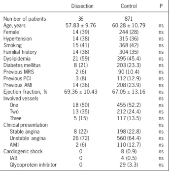
There was no statistically significant difference regarding the mean age of both groups (dissection=57.83±9.76 vs. con-trol=60.28±10.79; P=ns) or female frequency (dissection=39% vs. control=28%; P=ns). Regarding the presence of risk factors for ischemic heart disease, previous coronary intervention, and pre-vious acute myocardial infarction, a statistically significant difference did not occur between the 2 groups, nor did it occur in the mean left ventricular ejection fraction before the procedure, the number of involved vessels, and clinical presentation (tab. I).
1 3
group had significantly smaller vessels than did the control group(dissection = 3.06±0.27 mm vs. control. = 3.32±0.42 mm; P<0.0001); however, there were no differences regarding the severity of the stenosis before the procedure, and minimal luminal diameter before the procedure or lesion length. Regarding the coronary flow before the procedure, the patients in the dissection group had significantly less TIMI flow grade 2 or 3 (dissection = 75.7% vs. control. = 87.2 %; P=0.04). As for the characteristics of the lesion to be treated, the dissection group had type C lesions more frequently than did the control group (dissection = 40.5% vs. control. = 21.5%; P=0.01). There were no statistically sig-nificant differences between the 2 groups regarding the other characteristics of the lesions, such as thrombus, presence of calcium, ulcerations, eccentricity, and excessive tortuosity of the vessel (tab. II).
The indications for stent implantation were similar in both groups; however, the dissection group was treated with longer stents (dissection = 17.3±4.72 mm vs. = 15.76±4.48 mm; P=0.05), and the control group with second-generation stents (dissection = 16.2% vs. control = 43.7%; P=0.04). There were no differences regarding the mean of the pressures used, the size of the balloon, or the aggressiveness score, but the balloon/artery ratio was significantly greater in the dissection group (dissection = 1.07±0.09 vs. control = 1.00±0.09; P<0.0001). Luminal diameter at the end of the procedure was significantly lower in the dissection group (dissection = 3.15±0.35 mm vs. control = 3.34±0.40 mm; P<0.003), but there was no difference regarding residual stenosis after the implant (tab. III).
During the study period, 87 patients were identified with cli-nical or angiographic failure, or with major ischemic complications within the first 24 hours of the procedure. They were not included in the groups assessed but were described only for comparison
with the sample studied (tab. IV). Using multivariate analysis, only bailout stenting, cardiogenic shock, and angiographically vi-sible calcium were independently associated with failure or major ischemic complications (tab. V).
No differences were found between the 2 groups regarding in-hospital outcomes, because we only selected patients with suc-cessful procedures and no complications. Regarding the inciden-ce of major cardiovascular events at 1 year, there was no statis-tically significant difference between the 2 groups (dissection = 17.1% vs. control = 9.5%; P=0.14) (tab. VI). Survival analysis with Kaplan-Meier curves and comparisons with the log-rank test did not demonstrate statistically significant differences between the 2 groups (fig. 1). Regarding other 1-year outcomes, statistically significant differences were not observed between the 2 groups, although the dissection group tended to have adverse results more frequently (tab. VI).
The dissection group had a nonsignificant tendency towards worse outcomes, but it also more frequently had several clinical and angiographic characteristics associated with a worse prognosis. Multivariate analysis was performed with these unfavorable characteristics, analyzing their relationship with clinical outcomes. In this multiple logistic regression model, the incidence of major cardiovascular events at 1 year was the dependent variable; the dissection, reference diameter, postimplant residual stenosis, dia-betes mellitus, type of stent implanted, type of treated lesion, and extension of the lesion were the independent variables. Reference diameter (odds ratio = 0.33, confidence interval 0.17-0.65; P=0.001), residual stenosis (odds ratio=1.04, confidence interval 1.01-1.07; P=0.01) and lesion extension (odds ratio = 1.09, confidence interval 1.03-1.15; P=0.002) were independently associated with major cardiovascular events at 1 year rather than the presence of postimplant dissection (tab. VII).
Multivariate analysis was performed to identify factors inde-pendently associated with dissections. Reference diameter, balloon/ artery ratio, type of lesion, and stent length were the independent variables used; dissection was the dependent variable. In this analysis, only the reference diameter and the balloon/artery ratio were independently associated with dissection.
Discussion
Uncomplicated residual dissections were associated with smaller vessels and a greater balloon/artery ratio. One-year clinical follow-up of patients with residual dissections was not significantly different from the control group. The factors independently asso-ciated with major cardiovascular events in the multivaried analysis were reference diameter, residual stenosis, and lesion extension, rather than the presence of dissection.
Although atherosclerotic plaque rupture is a sine qua non con-dition for an effective coronary angioplasty, the incidence of angio-graphically visible dissections is 30% after balloon angioplasty and 5 to 10% after stent implantation 1-4,14. The factors associated with coronary dissections after conventional angioplasty are angio-graphic calcification 2, lesion extension 2,14, balloon/artery ratio 1,2, complex lesions 7-15, presence of other lesions in the same vessel 2, female gender 13, low cholesterol 14, stable angina 14, right coronary artery intervetion 14, lesions in a curve 14, postprocedure thrombus 14, high pressures 14, and noncompliant balloon 14. The predictive
fac-Table I - Clinical features of patients
Dissection Control P
Number of patients 36 871
Age, years 57.83 ± 9.76 60.28 ± 10.79 ns
Female 14 (39) 244 (28) ns
Hypertension 14 (38) 315 (36) ns
Smoking 15 (41) 368 (42) ns
Familial history 14 (38) 304 (35) ns
Dyslipidemia 21 (59) 395 (45.4) ns
Diabetes mellitus 8 (21) 203 (23.3) ns
Previous MRS 2 (6) 90 (10.4) ns
Previous PCI 3 (8) 112 (12.9) ns
Previous AMI 14 (36) 208 (23.9) ns
Ejection fraction, % 69.36 ± 10.43 67.05 ± 13.16 ns
Involved vessels ns
One 18 (50) 455 (52.2) ns
Two 13 (35) 212 (24.4) ns
Three 5 (15) 117 (13.5) ns
Clinical presentation ns
Stable angina 8 (22) 198 (22.8) ns
Unstable angina 26 (72) 560 (64.4) ns
A M I 2 (6) 110 (12.7) ns
Cardiogenic shock 0 8 (0.9) ns
IAB 0 4 (0.5) ns
Glycoprotein inhibitor 0 29 (3.3) ns
1 4
tors for severe ischemic complications secondary to dissections were balloon/artery ratio 1, dissection extension 5,7,8, presence of a significant residual stenosis 6,7,and the type of dissection according to the ACC 8 classification. Despite the importance of these angio-graphic data, most studies demonstrate that the clinical status of the patient at the time of angioplasty, that is, the presence of angina, electrocardiogram alterations, and/or hemodynamic invol-vement, is the greatest predictor of acute ischemic complications. In our study, we have demonstrated that the reference diameter of the treated vessel and the balloon/artery ratio rather than the lesion type, were independently associated with dissections. These results emphasize the importance of a proper balloon/artery ratio in the prevention of dissections, especially in narrower vessels. Also, the type of lesion is not associated with the dissections after stent implantation, unlike what is seen after conventional angioplasty.
Some studies demonstrate that no association exists between residual dissections and worse late clinical outcomes in patients undergoing coronary angioplasty. Leimgruber et al 13 reported si-milar rates of angiographic restenosis in 986 patients with or without dissection, with the exception of those with final trans-stenotic gradients < 15 mmHg, who experienced better evolve-ment. Hermans et al 14 studied a prospective series of 693 patients with angiographic follow-up in 94% of cases, demonstrating that a successful angioplasty with residual dissection does not increase restenosis. Cappelletti et al 15 demonstrated that most dissections disappear during angiographic follow-up and that patients with uncomplicated dissections have lower restenosis rates than do
those without dissections. They have also reported that patients with uncomplicated dissections treated with a stent have higher restenosis rates than do those without stent implantation to treat the dissection 15. However, these results are only applicable when angioplasty is successful, without significant residual stenosis, or significant contrast retention or flow compromise.
On the other hand, a few studies have assessed long-term clinical follow-up of patients treated with coronary stents. Even uncomplicated residual dissections are associated with subacute thrombosis after stent implantation, or implant of multiple stents 9 . Cutlip et al 24 assessed the clinical follow-up of patients with su-boptimal implants, either by the presence of residual dissections, multiple stents, flow involvement during the procedure, or residual thrombus. Through multivariate analysis, lower final luminal dia-meter, a higher number of stents, and the absence of treatment with ticlopidine were related to cardiovascular events in 30 days. Final luminal diameter, a higher number of stents, and diabetes mellitus were related to revascularization rates of the target vessel at 9 months. Dissections were not independently associated with adverse clinical outcomes in 30 days or 9 months. Alfonso et al 17 reported the clinical follow-up of 17 patients with residual dissections after stent implantation and without coronary flow compromise. In the control angiography, all dissections had disappeared, and even the patients with more extensive dissections did not have restenosis, concluding, therefore, that patients with residual dissections may have a good clinical follow-up, as long as no coronary flow invol-vement or significant residual stenosis are present.
Table II - Angiographic characteristics of the procedures
Dissection Control P
Number of procedures 37 923
Treated vessel
Left main 0 7 (0.8) ns
Anterior descendent 22 (59.5) 477 (51.7) ns
Circumflex 4 (10.8) 121 (13.1) ns
Right 10 (27.0) 271 (29.4) ns
Saphenous vein graft 1 (2.7) 46 (5.0) ns
Lesion site
Ostial 0 12 (1.3) ns
Proximal 25 (67.6) 566 (61.3) ns
Medial 12 (32.4) 302 (32.7) ns
Distal 0 43 (4.7) ns
Reference diameter, mm 3.06 ± 0.27 3.32 ± 0.42 <0.0001
Stenosis severity, % 82.49 ±11.99 84.21 ±10.62 ns
Minimum luminal diameter, mm 0.54 ± 0.36 0.53 ± 0.36 ns
Length of the lesion, mm 11.59 ± 5.94 10.15 ± 4.60 ns
Lesions greater than 10 mm 18 (48.6) 396 (42.9) ns
Vessel flow before the stent (TIMI)
0/1 9 (24.3) 118 (12.8) 0.07
2/3 28 (75.7) 805 (87.2) 0.04
Type of lesion (ACC classification)
A 1 (2.7) 28 (5.3) ns
B1 67 (13.5) 171 (18.5) ns
B2 16 (43.2) 500 (54.2) ns
C 15 (40.5) 198 (21.5) 0.01
Thrombus 30 (81.1) 723 (78.3) ns
Calcium 8 (21.6) 141 (15.3) ns
Ulceration 13 (35.1) 230 (24.9) ns
Branch involvement 13 (35.1) 375 (40.6) ns
Eccentricity 31 (83.8) 816 (88.5) ns
Excessive tortuosity 2 (5.4) 30 (3.3) ns
1 5
Table III - Aspects related to the procedure
Dissection Control P
Number of procedures 37 923
Indication
Elective 17 (47.2) 534 (57.9) ns
Suboptimal result 15 (41.7) 332 (36.0) ns
Bailout 4 (11.1) 56 (6.1) ns
Type of stent
First generation 15 (40.5) 262 (28.4) ns
Second generation 6 (16.2) 403 (43.7) 0.04
Silicon carbide 16 (43.2) 258 (28.0) ns
Stent length, mm 17.3 ± 4.72 15.76 ± 4.48 0.05
Pressure implantation, ATM 12.97 ± 3.01 13.21 ± 2.57 ns
Balloon diameter, mm 3.26 ± 2.50 3.30 ± 0.38 ns
Residual stenosis, % - 0.92 ± 8.94 - 0.33 ± 8.73 ns F i
-nal lumi-nal diameter, mm 3.15 ± 0.35 3.34 ± 0.40 0.003
Balloon/artery ratio, mm 1.07 ± 0.09 1.00 ± 0.09 <0.0001
Aggressiveness score, U 13.89 ± 3.42 13.19 ± 2.75 ns
Another PTCA
None 31 (83.8) 811 (87.9) ns
1 6 (16.2) 99 (10.7) ns
2 0 13 (1.4) ns
Stents/patient number
1 32 (86.5) 824 (89.3) ns
2 5 (13.5) 87 (9.4) ns
3 0 12 (1.3) ns
mm - millimeters; ATM - atmosphere; another PTCA (percutaneous transluminal coronary angioplasty) performed in lesion not treated with stent; U - units; categorical variables: number of procedures (percentages); continuous variables: mean ± standard-deviation
Table IV - Characteristics associated with procedure failure: unvariate analysis
Failure Success P
Number of patients 87 907
Age, years 61.67±10.66 60.14±10.75 Ns
Female 31 (35.6) 261 (28.8) Ns
Developing AMI 31 (35.6) 112 (12.4) 0.0001
Bailout starting 23 (26.7) 54 (6.0) 0.0001
Cardiogenic shock 12 (13.8) 7 (0.8) 0.0001
Left main lesion 4 (4.7) 6 (0.7) 0.01
Type C lesions (ACC) 39 (45.3) 197 (21.7) 0.0001
Calcified lesions 23 (26.4) 135 (14.9) 0.006
Diabetes mellitus 28 (32.4) 204 (22.5) 0.05
Severity of the lesion, % 87.22±11.22 84.25±10.65 0.01
Stent length, mm 17.21±4.71 15.84±4.46 0.01
mm: millimeters; categorical variables: number of patients (percentages); continuous variables: mean ± standard-deviation
Table V - Characteristics associated with procedure failure: multivariate analysis
Odds ratio Confidence interval P
Bailout starting 5.32 2.34 - 12.13 0.0001
Cardiogenic shock 10.23 3.10 - 33.80 0.0001
Calcium 2.10 1.13 - 3.89 0.02
Table VI - One-year follow-up
Dissection Control p
Number of patients 35 836
Coronary angioplasty 3 (8.6) 39 (4.6) 0.23 Revascularization surgery 2 (5.7) 26 (3.1) 0.307 Acute myocardium infarction 2 (5.8) 27 (3.2) 0.32
Death 1 (2.9) 25 (3.0) 1.0
Target vessel revascularization 5 (14.3) 63 (7.5) 0.182 Major cardiovascular events 6 (17.1) 79 (9.5) 0.14
Number of patients (percentages), cumulative In our study, we have also demonstrated that there was no
association between the presence of uncomplicated residual dis-sections after coronary stent implantation and adverse clinical events at 1-year follow-up. Although the dissection group has a tendency toward poorer outcomes, the presence of dissections was not independently associated with major cardiovascular events in the multivariate analysis. As the dissections were associated with smaller vessels, longer stents, and more complex lesions, and these factors are associated with greater restenosis rates, the differences in outcomes between the 2 groups may be explained by these differences rather than by the occurrence of dissections.
Studies with angiographic follow-up have demonstrated that most nonocclusive dissections “seal” in some days or weeks, which explains the absence of a correlation between dissection and restenosis.
1 6
1. Roubin FS, Douglas Jr. JS, King III SB, et al. Influence of balloon size on initial suc-cess, acute complications, and restenosis after percutaneous transluminal coronary angioplasty. A prospective randomized study. Circulation 1988; 78: 557-65. 2. Sharma SK, Israel DH, Kamean JL, Bodian CA, Ambrose JA. Clinical,
angiogra-phic, and procedural determinants of major and minor coronary dissection during angioplasty. Am Heart J 1993; 126: 39-47.
3. Baim DS. Coronary angioplasty. In: Baim DS, Grossman W. Grossman´s Cardiac Catheterization, Angiography, and Intervention. 6th ed. Philadelphia: Lippincott. Williams & Wilkins; 2000: 547-600.
4. Williams DO, Holubkov R, Yeh W, et al. Percutaneous coronary intervention in the current era compared with 1985-1986. The National Heart, Lung, and Blood Ins-titute Registries. Circulation 2000; 102: 2945-51.
5. Cripps TR, Morgan JM, Rickards AF. Outcome of extensive coronary artery dissec-tion during coronary angioplasty. Br Heart J 1991; 66: 3-6.
6. Agarwal R, Kaul U, Dev V, Sharma S, Venugopal P. The morphology of coronary ar-terial dissection ocurring subsequent to angioplasty and its influence on acute complications. Int J Cardiol 1991; 31: 59-64.
7. Waller BF, Orr CM, Pinkerton CA, Tassel JV, Peters T, Slack JD. Coronary balloon angioplasty dissections: “The good, the bad and the ugly.” J Am Coll Cardiol 1992; 20: 701-6.
8. Huber MS, Mooney JF, Madison J, Mooney MR. Use of a morphological classifi-cation to predict clinical outcome after dissection from coronary angioplasty. Am J Cardiol 1991; 68: 467-71.
9. Cutlip DE, Baim DS, Ho KKL, et al. Stent thrombosis in the modern era. A pooled analysis of multicenter coronary stent clinical trials. Circulation 2001; 103: 1967-71. 10. Bauters C, Hubert E, Prat A, et al. Predictors of restenosis after coronary stent
im-plantation. J Am Coll Cardiol 1998; 31: 1291-8.
References
11. Antoniucci D, Valenti R, Santoro G, et al. Restenosis after coronary stenting in cur-rent clinical practice. Am Heart J 1998; 135: 510-8.
12. Kobayashi Y, De Gregorio J, Kobayashi N, et al. Stented segment length as an in-dependent predictor of restenosis. J Am Coll Cardiol 1999; 34: 651-9. 13. Leimgruber PP, Roubin GS, Anderson V, et al. Influence of intimal dissection on
res-tenosis after successful coronary angioplasty. Circulation 1995; 72: 530-5. 14. Hermans WRM, Rensing BJ, Foley DP, et al. Therapeutic dissection after sucessful
coronary balloon angioplasty: no influence on restenosis or on clinical outcome in 693 patients. J Am Coll Cardiol 1992; 20: 767-80.
15. Cappelletti A, Margonato A, Rosano G, et al. Short- and long-term evolution of unstented nonocclusive coronary dissection after coronary angioplasty. J Am Coll Cardiol 1999; 34: 1484-8.
16. Alfonso F. Nonocclusive coronary dissections: to stent or not to stent? J Am Coll Cardiol 2000; 36: 303-4.
17. Alfonso F, Hernandez R, Goicolea J, et al. Coronary stenting for acute coronary dissection after coronary angioplasty: implications of residual dissection. J Am Coll Cardiol 1994; 24: 989-95.
18. Holmes DR, Hirshfield J, Faxon D, et al. ACC expert consensus document on coro-nary artery stents. Document of the American College of Cardiology. J Am Coll Cardiol 1998; 32: 1471-82.
19. Carrozza JP, Baim DS. Coronary stenting. In: Baim DS, Grossman W. Grossman´s cardiac catheterization, angiography, and intervention. 6th ed. Philadelphia: Lip-pincott Wiliams; 2000: 637-66.
20. The TIMI Study Group. The thrombolysis in myocardial infarction (TIMI) trial: Phase I findings. N Engl J Med 1985; 312: 932-6.
21. Ellis SG, Vandormael MG, Cowley MJ, et al. Coronary morphologic and clinical
de-Table VII - Multiple logistic regression of variables associated with major cardiovascular events at 1-year follow-up
Odds ratio Confidence interval Wald B P
Reference diameter 0.33 0.17-0.65 10.36 -1.11 0.001
Residual stenosis 1.04 1.01-1.07 6.16 0.036 0.01
Lesion length 1.09 1.03-1.15 9.97 0.08 0.002
Type of lesion 1.65 0.34-8.09 0.39 0.50 0.53
Type of stent 1.09 0.57-2.11 0.07 0.08 0.79
Diabetes melittus 1.67 1.00-2.78 3.84 0.51 0.05
Dissection 1.20 0.43-3.39 0.12 0.19 0.72
Constant 0.0492 -0.2945 0.8244
MACE-free survival
1.00
.90
.80
.70
.60
.50
0 90 180 270 360
Control
Dissection
p=0.22
Time (days)
Fig. 1 - Major cardiovascular events (MACE) at 1 year.
that these patients probably do not need a more strict angiographic or clinical follow-up after the first days of the procedure. The fac-tors independently associated with restenosis in our study are si-milar to those already described, such as reference diameter, resi-dual stenosis after the procedure, and lesion extension 10-12,25-28. We have also demonstrated that higher balloon/artery ratios and
narrower vessels are associated with dissections; this finding should direct technicians to optimize the stent size, especially in patients with smaller vessels. These recommendations are also corrobo-rated by previous studies, demonstrating that oversized balloons are more frequently used in patients with smaller vessels. Finally, another important observation to be considered is that morphologic characteristics of the treated lesion do not influence the occurrence of dissections after stent implantation, in contrast with that obser-ved after conventional angioplasty.
1 7
terminants of procedural outcome with angioplasty for multivessel coronarydisea-se: implications for patient selection. Circulation. 1990; 82: 1193–202. 22. Hoffmann R, Mintz G, Mehran R, et al. Tissue proliferation within and surrounding
Palmaz-Schatz stents is dependent of the aggressiveness of stent implantation and technique. Am J Cardiol 1999; 83: 1170-4.
23. Hosmer DW, Lemeshow S. Assessing the fit of the model. In: Hosmer DW, Lemes-how S. Applied Logistic Regression. 1st ed. New York: John Wiley; 1989: 135-175. 24. Cutlip DE, Leon MB, Ho KKL, et al. Acute and nine-month clinical outcomes after “subotimal” coronary stenting. Results from the Stent Anti-thrombotic Regimen Study (STARS) registry. J Am Coll Cardiol 1999; 34: 698-706.
25. Kuntz RE, Gibson M, Nobuyoshi M, Baim DS. Generalized model of restenosis
after conventional balloon angioplasty, stenting and directional atherectomy. J Am Coll Cardiol 1993; 21: 15-25.
26. Serruys PW, Kay P, Disco C. Periprocedural quantitative coronary angiography after Palmaz-Schatz stent implantation predicts the restenosis rate at six months. J Am Coll Cardiol 1999; 34: 1067-74.
27. Gottschall CAM, Miller V, Yordi LM, Cardoso CR, Rodrigues L. Detection of reste-nosis after percutaneous transluminal coronary angioplasty by an angiograhic score. J Invas Cardiol 1998; 10: 1-11.
28. Hsieh IC, Chien CC, Chang HJ, et al. Acute and long-term outcomes of stenting in coronary vessel > 3.0 mm, 3.0-2.5 mm, and < 2.5 mm. Cathet Cardiovasc Inter-vent 2001; 53: 314-22.
Centro Cultural Banco do Brasil - Rio de Janeiro - RJ Múcio Tavares de Oliveira Jr.
Editor da Seção de Fotografias Artísticas: Cícero Piva de Albuquerque