6 3
Although implantation of coronary stents has become the most common percutaneous procedure for myocardial revascularization in recent decades, the presence of restenosis remains the major limitation of the procedure, and its solution is one of the greatest challenges in current interventional cardiology 1. Recurrence of stenosis after coronary stent implantation ranges from 7 to 37% of the cases, depending on the clinical characteristics of the patient, the morphological characteristics of the lesion, and the techniques of the procedure 2-4. In-stent restenosis is mainly caused by focal or diffuse proliferation of the neointimal tissue 4,5. The incidence of major cardiac events, especially revascularization of the target vessel, after conventional treatment of in-stent restenosis is high (30%-80%), independently of the technique and instru-mentation used 6-9.
Brachytherapy is a promising treatment for the reduction of new in-stent restenosis, based on the potential of inhibition exerted by radiation on the proliferation of smooth muscle cells, which are the major constituents of neointimal tissue 10. It acts through an active isotope, released to block the mitotic phase of the cell cycle, causing a double rupture in the DNA strand of the smooth muscle cell 11,12.
Previous studies on balloon catheter angioplasty followed by in-tracoronary gamma-radiation treatment with iridium-192 9,13,14 or intracoronary beta radiation with strontium/yttrium 90 (Sr90/Y90) 15 reported a reduction in clinical and angiographic diffuse recurrences. Our objective was to assess the safety and feasibility of intra-coronary brachytherapy after balloon catheter angioplasty using the Beta-Cath systemTM (Novoste Corp., Norcross, Georgia, USA), as well as to analyze the clinical, angiographic, and intracoronary ultrasonographic results immediately after the intervention and at 180 days of late follow-up.
Methods
From August 2001 to September 2002, 30 patients with in-stent restenosis, antecedents of in-stent implantation in native coro-nary arteries, and symptoms of angina or silent ischemia were included in the study. The angiographic criteria of inclusion were as follows: stenosis ≥ 50% at the site of the in-stent lesion; reference diameter between 2.5 mm and 4.0 mm; extension of the lesion ≤ 25 mm; and angiographic success (residual stenosis < 50%) immediately after balloon angioplasty. Patients with the following characteristics were excluded from the study: significant left ventricular dysfunction (ejection fraction < 30%); recent acute
Original Article
Intracoronary Brachytherapy. Treatment of
In-stent Restenosis with the Beta-Cath System.
Initial Experience in Latin America
Juan Simon Muñoz, Fausto Feres, Alexandre C. Abizaid, Luiz A. Mattos, Rodolfo Staico,
Marinella Centemero, Luiz F. Tanajura, Ibraim Pinto, Amanda G.M.R. Sousa, J. Eduardo Sousa
São Paulo, SP - Brazil
Instituto Dante Pazzanese de Cardiologia
Mailing address: Juan Simon Muñoz – Instituto Dante Pazzanese de Cardiologia - Av. Dr. Dante Pazzanese, 500 – Cep 04012-909 São Paulo, SP, Brazil - E-mail: [email protected]
Received: 4/16/03 Accepted: 7/6/03
English version by Stela Maris Costalonga
Objective
To assess the safety and efficacy of intracoronary brachythe-rapy using the Beta-Cath systemTM for preventing recurrence of in-stent restenosis (ISR), by analyzing clinical, angiographic, and intracoronary ultrasound (ICUS) results.
Methods
This study assessed 30 patients with ISR in native coronary arteries who underwent balloon catheter angioplasty followed by intracoronary beta radiation with the Beta-Cath systemTM (90Sr/Y).
Results
The study comprised complex, extensive (18.66±4.15 mm) restenotic lesions, 77% of which were of the diffuse-proliferative type. Brachytherapy was successful in 100% of the cases. The mean radiation dose used was 20.7±2.3 Gy, released for a mean period of 3.8±2.1 minutes. On late follow-up, the in-stent minimum luminal diameter (MLD) slightly decreased (from 1.98±0.30 mm to 1.84±0.39 mm at 6 months; P=0.13), with a late loss of 0.14±0.18 mm. The intrasegmentary MLD was significantly smaller than the in-stent diameter (1.55±0.40 mm vs 1.84±0.39 mm; P=0.008), and was associated with a more significant late loss (0.40±0.29 mm vs 0.14±0.18 mm; P=0.0001). On ICUS, a mild increase of 6.8±14.3 mm3 in the neointimal tissue was observed at 6 months (P=0.19), and the percentage of volumetric obstruction increased by 4.7±7.5%. Binary restenosis and revascularization of the target vessel recur-red in 17% of the cases; late occlusion associated with myocardial infarction occurred in 1 case (3%). Event-free survival was 80%.
Conclusion
The management of in-stent restenosis with intracoronary beta radiation proved to be a safe and effective procedure, with a high rate of immediate success, representing a therapeutic option for inhibiting neointimal hyperplasia.
Key words
6 4
Intracoronary Brachytherapy. Treatment of In-stent Restenosis with the Beta-Cath System. Initial Experience in Latin America
myocardial infarction (< 72h); evidence of thrombi on angiography; presence of multiple lesions in the target vessel; and previous thoracic radiotherapy. During the process of inclusion, those who did not meet the angiographic criterion of success did not receive intracoronary treatment with beta radiation, but were not excluded from the records. All patients were previously informed about the characteristics of the procedure and signed a written informed consent prior to inclusion.
All patients received acetylsalicylic acid (325 mg/day) one day before the procedure. Intravenous heparin (100 UI/kg) was administered before the intervention to keep the activated coagu-lation time > 300 seconds. The therapeutic regimen after the intervention consisted of acetylsalicylic acid and thienopyridinic derivatives (ticlopidine: 500 mg/day or clopidogrel: 75 mg/day) for at least 4 months.
Prior to intervention, the extension and severity of the lesion, and the luminal diameter in the proximal and distal segments of reference were documented on angiography (≥ 2 projections).
The Beta-Cath systemTM available at our facility (Novoste Corp., Norcross, Georgia, USA) consists of a series of 16 independent, radiopaque, cylindrical seeds containing a pure beta strontium/ yttrium 90 (90Sr/90Y) emitter, limited by 2 radiopaque gold markers positioned 40 mm apart, constituting the extension of the source. The catheter’s profile is 5 French and the source system is not centralized. The release of the dose occurs 2 mm from the source, being adapted to the diameter of the vessel 15.
After balloon catheter angioplasty, intracoronary ultrasound with previous intracoronary administration of isosorbide dinitrate (0.2 mg) was performed. Then, the beta-railTM delivery catheter premounted on a guidewire was advanced and placed equidistantly from the center of the injured segment, with proximal and distal margins of at least 7 mm, through the angiographic vision of the markers of the source (40 mm). After removing the guidewire, the source was hydraulically transported to the distal portion of the delivery catheter, and its position was always angiographically documented. Immediately after finishing the radiation time cal-culated by the oncologist (tab. I), the source was withdrawn and the beta-railTM delivery catheter was removed. Angiographic as-sessment in the same orthogonal projections and intracoronary ultrasound were repeated, thus finishing the intervention.
Angiographic success was defined as the presence of residual stenosis < 50%, with no technical intercurrence during the ra-diation process. The success of brachytherapy was characterized by the complete delivery (> 90%) of the established radiation dose. Clinical success was defined as absence of major cardiac events [death, acute myocardial infarction documented according to electrocardiographic and enzymatic criteria (CK-MB > 10-13
U/L or > 5% of the CK activity, or elevation in troponin T-I, or both), and revascularization of the target vessel] during hospitali-zation. Success of the procedure after brachytherapy was defined as the association of the preceding 3 successes.
Analysis through quantitative coronary angiography was per-formed with the off-line method using the QCA-CMS system
version 5.1 (MEDIS - Medical Imaging Systems Inc. - AJ Leiden, The Netherlands) in the hemodynamic data and analyses laboratory of our institution. All angiographies were performed after previous intracoronary administration of nitrate, calculating the minimum luminal diameter, the percentage of stenosis (%DS), and the re-ference diameter. The acute luminal gain was determined by the subtraction of the initial minimum luminal diameter from the mi-nimum luminal diameter after treatment, and the late loss was calculated by the subtraction of the late minimum luminal diameter from the minimum luminal diameter after treatment. Angiogra-phically, the definition of aneurysm was based on a 25% increase in the luminal diameter of the segment treated compared with the luminal diameter of the adjacent segments of reference 16.
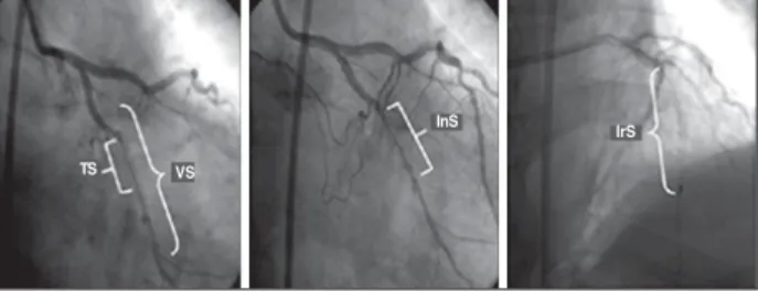
Still in regard to the minimum luminal diameter, the following parameters were assessed: 1) in-stent minimum luminal diameter, comprising the target segment and the distal and proximal margins of the lesion; 2) intrasegmentary minimum luminal diameter, compri-sing the injured segment located between the proximal and distal marks of the balloon. Other parameters assessed were the irradiated segment limited by the proximal and distal marks of the radiation catheter, and the segment of the vessel limited by 2 anatomic references (branches), in which the target segment, the injured segment, and the irradiated segment were included. This classifi-cation has been frequently used in other clinical studies 17 (fig. 1). Intracoronary ultrasound was routinely performed before the intervention after balloon catheter angioplasty, after brachytherapy, and in late follow-up. The studies were performed with the CVIS system (Boston Scientific Corp., Maple Grove, MN, USA) incor-porating a 30-MHz transducer with a rotation of 1800 rpm. For obtaining the sequence of images, the transducer of the ultrasound was pulled by an automated device at a velocity of 0.5 mm/s. The 3-dimensional reconstruction of the image was processed with a computed system (IndecSystems, Mountain View, CA, USA).
The volumetric analyses were calculated based on the formula of Simpson: Volume =Σ total area of the vessel, identified by the
external elastic membrane, stent, plaque (external elastic mem-brane – stent), neointimal hyperplasia (NIH), and lumen. To assess the volumetric changes in each element of the vessel at 6 months, the value of each measure was calculated as delta (∆volume),
resulting from the difference between the volumetric values ob-tained in late follow-up and after the intervention. Similarly, the
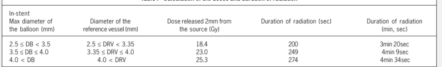
Table I - Calculation of the doses and duration of radiation
In-stent
Max diameter of Diameter of the Dose released 2mm from Duration of radiation (sec) Duration of radiation the balloon (mm) reference vessel (mm) the source (Gy) (min, sec)
2.5 ≤ DB < 3.5 2.5 ≤ DRV < 3.35 18.4 200 3min 20sec 3.5 ≤ DB ≤ 4.0 3.35 ≤ DRV ≤ 4.0 23.0 249 4min 9sec
4.0 < DB 4.0 < DRV 25.3 274 4min 34sec
6 5
percentage of volumetric obstruction was calculated [% of obs-truction = (NIH volume/stent volume) x 100] for the 3 periods assessed (pre-, postintervention, and at 6 months) 17,18. On intra-coronary ultrasound, aneurysm formation was defined as a 50% increase in the maximum luminal area in the place treated com-pared with the luminal area in the normal segment of reference16. Incomplete position of the stent was defined as the presence of 1 or more rods of the stent clearly separated from the wall of the vessel, with evident blood flow behind the rod on ultrasonography 19,20.
The occurrence of a defect in the target vessel (DTV) [death, acute myocardial infarction, restenosis according to the binary criterion (lesion > 50%), and revascularization of the target vessel] and the percentage of volumetric obstruction 180 days after bra-chytherapy were assessed.
The statistical analysis was performed using SPSS software, version 8.0 (SPSS Inc, Chicago, Illinois, USA). The continuous variables were expressed as means and standard deviation, while the categorical variables were presented as absolute frequencies and percentages. The categorical variables were compared using the chi-square test with continuity correction, or the Fisher exact test, when appropriate. The continuous variables were compared using the Student t test with correction using the Welch test, when necessary. Measures without a normal distribution were analyzed using the nonparametric Mann-Whitney test. The signi-ficance level adopted was P ≤ 0.05. The correlations between the changes in continuous variables were performed with the Pearson correlation test.
Results
The clinical and angiographic characteristics of the 30 patients studied are shown in table II. Ten (33%) patients had diabetes mellitus, 44% had stable angina, 4 (13%) had already undergone more than 1 intervention for the treatment of in-stent restenosis, and the mean time since the last intervention was 7.3±2.3 months. Complex lesions predominated as follows: 60% were diffuse, 17% were proliferative, 17% were focal, and 6% were occlusions 3.
Balloon catheter angioplasty was performed in all in-stent lesions. In 1 of the procedures, rotational atherectomy was per-formed prior to balloon dilation. In our study, no case of residual stenosis > 50% was observed immediately after the procedure. One type A dissection (nonflow-limiting) was evidenced, but required no additional treatment. In regard to brachytherapy, it was used in 100% of the patients, with no electrocardiographic alterations suggestive of myocardial ischemia during the procedure. A 40-mm extension source was used, with a mean radiation dose of 20.7±2.3 Gy applied for 3.8±2.1 minutes.
The extensions of the target segment and the injured segment were 18.6±4.1 mm and 23.3±3.8 mm, respectively. The in-stent minimum luminal diameter and the intrasegmentary diameter sig-nificantly increased after the intervention (both P < 0.0001), resulting in acute luminal gains of 0.91±0.41 mm and 0.85±0.40 mm, respec-tively (tab. III). At 6 months, angiographic follow-up was performed in all cases. The in-stent minimum luminal diameter decreased non-significantly (1.98±0.30 mm to 1.84±0.39 mm at 6 months; P = 0.13), associated with a late loss of 0.14±0.18 mm. The in-trasegmentary minimum luminal diameter was significantly smaller than the in-stent diameter at 6 months of treatment (1.55±0.40 mm versus 1.84±0.39 mm; P=0.008), associated with a greater late intrasegmentary loss (0.40±0.29 mm versus 0.14±0.18 mm; P=0.0001). Five cases of angiographic restenosis (17%) were obser-ved in late follow-up. One patient had restenosis in the distal extremity of the stent; another had 2 restenotic lesions (in-stent and in its distal extremity); another third patient had restenosis in the body of the endoprosthesis. Two cases had stenotic lesions in the proximal extremity of the irradiated segment.
TS VS
InS
IrS
Fig. 1 - TS (target segment): distal and proximal margins of the target lesion; InS (injured segment): between the proximal and distal marks of the balloon; VS (vascular segment): between anatomic references (branches); IrS (irradiated segment): between the proximal and distal marks of the radiation catheter.
Table II - Clinical and angiographic characteristics
Clinical characteristics n=30 Angiographic characteristics n=30
Age (years) 56.4 ±10.1 Target vessel
Male sex 18(60) AD 14(47)
Hypertension 16(53) Cx 10(33)
Dyslipidemia 16(53) Cx 4(14)
Diabetes mellitus 10(33) LCA 2(6)
Smoking 15(50) Class. of the lesion (ACC/AHA)
Asymptomatic 17(56) A/B1/B2/C 1(3)/9(30)/17(57)/3(10)
CCS 1 2(6) Extension of the lesion (mm) 18.66 ± 4.15
CCS 2 11(38) Extension of the stent (mm) 21.25 ± 3.26
≥ 2 previous ISR 4(13) Diameter of the balloon (mm) 3.18 ± 0.30 Time from the last intervention (months) 7.3 ± 2.3 Extension of the balloon (mm) 20.08 ± 3.80 Max inflation (Atm) 14.00± 4.20
Balloon/artery ratio 1.180± 0.40
6 6
Intracoronary Brachytherapy. Treatment of In-stent Restenosis with the Beta-Cath System. Initial Experience in Latin America
Intracoronary ultrasound was performed in 87% (26/30) of the patients immediately after the intervention and at 180 days of late follow-up. At 6 months, the neointimal tissue volume increased slightly (∆NIH = +6.75±14.3 mm3; P = 0.19), and a nonsigni-ficant reduction in the volume of the in-stent lumen was observed (∆lumen = -10.38±15.7 mm3; P = 0.18) (tab. IV). A quantitative increase both in the volume of the vessel and in the plaque was observed at 6 months, without reaching statistical significance. The percentage of volumetric obstruction was reduced from 61.1±8.1% to 26.8±7.2% after the intervention (P < 0.0001), but increased to 30.5±8.0% with 6 months of clinical follow-up (∆% obstruction = +4.7±7.5%; P = 0.09). Neither aneurysms, nor late incomplete apposition of the stents was observed.
At 6 months, the volume of the vessel showed a positive correlation with the changes in the volume of plaque (r = 0.963; P = 0.01).
Neither major cardiac events, nor cases of subacute thrombo-sis, occurred during the hospitalization period (48.8±6.2 h). Of the 30 cases analyzed on late clinical follow-up, 1 (3%) had late thrombosis (46 days after intervention), which evolved with non-Q-wave acute myocardial infarction, and 5 had revascularization of the target vessel (17%), of which 60% (3/5 cases) were perfor-med through a percutaneous route. The total percentage of defect in the target vessel in this series was 20%, resulting in an event-free survival of 80% at 6 months of clinical evolution (tab. V).
Discussion
In our study, the use of intracoronary brachytherapy with the Beta-Cath system suggests that it is a technically safe and feasible procedure, with favorable results in the management of in-stent restenosis 21,22 at 6 months of clinical follow-up (fig. 2).
Diffuse and proliferative restenoses, which represented 77% of our cases, have the worst prognosis after percutaneous mana-gement. In the study by Mehran et al 3, those cases had a new relapse in 50-80% of the cases after balloon catheter angioplasty, in contrast with our results, in which the relapse rate 6 months after brachytherapy was 17%. This suggests that brachytherapy is better than the conventional treatment with the balloon, con-firming the results of previous investigations 21-23.
The pre-established radiation dose was totally delivered in all lesions. All patients received antiplatelet therapy (acetylsalicylic acid indefinitely) and thienopyridinic derivatives for at least 4 months; no major cardiac events occurred during the first month. One (3%) late thrombotic occlusion was observed, manifested as non-Q-wave acute myocardial infarction, a percentage lower than that reported in the WRIST, Long-WRIST, SVG-WRIST, GAMMA-1, and BETA-WRIST studies (9.1%, 5 months after the procedure) 24 and similar to that reported in the WRIST-PLUS 26 and INHIBIT studies 26. Only 1 nonflow-limiting dissection was documented at the end of the procedure; although it did not require additional treatment, it later was associated with 1 of the restenoses observed (distal extremity of the stent). The impact of nonflow-limiting dissections on the prognosis of patients undergoing brachytherapy is still unknown. In 2 series of 16 patients each, the presence of acute dissection after balloon angioplasty and beta radiation persisted in approximately 50% of the patients after 6 months of clinical follow-up 27,28, and it showed no association with the recurrence of angina or any other clinical event in particular 28.
In regard to the clinical events here documented, our results were similar to those found in previous studies using intracoronary beta radiation, in which the incidence of major cardiac events ranged from 18% to 33% as follows: START (18%) 29, INHIBIT (22%) 26, and BETA-WRIST (33%) 30. Similarly, the restenosis rate in these studies ranged from 15% to 29% 22,26,30.
Comparing the late loss in the segments angiographically de-fined, the intrasegmentary minimum luminal diameter showed a statistically greater reduction than the in-stent minimum luminal diameter, due to the lesion caused by the balloon in the segments adjacent to the stent. This phenomenon, reported as “geographical loss,” observed in 13% of new cases, is mainly characterized by inadequate radiation in the injured segment 31. Theoretically, this condition occurs when the extremities of the irradiated segment are damaged, in which, by definition, the dose emitted is very
Table IV - Results obtained on intracoronary ultrasound
Volume Pre-intervention Post-intervention 6 months ∆ P
(mm3) (n=26) (n=26) (n=26) (6 months - Post) (6 months vs Post)
Stent 141.17 ± 36.20 155.01 ± 37.95 151.36 ± 38.43 -3.63 ± 7.75 0.73 Lumen 56.00 ± 22.86 114.62 ± 30.03 104.24 ± 25.17 -10.38 ± 15.73 0.18 NIH 85.17 ± 19.21 40.38 ± 16.27 47.13 ± 20.01 6.75 ± 14.27 0.19 EEM 272.31 ± 64.48 285.24 ± 58.20 306.64 ± 66.62 21.41 ± 27.75 0.22 Plaque 131.13 ± 48.20 130.23 ± 46.74 155.28 ± 49.48 25.04 ± 24.61 0.07
The values are expressed as mean ± standard deviation or n (%). ∆: Delta; NIH: neointimal hyperplasia; EEM: external elastic membrane, plaque; EEM: Stent
Table III - Results obtained on quantitative coronary angiography
Angiographic Beta radiation P vs. calculations after intervention
Before intervention (n=30)
Reference diameter (mm) 2.66 ± 0.33 0.53 In-stent MLD (mm) 1.06 ± 0.30 <0.0001 Intrasegmentary MLD (mm) 1.10 ± 0.29 <0.0001 %DS 69.60± 11.40 <0.0001
After intervention (n=30) Reference diameter (mm) 2.72 ± 0.37 In-stent MLD (mm) 1.98 ± 0.30 Intrasegmentary MLD (mm) 1.95 ± 0.30
%DS 24.40± 7.90
Late follow-up (6 months) (n=30)
Reference diameter (mm) 2.69 ± 0.35 0.80 In-stent MLD (mm) 1.84 ± 0.39 0.13 Intrasegmentary MLD (mm) 1.55 ± 0.40 <0.0001
%DS 31.90± 17.20 0.04
Binary restenosis (in-stent) 3/30 (10) Binary restenosis 5/30 (17) (intrasegmentary)
6 7
In-stent lesionPRE
POST
Source
6 MONTHS
Fig. 2 - Diffuse in-stent lesion in the right coronary artery treated with intracoronary beta radiation. The 2 arrows show the position of the stent.
Table V - Clinical events at 6 months
Major clinical events Patients (n=30)
Death 0/30 (0)
Q-wave AMI 1/30 (3)
Non-Q-wave AMI 0/30 (0)
Late thrombosis 1/30 (3)
RTV 5/30 (17)
RTL 3/30 (10)
BCA 3/30 (10)
DTV 6/30 (20)
MCE - free survival 24/30 (80)
The values are in number of patients (%); AMI - acute myocardial in-farction; RTV - revascularization of the target vessel; RTL - revascularization of the target lesion; BCA - balloon catheter angioplasty; DTV - defect in the target vessel (Death, AMI, restenosis, RTV); MCE - major clinical event
low. In a retrospective analysis 31 of 50 consecutive patients treated with beta radiation after balloon catheter angioplasty or stent, the incidence of that phenomenon was 32%.
The volumetric analysis on intracoronary ultrasound showed that the growth of neointimal tissue was mild after 6 months of clinical follow-up, a result similar to that of other studies 32. The growth of the vessel (+21.4±27.8 mm3) observed after 180 days of treatment suggests that intracoronary beta radiation induced a positive remodeling of the arterial wall, characterized by an “accommodative” process of the plaque (correlation of the increase in the ∆ external elastic membrane vs D plaque, r=0.963, P < 0.01), preserving the volume of the in-stent lumen. Experimental data have suggested that radiation has an effect on the remodeling of the vessel, due to the change in cell response in the adventitia layer 33,34. Positive remodeling, accommodating the neointimal growth after 6 months,
has been shown on ultrasonographic analyses 35, considering that other studies did not report changes in the total area of the vessel or of the plaque behind the stent 27.
Compared with gamma radiation, beta radiation has a smaller depth of penetration, minimizing the exposure to radiation for patients and operators, requiring, therefore, less radioprotection. However, the rapid decrease in the dose of beta radiation within 2-5mm of depth is associated with a less homogeneous release of the dose 32. In addition, the “shield effect” produced by the rods of the stent could be associated with an attenuation in the radiation dose through its rods greater than 15% 36. Comparing the results on intracoronary ultrasound in 2 large studies 13,15, beta radiation showed a greater benefit than gamma radiation did in preventing neointimal hyperplasia inside the stent 37,38.
Finally, it is worth emphasizing that the Latin-American par-ticipation in the development of this innovative therapeutic option in managing stenotic lesions has been paramount. The first clinical experience with radiation in human coronary arteries was reported by Condado et al 39 in Venezuela. Those authors introduced an iridium 192 guide inside a catheter, which was not centralized in the lumen of a vessel, using a manually deployed device. They, therefore, showed that the administration of radiation inside the coronary artery was feasible and safe, although followed by high restenosis rates in that initial experience.
Our study consisted of an initial series of consecutive cases of in-stent restenosis treated at our facility, with a late clinical follow-up limited to 6 months. Due to the relatively reduced number of patients assessed and to the design of the study (nonrandomized, with no control group, and with the exclusion of more complex cases), the results here reported should not be extrapolated to all cases of this affliction.
Although the clinical events observed in the late follow-up of our patients did not differ from those reported in the large rando-mized studies, one cannot say they are clinically equivalent, because our sample size does not allow this conclusion. Independently of the already cited observations, this study provided a satisfactory initial clinical experience with the use of this technique. The patients studied continue to be followed up, and new cases have been progressively incorporated.
6 8
Intracoronary Brachytherapy. Treatment of In-stent Restenosis with the Beta-Cath System. Initial Experience in Latin America
1. Kastrati A, Schoming A, Elezi S et al. Predictive factors of restenosis after coronary stent placement. J Am Coll Cardiol 1997; 30: 1428-36.
2. Van Belle E, Baurers C, Hurbert E et al. Restenosis rates in diabetic patients: a comparison of coronary stenting and balloon angioplasty in native coronary ves-sels. Circulation 1997; 96: 1454-60.
3. Merhan R, Dangas D, Abizaid AS et al. Angiographic patterns of in-stent resteno-sis: classification and implications for long-term outcome. Circulation 1999; 100: 1872-8.
4. Dussaillant GR, Mintz GS, Pichard AD et al. Small stent size and intimal hyperpla-sia contribute to restenosis: a volumetric intravascular ultrasound analysis. J Am Coll Cardiol 1995; 26: 720-4.
5. Hoffman R, Mintz GS, Dussaillant GR et al. Patterns and mechanisms of in-stent restenosis: a serial intravascular ultrasound study. Circulation 1996; 94: 1247-54. 6. Baim DS, Levine MJ, Leon MB, Levine S, Ellis SG, Schatz RA. Management of res-tenosis within the Palmaz-Schatz coronary stent (the US multicenter experience). Am J Cardiol 1993; 71: 364-6.
7. Merhan R, Mintz GS, Satler LF et al. Treatment of in-stent restenosis with exci-mer laser coronary angioplasty: mechanisms and results compared to PTCA alo-ne. Circulation 1997; 96: 2183-9.
8. Sharma SK, Duvvuri S, Dangas G et al. Rotational atherectomy for in-stent reste-nosis: acute and long-term results of the first 100 cases. J Am Coll Cardiol 1998; 32: 1358-65.
9. Teirstein PS, Massullo V, Jani S et al. Catheter-based radiotherapy to inhibit res-tenosis after coronary stenting. N Engl J Med 1997; 336: 1697-703.
10. Casterella PJ, Teirstein PS. Prevention of coronary restenosis. Cardiol Rev 1999; 7: 19-31.
11. Hall EJ. DNA strand breaks and chromosomal aberrations. In: Hall EJ. Radiobiology for the Radiologist. Philadelphia, Penn: J.B. Lippincott Company; 1994: 15-73. 12. Rubin P, Soni A, Williams JP. The molecular and cellular biologic basis for the
radia-tion treatment of benign proliferative diseases. Semin Radiat Oncol 1999; 9: 203-14. 13. Waksman R, White LR, Chan RC et al. Intracoronary g-radiation therapy after an-gioplasty inhibits recurrence in patients with in-stent restenosis. Circulation 2000; 101: 2165-71.
14. Leon MB, Teirstein PS, Moses JW et al. Localized intracoronary gamma radiation therapy to inhibit the recurrence of restenosis after stenting. N Engl J Med 2001; 344: 250-6.
15. Waksman R, Bhargava B, White L et al. Intracoronary beta-radiation therapy inhi-bits recurrence of in-stent restenosis. Circulation 2000; 101: 1895-8. 16. Akiko Maehara, Gary S Mintz, Javed M Ahmed et al. An Intravascular Ultrasound
Clas-sification of Angiographic Coronary Artery Aneurysms. Am J Cardiol 2001; 88: 365-70. 17. Sabaté M, Costa MA, Kozuma K et al. Methodological and clinical implications of the relocation of the minimal luminal diameter after intracoronary radiation the-rapy. J Am Coll Cardiol 2000; 36: 1536-41.
18. Kearney PP, Ramo MP, Shaw TR et al. Analysis of reproducibly of reference lumen quantification with intravascular ultrasound in stented coronary arteries. Cathet Cardiovasc Diagn 1997; 40: 1-7.
19. Kozuma K, Costa MA, Sabaté M et al. Three-dimensional intravascular ultra-sound assessment of noninjured edges of b-irradiated coronary segments. Circu-lation 2000; 102: 1484-9.
20. Honda Y, Grube E, de la Fuente L et al. Novel drug-delivery stent: intravascular ul-trasound observations from the first human experience with the QP2-eluting po-lymer stent system. Circulation 2001; 104: 380-3.
References
21. Kobayashi Y, Honda Y, Christie LG et al. Long-term vessel response to a self-expan-ding coronary stent: a serial volumetric intravascular ultrasound analysis from the ASSURE trial. J Am Coll Cardiol 2001; 1329-34.
22. Regar E, Kozuma K, Sianos G et al. Routine intracoronary beta-irradiation: Acute and one year outcome in patients at high risk for recurrence of stenosis. Eur Heart J 2002; 23: 1038-44.
23. King SB, 3rd, Williams DO, Chougule P et al. Endovascular beta-radiation to reduce
restenosis after coronary balloon angioplasty: results of the beta energy restenosis trial (BERT). Circulation 1998; 97: 2025-30.
24. Urban P, Serruys PW, Baumgart D et al. Clinical application of intracoronary beta brachytherapy using sr/Y90 source trains the European surveillance registry with the novoste beta-cath system. Eur Heart J 2001; 22: 4.
25. Waksman R, Bhargava B, Mintz GS et al. Late total occlusion after intracoronary bra-chytherapy for patients with in-stent restenosis. J Am Coll Cardiol 2000; 36: 65-8. 26. Waksman R, Ajani AE, White RL et al. Prolonged antiplatelet therapy to prevent
late thrombosis after intracoronary g-radiation in patients whit in-stent restenosis: Washington Radiation for In-Stent Restenosis Trial Plus 6 Months of Clopidogrel (WRIST PLUS). Circulation 2001; 103: 2332-5.
27. Waksman R, Raizner AE, Yeung AC et al. Localized intracoronary b radiation the-rapy to inhibit recurrence of in-stent restenosis. Lancet 2002; 359: 551-7. 28. Meerkin D, Tardif JC, Crocker IR et al. Effects of intacoronary beta-radiation
the-rapy after coronary angioplasty: an intravascular ultrasound study. Circulation 1999; 99: 1660-5.
29. Kay IP, Sabaté M, Van Langenhove G et al. Outcome from balloon induced coro-nary artery dissection after intracorocoro-nary beta radiation. Heart 2000; 83: 332-7. 30. Ajani A, Kim H-S, Waksman R. Clinical trials of vascular brachytherapy for in-stent
restenosis: update. Cardiovasc Radiat Med 2001; 2: 107-13.
31. Salame MY, Verheye S, Crocker IR et al. Intracoronary radiation therapy: Review Article. Eur Heart J 2001; 22: 629-47.
32. Sabaté M, Costa MA, Kozuma K et al. Geographic miss: a cause of treatment fai-lure in radio-oncology applied to intracoronary radiation therapy. Circulation 2000; 101: 2467-71.
33. Hong M-K, Park S-W, Moon D-H et al. Intravascular ultrasound analysis of beta radiation therapy for diffuse in-stent restenosis to inhibit intimal hyperplasia. Cathet Cardiovasc Intervent 2001; 54: 169-73.
34. Waskman R, Rodriguez JC, Robinson KA et al. Effect of intravascular irradiation on cell proliferation, apoptosis, and vascular remodeling after balloon overstretch injury of porcine coronary arteries. Circulation 1997; 96: 1944-52.
35. Wilcox JN, Waksman R, King SB et al. The role of adventicia in the arterial respon-se to angioplasty: the effect of intravascular radiation. Int J Radiat Oncol Biol Phys 1996; 36: 789-96.
36. Sabaté M, Serruys PW, Van der Giessen WJ et al. Geometric vascular remodeling after balloon angioplasty and beta-radiation therapy: A three-dimensional intra-vascular ultrasound study. Circulation 1999; 100: 1182-8.
37. Amols HI, Trichter F, Weinberger J. Intracoronary radiation for prevention of res-tenosis: dose perturbations caused by stents. Circulation 1998; 98: 2024-9. 38. Bhargava B, Mintz GS, Mehran R et al. Serial volumetric intravascular ultrasound
analysis of the efficacy of beta irradiation in preventing recurrent in-stent resteno-sis. Am J Cardiol 2000; 85: 651-3.