Great Arteries Contribution in Orthostasis Cardiovascular
Adaptation
Jorge Elias Neto
Departamento de Ciências Fisiológicas - Universidade Federal do Espírito Santo – UFES - Vitória, ES - Brazil
M a i l i n g A d d r e s s : J o r g e E l i a s N e t o • R u a A l a ô r d e Q u e i r o z , 1 1 5 / 1 0 1 - 2 9 0 5 0 - 0 0 0 – V i t ó r i a , E S - B r a z i l E-mail: [email protected], [email protected] Received on 05/14/05 • Accepted on 12/08/05
The capacity of adopting an orthostatic posture is, undoubtedly, a milestone in the evolution of the human species and has motivated extensive research in the physiological and anthropological areas. The physiological mechanisms involved in adopting and maintaining an orthostatic posture and its complications have been investigated through the centuries. The anatomists do not consider the postural adaptation of the human being a peculiar transformation that occurred to mankind, but the culmination of a series of evolutionary phases that might have started in the body of the primate ancestors1.
It is believed that the human cardiovascular system is adapted to maintain cerebral perfusion during the orthostatic posture. Although the gravitational force creates a gradient of pressure at the level of the circulatory system, the human being is capable of maintaining an upright posture because the gravitational pressure is partially neutralized by mechanisms that prevent the accumulation of fl uids in the lower limbs2.
Despite that, syncope, defi ned as a transient loss of consciousness and postural tonus due to cerebral hypoperfusion, has a considerable medical, social and economical impact. This clinical syndrome can be the consequence of not only refl ex mechanisms3, but also a clinical manifestation of a broad range of distinct clinical conditions4.
A perspective of the cardiovascular adaptation to orthostatism is presented herein, in an attempt to assess evolutionary and physiological issues regarding the maintenance of circulatory homeostasis during orthostasis, stressing the role of the great arteries.
O
RTHOSTATIC POSTURE AND GRAVITATIONAL STRESSYears of experiments carried out in quadrupeds or models constructed in the horizontal plane have helped researchers of the cardiovascular system to develop a type of “horizontal” idea about the cardiovascular system, completely neglecting the effects of gravity because the “vertical” gravity force exerts a minimal effect on circulation in the horizontal plane. However,
the human cardiovascular system works in the orthostatic posture two-thirds of the time, in which the classical experiments conducted by the Guyton group, for the study of cardiovascular hemodynamics, present large limitations5.
The change from a quadrupedal posture to a bipedal one created diffi culties that can be interpreted through several signs, which are representative of an inadequate adaptation to orthostatic stress6. Bipedism presupposes a complete reorganization of the orthodynamics of the human body when compared to other mammals. This implicates in a different reorganization of the energetic supply for the body (oxygen and nutrients). As oxygen and the nutrients are carried by the blood, a complete reorganization and rearrangement of the blood supply between the different parts of the body is expected when there is a change from the quadrupedal posture to the bipedal one7. Thus, the hemodynamics must also be different.
The control of blood fl ow distribution in man is more complex due to the fact that the human brain has larger proportions compared to the other species. The brain represents 2% of the total body weight in man, demands 15% of the cardiac output at rest and requires the maintenance of a stable output regardless of posture or locomotion8. The change from a supine posture to the orthostatic one results in a displacement of blood at the thorax level to the subdiaphragmatic venous system, a phenomenon usually called venous pooling9. If compensatory adjustments do not occur promptly after the change in posture, the arterial pressure decreases, and the individual can present from milder symptoms related to orthostatic intolerance to the sudden loss of consciousness10.
variable capable of affecting circulatory homeostasis. It is gravity (+Gz), acting at a mean velocity of 9.8 m/s2, the variable responsible for the blood redistribution present through the adoption of orthostasis.
The hydrostatic opposition to the venous return, the third-space loss of fl uid, venous return decrease and cardiac output reduction, all of which are hemodynamic alterations secondary to the gravitational effect, act as multiple stimuli, generating a variety of compensatory mechanisms16,17. Additionally, the gravitational stress can be aggravated by the level of physical activity, environmental temperature, and drug use and by several physiopathological processes, all capable of augmenting the disparity between the vascular volumetric capacity and blood fl ow volume 18-20.
M
AN AND MICROGRAVITYIf gravity did not exist, man would not need such a complex cardiovascular system, particularly the presence of protection mechanisms to maintain a circulatory homeostasis during orthostatism. In general, the adaptation to gravity is, in part, acquired, and needs to be continuously trained.
For astronauts, the possibility of absence of weight due to microgravity causes major alterations in the cardiovascular system. The new pattern of blood distribution is interpreted as a volemia increase and compensatory mechanisms are promptly initiated (increase of natriuretic hormone secretion, inhibition of the rennin-angiotensin-aldosterone system), leading to a plasmatic volume decrease. A decrease in the sensitivity of the barorefl ex and vestibulosympathetic refl ex, as well as alterations in the autonomic nervous system, increase of vessel compliance in the lower limbs and disorders of arteriolar vasoconstriction are also observed21-23.
All these alterations can be considered as a cardio-vascular “deconditioning” capable of generating intolerance to orthostatism when astronauts return to the Earth’s environment. Similar cardiovascular system alterations have been observed after long periods of bed rest (a situation that simulates microgravity), indicating that the orthostatic posture has a fundamental role in gravitational stress. The studies that seek the functional “understanding” of the organism in space are extended to high-prevalence clinical situations in the Earth’s environment, such as arterial hypertension, orthostatic hypotension, and fi nally, syncope.
T
HE FORCE OF GRAVITY ANDGRAVITATIONAL STRESS
– G
ENERALA
SPECTSThe effect of the Earth’s gravitational fi eld on the human cardiovascular system is similar to the effect of an electric fi eld in a conduit. Similarly to an electric fi eld
that moves free electrons along a conduit, the gravitational fi eld moves blood (free particles) along the cardiovascular system. The effect of the gravitational fi eld is measured by the gradient of the gravitational potential (Ugr), which is equal to the work performed by the fi eld to move the mass of blood unit through the system towards Earth (Ugr=gz x r). The direction of the Earth’s gravitational fi eld close to the surface is vertical, and the distance r is equal to the system height: Ugr=gz x h. That explains why the effect of gravity is signifi cant only in the orthostatic posture. In the horizontal posture, the height of the human body, h, is too little, making Ugr negligible, but it increases several fold during orthostasis24.
The gravitational stress can be defi ned as follows: gravity causes a signifi cant displacement of blood to the infradiaphragmatic territory when the cardiovascular system is in the orthostatic posture. For the adequate maintenance of circulation to the supradiaphragmatic territory during orthostasis, the cardiovascular system must perform an additional work to return blood to the heart and neutralize the effect of gravity. The sum of this work (W), equal to the product of the gradient of the gravitational potential and the displaced mass of blood (M) represents the gravitational stress on the cardiovascular system: GS = W = Ugr x M. It is unnecessary to mention that this additional overload on the cardiovascular system during orthostasis remains unknown in the traditional studies on the human circulatory hemodynamics. Nonetheless, the gravitational stress can increase with no gravity force augmentation. The natural action of the gravitational stress is to generate a redistribution of blood to the lower limbs in the cardiovascular system.
Contrary to water in a glass, the blood contained in an elastic and complex vascular system is redistributed slowly, and the change of blood does not happen instantly, but increases proportionally to the time the individual remains in orthostasis. Thus, based on the equation established for the gravitational stress, the mass of blood displaced by infl uence of gravity, M, is a function of time, GS=Ugr x M (t) = g x h x M (t). This means that gravitational stress can increase not only through the gravity force increase, but also depending on the duration of the orthostatic posture period24. Hence, remaining an excessive period of time in orthostasis requires a more advanced stage of anti-gravitational response with higher sympathetic hyperactivity, hypervolemia and arterial pressure increment, similarly to what would occur with an increment of the gravitation force.
D
YNAMIC CARDIOVASCULAR RESPONSE TO THEORTHOSTATIC POSTUREreference in which the pressure of the venous blood column is not altered by the postural rearrangement, located at the level of the right atrium in supine decubitus, going to the infradiaphragmatic aortic territory in orthostasis25. However, due to higher vessel compliance, the main effect is the redistribution of the venous volume, which thus interferes with the global blood pressure. The accumulation of blood in the venous territory starts immediately during orthostasis and is completed in 3 to 5 minutes26. The venous volume of the lower members increases in approximately 500 to 700 ml27. Around 200 to 300 ml of blood is transferred to the veins below the abdomen. This translocation of blood volume is mainly derived from the intrathoracic compartment and results in the decrease of cardiac fi lling pressure and systolic volume28-31.
In addition to the blood transference from the thoracic compartment, the central blood volume is affected by the transcapillary fi ltration of fl uid into the interstitial space in some body parts, in response to the high capillary pressure associated to the low contraposition of the interstitial pressure. Additionally, the continuous fi ltration into the interstitial space further reduces the circulating volume32. The loss of transcapillary fl uid is stabilized after approximately 20 to 30 minutes, with a plasmatic volume decrease of around 15%, although this process remains present with the maintenance of orthostasis33. Therefore, the distribution of blood volume is carried out through a complex system, by the intrinsic characteristics of the venous system, to the pumping effect of the skeletal musculature of the lower limbs (antigravity musculature) and, as mentioned previously, to the displacement of the hydrostatic indifferent point12.
The capacity to maintain the cardiac output and the mean arterial pressure during the orthostatic “challenge” can be infl uenced by the behavior of the venous return. Amongst other factors, the venous return decrease can be the result of a combination of the decrease of the total blood volume with the accumulation of blood in the lower limbs, which subsequently may cause a subnormal ventricular fi lling pressure and deviate the curve of Frank-Starling to a segment in which the capacity of compensating for the orthostatic hypotension is limited14,34,35. Nonetheless, the human cardiovascular system has such a powerful “built-in” defense mechanism against the gravitational stress that it can completely neutralize the gravity-induced blood redistribution imposed by the orthostatic posture. The mechanism is the peripheral pumping.
The lower limbs are equipped with a specifi c system of blood pumping, the calf musculature, which is many times called “the peripheral venous heart”. The deep veins of the calf, which are found inside a compartmented musculature, present valves that, similar to the cardiac valves, prevent blood refl ux and are able to resist to pressures of several hundreds of mmHg. The compression of the venous segment during muscular contraction
increases the pressure inside the muscular compartment, which leads to the closing of the valves and causes an ascendant blood fl ow. Through the regular contraction of the calf muscles during the orthostasis, the venous blood is pumped out of the lower limbs against gravity, reversing the gravitational action in blood distribution. Thus, the incapacity of the heart in “drawing” blood from the venous segment of the cardiovascular circuit is compensated by the presence of the additional pump in the venous system. This system, which consists of the calves, generates an effective pumping action that is not dependent on the fi lling pressure, helping to successfully maintain circulation during orthostasis.
Therefore, the fi nal equation of gravitational stress can be expressed as follows: GS = g x h x M (t) / Rp x Hper, where Rp is the peripheral resistance and Hper is the work of the “peripheral venous heart”24 .
Although initial studies have demonstrated a decrease in the brain blood fl ow during the orthostatic posture36, it is currently known that cerebral perfusion in humans is maintained by a cerebrovascular self-regulation, which allows an effective cerebral functioning and a satisfactory distribution of regional fl ow within a broad variation of perfusion pressure37. In comparison to what occurs with quadrupeds, human beings present a more constant distribution of total blood fl ow in the several body segments during the orthostatic posture, including for the great arteries, suggesting a better functional adaptability to gravitational stress8.
The redistribution of blood volume, fl ow and pres-sure, caused by the upright posture, can endanger pre-load, arterial pressure and organic perfusion, and it is compensated by a compensatory sequence of interconnected neurohumoral mechanisms that are opposed to the continuous circulatory caudal direction imposed by the hydrostatic pressure15. As early as 1904, Erlanger and Hooker observed, using the passive inclination test (IT), that the acceleration of the cardiac frequency and the decrease of the pulse pressure occurred in the upright posture. They considered this pattern of behavior similar to that observed during a hemorrhagic event and concluded it was the normal response of the human being to postural stress1. Turner emphasized that the circulatory adjustment would be specifi c for each individual, according to his own personal pattern, and that some patterns of individual response would be higher than others1.
present a complex inhibitory interaction between the cardiopulmonary and the sinoaortic barorefl exes, which causes the reduction in the cardiopulmonary afferent input to increase the gain of sinoaortic barorefl ex39. Finally, the decrease in the activation of the cardiopulmonary receptors produces a peripheral vasoconstriction for normotension maintenance, due to the decrease of the central venous pressure and cardiac output40.
The primary objective of the arterial barorefl ex is to maintain the arterial pressure close to a point of reference for a relatively short period of time. The rapid regulation of the arterial baroreceptors, regardless of the new pressure level, is due to the fact that this refl ex acts as a “buffer” system, which is effective on the arterial pressure fl uctuations that occur in everyday life. The arterial baroreceptors also play a smaller role in the long-term control of the arterial pressure level41.
These aorticocarotid baroreceptors tonically inhibit the vasomotor centers of the brainstem. A decrease in the blood pressure removes this inhibitory stimulus, resulting in an increase of the sympathetic tonus and decrease of the parasympathetic tonus42. From a functional point of view, the two components of the arterial barorefl ex (carotid and aortic) are not equivalent. It has been suggested that the aortic barorefl ex has a higher threshold and lower sensitivity than the carotid one38. The information of the baroreceptors is transmitted through several pathways that accompany the route of the ninth and tenth cranial pairs until the brainstem centers. These two nerves are the afferent pathway of the refl ex, whereas the efferent pathway consists of the vagus nerve and the sympathetic pathways that run at the level of the intermediolateral spinal cord6. The changes in the neural discharge can result in changes of vasomotor tonus, systolic volume and cardiac frequency.
The vestibular system also seems to contribute to the sympathetic activation through the recruiting of otolytic organs. This is achieved through the vestibulosympathetic reflex. This reflex is a feedback neural system that regulates the sympathetic neural discharge through nerve afferences from the vestibular system43.
During a prolonged orthostatic period, in addition to the baroreceptors and the vestibular system, delayed-action regulatory mechanisms based on additional refl exes of the humoral system that include the natriuretic hormone secretion, rennin and aldosterone, are also involved with the maintenance of arterial pressure15,27,44.
Regarding the hemodynamic parameters, orthostatism causes, in the individual chronically adapted to gravitational stress, a slight decrease in the systolic arterial pressure, in general lower than 20 mmHg. The diastolic arterial pressure remains stable or is slightly elevated, and the mean arterial pressure is not signifi cantly altered. The telediastolic pressure in the right ventricle diminishes, the cardiac output decreases around 20%, and the cardiac frequency and peripheral vascular resistance increase 10% to 30% of the initial value6.
When, however, the head-lower limb acceleration vector +Gz is chronically diminished, such as during long supine rest periods, human beings demonstrate a reduction in the orthostatic tolerance, associated to a compromised regulation of the arterial pressure. These observations include an increase in the compliance of the lower limbs, alteration of the cardiovagal refl ex response, contraction of the circulating blood volume and decrease of the systolic volume14.
On the other hand, Muenter et al observed that, individuals submitted to sleep restriction periods (approximately 4 hrs/night for 4 consecutive days), showed a better blood pressure regulation in an orthostatic situation45. The individuals presented an increase in systolic arterial pressure and a decrease in cardiac frequency during orthostatic stress, simulated through the use of sub-atmospheric pressures applied to the lower part of the body, called “lower body negative pressure” (LBNP), reinforcing the hypothesis that, in normal individuals, the continuous exposition to gravitational stress triggers a better circulatory adaptation. The same can be said of patients with neuromediated syncope, who, when submitted to daily orthostatic training through a technique called “tilt training”, presented remission of clinical symptoms, even when the drug therapy was ineffective47. Newman et al (1998) used IT to assess plane pilots, routinely exposed to high levels of gravity (+Gz) and normal individuals47. These authors observed that the pilots presented a signifi cant increase in the systolic arterial pressure, mean arterial pressure and pulse pressure during the IT with 75o, when compared to the control group (p< 0.05). No signifi cant alterations were observed between the two subgroups of this study regarding the response of cardiac frequency to orthostatic stress.
When analyzed at a systemic level, cardiac output, more than cardiac frequency, represents the fi nal organic refl ex response to the stimulation of the baroreceptors by orthostatic stress33. Based on this fact, Convertino14 analyzed cardiac output alterations that could occur in response to arterial pressure and cardiac frequency variation in individuals repeatedly exposed to high levels of gravitational acceleration. This author observed the importance of not only cardiac frequency analysis but also of the systolic volume, in the assessment of the effective gain of cardiac barorefl ex integrity in maintaining arterial pressure during the orthostatic hypotensive “challenge”. The better orthostatic performance observed in these individuals supports the hypothesis that a repeated exposition to gravitational acceleration is associated to changes in blood volume and autonomic function, in opposition to what occurs with bedridden individuals14.
to the postural variations. The analysis of the response of the blood volume and the carotid-cardiac barorefl ex in adapting to changes caused by the gravitational stimulation can have signifi cant importance, because the blood volume and the responsiveness of the carotid-cardiac reflex are two primary factors (in addition to height) when predicting the evolution for arterial hypotension and syncope48.
The comparison of the cardiocirculatory functional behavior when facing gravitational oscillations allows two conclusions: 1) the human being is capable of undergoing continuous adaptation to gravitational stress; 2) the mechanisms that allow us to tolerate the gravitational effect are highly malleable and trainable, or acquirable through training14. Therefore, it has been established that an autonomic nervous system, capable of maintaining a dynamic adaptive behavior that allows an adequate cardiac output in response to the blood volume oscillations caused by the orthostatic posture, is fundamental39,49. Thus, clinical conditions that interfere in the responsiveness of the barorefl ex system can somehow interfere in the circulatory homeostasis50-53.
F
ACTORS THAT INFLUENCE THEBAROREFLEX SENSITIVITYIt is known that alterations at activity and/or balance level of the several branches of the autonomic nervous system can be considered one of the main factors responsible for the development of essential chronic arterial hypertension. The hypertensive individuals present an increase in the variability of arterial pressure fl uctuation and a decreased sensitivity of the baroreceptors9,54. The hypertensive patient’s gender also seems to be an important determinant in barorefl ex sensitivity, as the autonomic function shows to be more compromised in the female sex54.
Hypertensive individuals are less responsive to induced sympathetic stimulation55,56. In fact, the analysis of the autonomic response of hypertensive individuals to postural change, when compared to that of normal individuals, shows that, although there is a basal increase of sympathetic activity during the orthostatic posture, the sympathetic response is signifi cantly decreased, suggesting an impairment of the mean arterial pressure control by the barorefl ex in these individuals9.
The refl ex hemodynamic responses to orthostatic stress are also attenuated by aging. When a young healthy individual rises from a sitting or supine posture, there is a variable change in the systolic arterial pressure of around 10 mmHg, an increase in the diastolic arterial pressure of 10 to 15 mmHg and an increase in the cardiac frequency of 25 to 30 bpm. This response occurs within approximately 30 seconds. In contrast, when an elderly healthy individual rises, a trend towards a decrease in the systolic arterial pressure (0 to 5 mmHg), as well as
the maintenance of the diastolic arterial pressure and the absence or attenuation of the cardiac frequency positive response (10 to 15 bpm) are observed. Around 6% to 30% of elderly individuals present orthostatic hypotension (decrease in the systolic arterial pressure
≥ 20 mmHg, or in the diastolic arterial pressure ≥ 10 mmHg), when standing upright58,59. Contrarily to what could be predicted, the majority of elderly individuals who present inadaptation to orthostatic stress do not have well-defi ned pathological alterations, such as central or peripheral degeneration of the autonomic pathways or adrenal failure. These individuals possibly represent an extreme condition of the distribution of age-related physiological changes to posture27.
The best-known biological change in arterial pressure control that occurs with age is an attenuation of the baroreceptor responsiveness, which is represented by the baroreceptor-heart refl ex3,60,61. This fi nding is commonly considered as indicative of an age-related primary abnormality in cardiovascular control, as the barorefl ex is the main factor that infl uences a rapid adaptive response to orthostasis53.
There are, however, many other age-related alterations in the cardiovascular function, which can contribute signifi cantly to an inadequate response to orthostasis, such as reduction in arterial27,62 and myocardial63 compliance, in the activation of the rennin-angiotensin-aldosterone system64, in renal sodium retention65, a defect in the brain self-regulation and the presence of systolic arterial hypertension27.
The location of arterial baroreceptors in the compliant great arteries means that changes in the natural compliance, which can be caused simply by aging, disease or physical activity, also have the potential of infl uencing the afferent activities of the baroreceptors and consequently affect the autonomic refl ex activity66.
The arterial compliance has a fundamental role in the cardiac energy demand, in maintaining the diastolic arterial pressure and the coronary blood flow66,67. Probably, the least evident is the infl uence of the vascular mechanical properties on the autonomic function by alterations in the afferent activity of the baroreceptors. Although it is difficult to investigate the extension to which these two phenomena are interconnected, Kingwell et al, analyzing arterial compliance and its possible infl uence on the barorefl ex in hypertensive individuals and athletes, observed not only a direct relation between arterial compliance and higher blood pressure due to refl ex activation in athletes, but also a baroreflex deficit in hypertensive individuals66. Nevertheless, basal assessments of vascular stiffness might not represent the mechanical stress that affects the great arteries with barosensory function during dynamic changes in pressure that characterize the barorefl ex
role68,69. Additionally, a compromised neuronal function
reduction of the barorefl ex gain with the decrease of the vascular mechanical function70.
Through the measurements of the integrated gain of the cardiovagal barorefl ex and its mechanical and neural components, analyzed dynamically (during parenteral administration of sodium nitroprussiate and phenylephrine), Hunt et al observed that the integrated gain in sedentary male elderly individuals was lower than half of that found in young, equally sedentary, individuals70. This fi nding was derived from a decrease in the mechanical as well as neural transduction. Another signifi cant observation of this study was that physical activity was able to compensate these results, making the physically active elderly individual to present duplicated barorefl ex gain values in relation to sedentary individuals of the same age and comparable to that of young sedentary individuals. A low neural transduction in sedentary elderly individuals is compatible with the evidence of an altered central autonomic integration40,71, of a decreased vagal output72 and a lower density of muscarinic receptors in the sinus node73, which occur with aging. The strong relation between the cardiovagal barorefl ex gain and the neural component can refl ect some of these declines.
Considering that age not only decreases the barorefl ex
gain53,60,61, but also causes a hardening of the great
arteries67, it is possible to establish that the dynamic function of the arteries with a barosensory role is important in the age-related cardiovagal barorefl ex decline. Another fundamental aspect is that the analysis of the behavior of the great arteries and the barorefl ex in a supine resting position can provide limited information regarding the age-related difference in vascular mechanisms during changes in the arterial pressure, which characterize the barorefl ex role70. The importance of the interference of the arterial pressure dynamic behavior on the barorefl ex sensitization in the hypertensive patient was confi rmed by Siché et al, who observed that the major determinant of the barorefl ex sensitivity was the 24-hour arterial pressure level, and that the latter was not signifi cantly determined by age41.
A decrease in the carotid arterial compliance can be an important mechanism responsible for the age-related cardiovagal baroreflex reduction. However, it has been suggested that the association between cardiovagal barorefl ex/arterial compliance is not simply due to a co-linearity with aging74. It is known that the vascular structure of the carotid and aortic sinus area determines the distortion and tension at the level of the arterial baroreceptors during acute changes in the blood arterial pressure75. In this context, an age-related decrease in the compliance of the carotid artery can restrict the capacity of its sensitive mechanical segments to adequately identify and translate changes in the intravascular pressure into afferent nervous signals to the central nervous system76,77.
Among the several theories proposed for the physiopathology of the vasovagal syncope, one of the most studied is the barorefl ex dysfunction44. Mosqueda-Garcia et al investigated the barorefl ex sensitivity in individuals with vasovagal syncope and positive IT and observed a decreased response of the barorefl ex cardiovagal and sympathetic components when compared to the control group78. Some authors have suggested that the function of the barorefl ex is preserved, but it undergoes a sudden suppression due to a depressor refl ex that originates from the heart79. In general, all studies that deal with this issue report some type of barorefl ex dysfunction that results in the incapacity to feel or compensate the hemodynamic alterations triggered by the gravity force in patients with neuromediated syncope44.
Thus, it is possible to consider that the analysis of the infl uence of aortic compliance on the barorefl ex can allow a further elucidation of the adaptation to orthostatism and the physiopathology of neuromediated syncope.
G
REATARTERIES AND ADAPTATION TO ORTHOSTATISMA typical characteristic of living tissues is their capacity to respond to overload changes, altering their geometry, structure and/or mechanical properties80.
As previously established, the increase of stiffness of the great arteries, caused by arterial hypertension and aging, for instance, also results in a compromised sensitivity of the barorefl ex, which can interfere in the adequacy of the cardiocirculatory response to orthostatic stress42,81.
Kingwell et al evaluated the infl uence of arterial compliance in the barorefl ex function in normotense athlete and hypertensive individuals. These authors suggested that, due to the decreased arterial compliance in hypertensive patients, broad arterial pressure variations do not cause adequate changes in vascular circumference, thus interfering with the baroreceptor fi ring66. A decreased arterial compliance can thus explain the behavior of in
plateau bradycardia in hypertensive subjects.
baroreceptors during acute changes in arterial pressure. In this context, a decrease in arterial compliance76 can be expected to restrict the capacity of its sensitive mechanical segments to identify and adequately translate changes in the intravascular pressure into afferent nervous signals to the central nervous system74.
As it can be observed, most data regarding the central circulatory hemodynamics in human beings were formulated based on hydraulic models, which are predominantly known in the supine posture, due to technical and ethical issues.
The first studies performed with the objective of assessing the effects of gravitational stress on hemodynamic parameters and its infl uence on the contour of the aortic pulse wave were carried out in primates 82-84. It was observed that the central hemodynamics
presented a pattern of response to gravitational stress that had well-defi ned phases. Immediately after the start of gravitational stress, there would not be a barorefl ex yet, due to a delay in the physiological response time. In this phase, in comparison to what was observed in the basal phase, a later systolic peak would not occur in the contour of the ascending aorta pulse wave, resulting from a refl ected pulse wave returning to the heart. Therefore, the refl ected wave would later appear in the diastole, suggesting a decrease in the pulse wave velocity, likely due to an increase of aortic compliance together with a mean pressure decrease.
Immediately after that, the so-called compensatory phase would ensue, in which the barorefl ex response would be present. In this new phase, the pulse wave contour would return to the basal patterns, with an increase in the mean arterial pressure. The refl ected wave would appear prematurely, suggesting a stiffer aorta at this phase, compared to the anterior one. An increase in the systolic volume and peripheral vascular resistance could also contribute to an increase in wave refl ection. According to the authors, the aim of this response would be to maintain the effi ciency of the ventricular/vascular coupling, in opposition to the gravitational effect83.
In spite of the importance of using experimental models that utilize animals that are phylogenetically close to humans, such as baboons, it is also important to bear in mind the specifi c characteristics of the circulatory system architecture, which depend on postural body habits and affect the pulse wave contour due to the variation of amplitude, the summation and/or cancellation of arterial refl exions84.
The analysis of the characteristics of pulse transmission and pulse wave shape in baboons was performed by Lathan et al84. These authors observed that the wave shape, the aortic impedance and the PWV in these primates were different when compared to those of humans84. A signifi cant difference demonstrated in this study was that the dimension of the abdominal aorta was only slightly smaller than that of the thoracic aorta
in comparison with those of humans, in whom the aorta presents a cuneiform aspect, with its abdominal segment, located below the renal arteries, presenting a markedly decreased diameter when compared to that of the lower portion of the thoracic aorta85,86. This fi nding would be responsible for a low coeffi cient of aortic refl ection at the level of the renal arteries in baboons, a fi nding that could be accountable for the differences observed in humans. The current evidence points out to the arterial stiffening, calculated by the PWV measurement, as an important risk factor for cardiovascular morbidity and mortality, and emphasize its infl uence in the adaptive control of arterial pressure87-89. Thus, the measurement of PWV might contribute to a better understanding of the dynamic behavior of the great arteries against the gravitational stress.
A
NEWHYPOTHESIS FOR THEUNDERSTANDING OF CARDIOVASCULAR ADAPTATIONTO ORTHOSTATISM
— T
HE ROLEOF THEGREAT ARTERIESRecent studies demonstrate that the great arteries are no longer seen merely as passive blood conduits that have the function of transporting and distributing blood, but are now seen as having a fundamental and complex role in maintaining the circulatory physiology, as well as in the origin of cardiovascular disease66,87,90,91.
The technical limitations, the structural complexity of the arterial wall and the dynamic behavior of the great arteries, however, have hindered the study of the arterial tree under very common situations, such as the response and accommodation to postural stress92-94.
in the orthostatic posture, the author considered that the PWV increase was probably caused by alterations in the circulatory dynamics due to the gravitational action, in association to the structural and geometric characteristics of the aorta95. This hypothesis was supported by the knowledge that the PWV, based on the Moens-Korteweg formula, depends on the vascular radius and thickness, as well as on its elastic modulus.
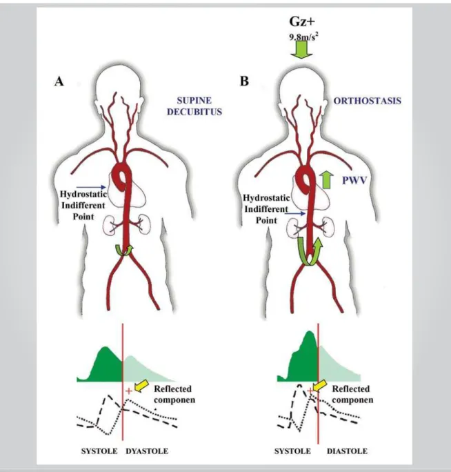
It is known that, structurally, the ratio of elastic/ collagen fibers varies from 3.1:1 in the proximal descending aorta, to 2.8:1 in the region of the middle thoracic aorta, and to 0.8:1 in the abdominal aorta96. The collagen elastic module is much superior to that of elastin, which means that, the farther from the heart, the stiffer the artery becomes, and also that its elastic module and its PWV increase. In other word, the vascular distension is limited by the collagen fi bers, and by the elevated Young’s elastic modulus97,98. The fact that PWV was obtained during the IT places the arterial segments differentially exposed to the gravitational action, somehow mimicking what happens during the active orthostatic posture. While the segment located between the aortic root and the aortic arch up to the level of the carotid artery is under the direct contraposition of gravity, the remaining aortic segments, up to the level of the femoral arteries, present the circulating blood fl ow going along with the vector of gravitational action. The immediate result of this is that gravity promotes a progressive increase in arterial pressure in the segments below the cardiac level in the orthostatic posture15,30,99.
Given the hydrostatic pressure generated by the gravitational action and the consequent change in the hydrostatic indifferent point, there is a higher blood fl ow to the arterial segments with a bigger elastic modulus and smaller radius, disclosing the increment of the carotid-femoral PWV measured during orthostasis100,101. This PWV increase is responsible for the premature return of the refl ected waves from the peripheral sites to the ascending aorta. These prematurely refl ected waves (during the ventricular ejection period) are added to the refl ected component of the pulse wave caused by the left ventricular ejection and infl uence the contour of the pressure and fl ow waves102. Hence, the early return of the refl ected component that occurs during the systolic component of the pulse wave leads to a consequent increase in pulse pressure103,104 (Fig. 1). It is likely that this increase, caused by the refl ected wave in the initial portion of the arterial pulse wave, is due to a complex evolutional functional adaptation of the vascular system with the objective of maintaining an effective blood fl ow to the brain in response to bipedism95.
The role of the wave refl ection in the circulatory homeostasis during orthostasis is reinforced by the observation that, when nitroglycerin, utilized in the sublingual route for IT sensitization, generates a peripheral vasodilation, it leads to a delay in the refl ected component
of the pulse wave and the consequent reduction in the proximal systolic pressure, resulting in symptoms of low brain fl ow in patients with neuromediated syncope105,106. In fact, although a decrease in the coeffi cient of refl ection is verifi ed with the use of nitroglycerin, paradoxically, an increment in aortic stiffness is also observed. Soma et al107 ascribe this secondary aortic stiffness to a possible refl ex activation of the sympathetic nervous system. These authors state that the cardiac frequency increase, which is a consequence of sympathetic activation, can be unfavorable as it reduces ventricular ejection time, time for the aortic root peak fl ow, the systolic volume and the cardiac output107.
Other aspects that deserve to be analyzed are the cardiac frequency behavior associated to postural change and the potential role of the alterations in the hemodynamics and structure of the great arterial vessels in their response patterns97. Initial experimental studies of humans in supine decubitus showed a positive correlation between the cardiac frequency increase and an augmented aortic stiffness108-111. By observing that the effect of the cardiac frequency increase, causing the stiffening of the elastic arteries, occurred independently from the sympathetic modulation, Mircoli et al112 suggested that the decrease of frequency-dependent arterial distensibility would primarily occur because of the viscous characteristic of the vascular wall and its response inertia to the intravascular pressure changes112. However, Wilkinson et al113, in a study that assessed the behavior of PWV in healthy individuals submitted to atrial stimulation, did not observe signifi cant alterations in aortic distensibility in the face of cardiac frequency increase.
In opposition to the findings obtained in supine decubitus, Elias95 observed a negative correlation between cardiac frequency and PWV in orthostatic posture (r= -0.37, P < 0.001). Age was the main variable responsible for the cardiac frequency behavior in the orthostatic posture (r= -0.52, P < 0.001). In search for a better understanding of this fi nding, the author also analyzed the correlation between the cardiac frequency, obtained during orthostatic stress, and the basal measurement of the PWV, having observed a negative correlation between the cardiac frequency and the basal PWV (r= -0.30, P < 0.01). These results showed the possibility of an association of the basal pattern of arterial compliance with the cardiac frequency response level, during orthostasis95.
that the resting cardiac frequency is modulated, in part, by the equilibrium between the sympathetic and the parasympathetic tonus, with a predominance of the latter. The intervention of age and posture on cardiac frequency suggest that age-related alterations occur in the regulatory mechanisms of cardiac frequency101. The acute cardiac frequency increase in response to the rapid change into the orthostatic posture, in general occurring within the fi rst seconds after standing upright, although present in all individuals, decreases in magnitude with age, and can be delayed118. This decrease in the variability of the cardiac frequency observed in elderly patients, when compared to young individuals in the orthostatic versus
the supine posture, has been attributed to a decrease in the recruitment of baroreceptor activity when the upright position is adopted17,117,118.
postural stress in elderly individuals when compared to young ones, the systolic volume reduction tends to be smaller in the healthy elderly individual than in the young one; consequently, the cardiac output does not signifi cantly vary with age, as the smaller increment of cardiac frequency is compensated by a smaller systolic volume reduction101.
Considering the dynamic behavior of the aortic compliance when facing postural change, one could presume that a higher systolic volume decrease would lead to a decrease in the aortic pulsatile deformation, with a subsequent decrease of baroreceptor stimulation and cardiac frequency increase.
The maintenance of the systolic volume in elderly individuals is attributed to a lower venous compliance in this group, which would allow the preservation of the cardiac fi lling volume, and consequently, of the systolic volume101. However, an age-related lower cardiac frequency could also mean an adaptation mechanism of man to bipedism95. It is known that the cardiac frequency affects pre-load through its effect on the diastolic fi lling time. The cardiac frequency also modulates the state of myocardial contractility through its effect, by altering the myocardial concentration of Ca2+ and Na+. As a consequence of the increase of myocardial contractility, the cardiac frequency also modulates the fi nal systolic volume, the systolic volume and the ejection fraction101. Thus, a lower cardiac frequency could, at fi rst, allow a better cardiac performance when facing a stiffer arterial system. Further large-scale longitudinal studies will defi ne whether this recent observation in the cardiac frequency behavior is caused by structural alterations of the great arteries, which are inherent to the aging process, and to some degree of interference in the mechanical component of the barorefl ex.
F
UNCTIONAL BEHAVIOR OF THE GREAT ARTERIES– P
OSSIBLE CLINICALIMPLICATIONS
Considering what was presented here, the following hypothesis can be formulated: the return of the refl ected component of the pulse wave is essential for the immediate adaptation to orthostatism, as well as in the activation, at an adequate degree, of the baroreceptors. Assuming that such hypothesis is correct, it is possible to consider the participation of these recent fi ndings in some very common clinical conditions associated to the orthostatic posture. The more frequent occurrence of neuromediated syncope in young individuals is well established120. Another aspect is that such events occur more commonly with taller individuals. As the aortic length is related to body height, the arterial refl ection wave occurs later in taller individuals121, and it would not be incongruous to suppose that, in young individuals, especially females, those who present lower basal levels of
arterial pressure, the presence of a more elongated aorta could interfere with the return of the refl ected component to the proximal aortic portions in an optimized way, which could result in the activation of refl exes related to the genesis of the syncope (for instance, the Bezold-Jarisch refl ex). Another aspect that supports this hypothesis is, as previously discussed, the importance of height (h) in gravitational stress24. Therefore, in young individuals, the premature return of the refl ected component of the pulse wave caused by the increment of the PWV during orthostasis would act together with the barorefl ex and other regulatory mechanisms of the arterial pressure, allowing an immediate adaptation to orthostatism95.
Based on the recent data, the hypothesis of the participation of the great arteries in the maintenance of circulatory homeostasis as well as in the syncope-triggering mechanism must be taken into account, aiming at a new theory, to be incorporated to the several existing ones, for the physiopathology of neuromediated syncope44.
As for the analysis of the orthostatic adaptation in elderly individuals, who present higher susceptibility to morbid cardiovascular events and postural hypotension, the application of these recent fi ndings is still questionable.
Literature data have shown a broad diurnal variation in the occurrence of cardiovascular events, with a peak incidence of myocardial infarction, sudden cardiac death and ischemic and hemorrhagic stroke occurring in the fi rst hours of the morning122-124. A more detailed analysis of this circadian pattern has identifi ed the hour that follows awakening, more than any other time of the day, as the one during which more cardiovascular events occur122,123. Although it has not been established whether the additional increase of PWV during orthostasis is just a structural adaptive process inducing mechanism or a marker of morbid events, this fi nding must be taken into account.
One must consider that, although a premature return of the refl ected component of the pulse wave can contribute to a lower cardiac frequency during orthostatic stress, generating potential advantages in the postural adaptation of the elderly, chronic abnormalities in the aortic distensibility, such as those possibly generated by the orthostatic posture, create an incompatibility between the ventricular ejection and the aortic fl ow energy. Hence, a more premature return of wave refl ection caused by the increase of the PWV can create an increase in the hydraulic load for the left ventricle125.
Therefore, a combined ventricle-vascular stiffening can potentially have important consequences for the cardiac response to the variation of fi lling volume, as a stiffer heart-arterial system generates higher alterations in systolic pressure for a given change in ejected systolic volume or in the ventricular volume126,127.
the arterial pressure fl uctuations related to postural and postprandial stress126,127. This postural increase of PWV can add to the sympathetic nervous activity augmentation, particularly the adrenergic activity and other acute risk factors such as platelet hyperactivity, hypercoagulability and hypofi brinolysis, blood viscosity and vasospasm increase130, contributing for the increase of cardiovascular morbidity and mortality in the morning period. Finally, the volemic oscillations caused by the upright posture are potentially more likely to be experienced by this population. This puts the elderly individual at higher risk, not only because a transient orthostatic hypotension generates the decompensation of a cardiac structural disease (for instance, coronary failure), but also because in situations of additional volemia decrease (for instance, infectious processes, use of vasodilator or diuretic agents, diarrhea and vomiting) there may be symptoms of orthostatic intolerance or even syncopal events53,131.
Hence, a longitudinal investigation of a large, unselected population group is necessary, aiming at assessing the independent contribution of the postural variation of the PWV for the individual cardiovascular risk.
C
ONCLUSIONThe exact mechanisms responsible for the
cardiovascular adaptation to orthostatism need further clarification. Several theories have been proposed as being fundamental in maintaining the circulatory homeostasis against the gravitational stress. However, a better understanding of the human adaptation to orthostasis must take into account the gravitational gradient generated by such posture.
Possibly, the hypothesis presented herein will contribute to the understanding of the participation of the great arteries in the cardiovascular adaptation to orthostasis and will enable a better analysis of the response of the great arteries to gravitational stress, of the physiopathology of neuromediated syncope and orthostatic hypotension.
Another aspect is how much the anti-gravitational adaptive phenomena can contribute to cardiovascular morbidity and mortality, when added to the inherent degenerative alterations such as arterial hypertension, aging and pre-determined genetic markers.
Acknowledgements
To Prof. Dr. Roberto Sá Cunha, for the technical assistance and suggestions. To Prof. Dr. Dalton Vassalo and Dr. Márcio Augusto Silva, for text review.
R
EFERENCES1. Haynes FW, Ellis LB, Weiss S. Pulse wave velocity and arterial elasticity in arterial hypertension, arteriosclerosis, and related conditions. Am Heart J. 1936; 11: 385-401.
2. Smit AA, Halliwill JR, Low PA, Wieling W. Pathological basis of orthostatic hypotension in autonomic failure. J Physiol. 1999; 519: 1-10.
3. Sheldon R, Morillo C, Krahn A. Management of vasovagal syncope: Expert Ver Cardiovasc Ther. 2004; 2: 915-23.
4. Kapoor WN. Syncope. N Engl J Med. 2000; 343 (25): 1856-62. 5. Guyton AC, Jones CE, Coleman TG. Circulatory Physiology: Cardiac
Out-put and Its Regulation. Philadelphia: Saunders WB Co.; 1973. 6. Goulon M, Raphael JC, Hervé F, Janier M. Adaptation et inadaptation
posturale de la pression artérielle. Ann Méd Interne. 1980; 131: 475-82.
7. Abitbol MM. Effect of posture and locomotion on energy expenditure. Am J Phys Anthropol. 1988; 77: 191-99.
8. Abitbol MM. Variations in blood supply allocations for quadrupedal and for bipedal posture and locomotion. Am Phys Anthropol. 1989; 80: 239-58.
9. Akselrod S, Oz O, Greenberg M, Keselbrener L. Autonomic response to change of posture among normal and mild-hypertensive adults: investigation by time-dependent spectral analysis. J Autonomic Nervous System. 1997; 64: 33-43.
10. Streeten DHP, Scullard TF. Excessive gravitational blood pooling caused by impaired venous tone is the predominant non-cardiac mechanism of orthostatic intolerance. Clinical Science. 1996; 90: 277-85. 11. Hicks JW, Badeer HS. Gravity and the circulation: “open” vs. “closed”
systems. Am J Physiol. 1992; 262: R725-32.
12. Akkerman EM, Karemaker JM, Strackee J. Computer modelling
the dynamics of the circulation under gravitational stress. Compact Analysis of Cardiovascular Signals. Ed. M. Di Rienzo; Amsterdam: IOS Press, 1995.
13. Naschitz JE, Mazov I, Eridzhanyan K, Keren D, Rennert HS, Yeshurun D. Hypotensive reactions on passive head-up tilt testing of hypertensive patients. J Human Hypertension. 1996;10: 369-73. 14. Convertino VA. Mechanisms of blood pressure regulation that differ in
men reatedly exposed to high-G acceleration. Am J Physiol Regulatory Integrative Comp Physiol. 2001; 280: R947-R58.
15. László Z, Rössler A, Hinghofer-Szalkay HG. Cardiovascular and humoral readjustment after different levels of head-up tilt in humans. Aviat Space Environ Med. 2001; 72: 193-201.
16. Schutzman J, Jaeger F, Maloney J, Fouad-Tarazi F. Head-up tilt and hemodynamic changes during orthostatic hypotension in patients with supine hypertension. J Am Coll Cardiol. 1994; 24: 454-61. 17. Gabbett TJ, Weston SB, Barrett RS, Gass GC. Cardiovascular
regulation during head-up tilt in healthy 20-30-year old and 70-75-year-old men. Clin Sci. 2001; 100: 199-206.
18. Tripathi A, Shi X, Wenger B, Nadel ER. Effect of temperature and baroreceptor stimulation on refl ex venomotor responses. J Appl Phys. 1984; 57: 1384-92.
19. Sawka MN, Convertino VA, Eichner ER, Schnieder SM, Young AJ. Blood volume:importance and adaptations to exercise training, environmental stresses, and trauma/sickness. Med Sci Sports Exerc. 2000; 32: 332-48.
20. Fadel PJ, Stromstad M, Hansen J, et al. Arterial barorefl ex control of sympathetic nerve activity during acute hypotension: effect of fi tness. Am J Phys Heart Circ Phys. 2001; 280: H524-H32.
22. Custaud MA. Ne pas oublier la gravité. Rev Prat. 2001; 51: 1745-47.
23. Convertino V, Hoffler GW. Cardiovascular physiology. Effects of microgravity. J Fla Med Assoc. 1992; 79: 517-24.
24. Pekarski SE. A gravitational hypothesis of essential hypertension as a natural adaptation to increased gravitational stress caused by regular, prolonged sitting typical of modern life. Med Sci Monit. 2004; 10: HY27-32.
25. Montastruc JL, Senard JM, Verwaerde P, Montastruc P. Physiological mechanisms of cardiovascular adaptation to orthostatism. Role of the sympathetic nervous system and pharmacological implications. Therapie. 1994; 49: 81-7.
26. Benditt DG, Ferguson DW, Grubb BP, et al. Tilt table testing for assessing syncope. J Am Coll Cardiol. 1996 Jul; 28: 263-75. 27. Kaplan NM. Southwestern internal medicine conference: two faces
of sympathetic nervous activity – hypotension and hypertension. Am J Med Sci. 1992; 303: 271-9.
28. Wang Y, Marshall RJ, Shepherd JT . The effect of changes in posture and of graded exercise on stroke volume in man. J Clin Invest. 1960; 39: 1051-61.
29. Stevens PM. Cardiovascular dynamics during orthostasis and the infl uence of intravascular instrumentation. Am J Cardiol. 1966; 17: 211-18.
30. Wieling W, Shepherd JT. Initial and delayed circulatory responses to orthostatic stress in normal humans and in subjects with orthostatic intolerance. Intern Angiol. 1992; 11: 69-82.
31. Montastruc JL, Senard JM, Verwaerde P, Montastruc P. Mécanismes physiologiques de l’adaptation cardiovasculaire à l’orthostatisme. Therapie. 1994; 49: 81-7.
32. van Lieshout JJ, Secher NH. Refl ex control of sympathetic vaso-constrictor activity in vasovagal syncope. Clin Auton Res. 2003; 13: 175-7.
33. Madsen P, Svendsen LB, Jorgensen LG, Matzen S, Jansen E, Secher NH. Tolerance to head-up tilt and suspension with elevated legs. Aviat Space Environ Med. 1998; 69: 781- 4.
34. Levine B. Regulation of central blood volume and cardiac fi lling in endurance athletes. The Frank-Starling mechanism as a determinant of orthostatic tolerance. Med Sci Sports Exerc. 1993; 25: 727-32. 35. Olsen H, Vernersson E, Länne T. Cardiovascular response to acute
hypovolemia in relation to age. Implications for orthostasis and hemorrhage. Am J Physiol Heart Cir Physiol.2000; 278: H222-H32. 36. Scheinberg P, Stead E. The cerebral blood fl ow in male subjects as measured by the nitrous oxide technique. Normal values for blood fl ow, oxygen utilization and peripheral resistence with observations on the effects of tilting and anxiety. J Clin Invest.1949; 28: 1163. 37. Ouchi Y, Okada H, Yoshikawa E, Futatsubashi M, Nobezawa S .
Absolute changes in regional cerebral blood flow in association with upright posture in humans: an orthostatic PET study. J Nucl Med.2001; 42: 707-12.
38. Sander-Jensen K, Secher NH, Astrup A, et al. Hypotension induced by passive head-up tilt: endocrine and circulatory mechanisms. Am J Physiol.1989; 251: R742-R48.
39. Jacobsen TN, Morgan BJ, Scherrer U, et al. Circulation Research 1993; 73: 367-78.
40. Xiangrong S, Gallagher KM, Welch-Oconnor RM, Foresman BH. Arterial and cardiopulmonary barorefl exes in 60- to 69- vs. 18- to 36-yr-old humans. J Appl Physiol.1996; 80: 1903-10.
41. Siché JP, Chevallier M, Tremel F, et al. Sensibilité baroréflexe et retentissement vasculaire chez l´hypertendu. Arch Mal Coeur.1995; 88: 1243-46.
42. Mancia G, Mark AL. Arterial barorefl exes in humans. In: Handbook of Physiology, section 2, The Cardiovascular System, Vol III, Peripheral Circulation and Organ Blood Flow, part 2, eds. Shepherd JT & Abboud FM 1983, pp755 – 813. American Physiological Society: Bethesda, Md, USA.
43. Ray CA, Monahan KD. Aging attenuates the vestibulosympathetic refl ex in human. Circulation.2002; 1059: 956-61.
44. Mosqueda-Garcia R, Furlan R, Tank J, Fernandez-Violante R. The elusive pathophysiology of neurally mediated syncope. Circulation.2000; 102: 2898-906.
45. Muenter NK, Watenpaugh DE, Wasmund WL, Wasmund SL, Maxwell SA, Smith ML. Effect of sleep restriction on orthostatic cardiovascular control in humans. J Appl Physiol. 2000; 88: 966-72.
46. Di Girolano E, Di Iorio C, Leonzio L, Sabatini P, Barsotti A . Usefulness of tilt training program for the prevention of refractory neurocardiogenic syncope in adolescents. A controlled study. Circulation. 1999; 100: 1798-801.
47. Newman DG, White SW, Callster R. Evidence of barorefl ex adaptation to repetitive +Gz in fi ghter pilots. Aviat Space Environ Med. 1998; 69: 446-51.
48. Ludwig DA, Convertino VA. Predicting orthostatic intolerance: physics or physiology? Aviat Space Environ Med. 1994; 65: 404-11. 49. Rossberg F, Penaz J. Does the arterial barorefl ex control the initial
heart rate response to standing? Biomed Biochim Acta. 1987; 46: K9-K12.
50. Smith AS, Fasler JJ. Age-related changes in autonomic function: Relationship with postural hypotension. Age and Ageing. 1983; 12: 206-10.
51. Jingu S, Takeshita A, Imaizumi T, Sakai K, Nakamura M. Age-related decreases in cardiac receptor control of forearm vascular resistance in humans. Clin Exp Hypertens A. 1989; 11(Suppl 1): 211-6. 52. Cleroux J, Giannattasio C, Bolla G, et al. Decreased cardiopulmonary
refl exes with aging in normotensive humans. Am J Physiol. 1989 Sep; 257 (3 Pt 2): H961- 8.
53. Wing LM, Tonkin AL. Orthostatic blood pressure control and ageing. Aust N Z J Med. 1997; 27(4): 462-6.
54. Sevre K, Lefrandt JD, Nordby G, et al. Autonomic functions in hypertensive and normotensive subjects. The importance of gender. Hypertension. 2001; 37: 1351-56.
55. Maliani G, Ferrari A, Gregorini L, et al. Spectral analysis to assess increased sympathetic tone in arterial hypertension. Hypertension. 1991; 17 (suppl III): 36-42.
56. Yo Y, Nagano M, Nagano N, et al. Effect of age and hypertension on autonomic nervous regulation during passive head-uptilt. Hypertension 1994; 23(suppl. I): 182-86.
57. Tonkin A, Wing L. Aging and susceptibility to drug-induced orthostatic hypotension. Clin Pharmacol Ther. 1992; 52: 277-85.
58. James MA, Potter JF. Orthostatic blood pressure changes and arterial barorefl ex sensitivity in elderly subjects. Age and Ageing. 1999; 28: 522-30.
59. Rutan GH, Hermanson B, Bild DE, Kittner SJ, LaBaw F, Tell GS. Orthostatic hypotension in older adults. Cardiovascular Health Study. Hypertension. 1992; 19: 508-19.
60. Gribbin B, Pickering TG, Sleight P, Peto R. Effect of age and high blood pressure on barorefl ex sensitivity in man. Circ Res. 1971; 29: 424-31. 61. Korner PI, West MJ, Shaw J, Uther JB. “Steady-state” properties of
the baroreceptor-heart rate refl ex in essencial hypertension in man. Clin Exp Pharmacol Physiol. 1974; 1: 65-76.
621-37.
63. Lipsitz LA, Jonsson PV, Marks BL, Parker JA, Royal HD, Wei JY. Reduced supine cardiac volumes and diastolic fi lling rates in elderly patients with chronic medical conditions. Implications for postural blood pressure homeostasis. J Am Geriatr Soc. 1990; 38: 103-7. 64. Weidmann P, De Myttenaere-Bursztein S, Maxwell MH, de Lima J.
Effect of aging on plasma renin and aldosterone in man. Kidney Int. 1975; 8: 325-33.
65. Epstein M, Hollenberg NK. Age as a determinant of renal sodium conservation in normal man. J Lab Clin Med. 1976; 87: 411-17. 66. Kingwell BA, Cameron JD, Gillies KJ, Jennings GL, Dart AM. Arterial
compliance may influence baroreflex function in athletes and hypertensives. Heart Circ. Physiol. 1995; 37: H411-H418. 67. Nichols WW, O’Rourke MF. McDonalds Blood Flow in Arteries:
Theoretical, Experimental, and Clinical Principles. 4rd Ed. London: Melbourne, Auckland; Edward Arnold, 1998.
68. Glaser E, Lacolley P, Boutouyrie P, et al. Dynamic versus static compliance of the carotid artery in living Wistar-Kyoto rats. J Vasc Res. 1995; 32: 254-65.
69. Lenard Z, Fulop D, Visontai Z, et al. Static versus dynamic distensibility of the carotid artery in humans. J Vas Res. 2000; 37: 103-11. 70. Hunt EB, Farquhar WB, Taylor JA. Does reduced vascular stiffening
fully explain preserved cardiovagal baroreflex function in older, physically active men? Circulation. 2001; 103: 2424-27. 71. Itoh H, Bunag RD. Aging reduces cardiovascular and sympathetic
responses to NTS injections ofserotonin in rats. Exp Gerontol. 1992; 27: 309-20.
72. Dauchot P, Gravenstein JS. Effects of atropine on the electrocardiogram in different age groups. Clin Pharmacol Ther. 1971; 12: 274-80. 73. Brodde OE, Konschak U, Becker K, et al. Cardiac muscarinic
receptores decrease with age. In vitro and in vivo studies. J Clin Invest. 1998; 101: 471-8.
74. Monahan KD, Dinenno FA, Seals DR, Clevenger CM, Desouza CA, Tanaka H. Age-associated changes in cardiovagal barorefl ex sensitivity are related to central arterial compliance. Am J Phys Heart Circ Phys. 2001; 281: H248-H249.
75. Brown AM. Receptors under pressure: an update on baroreceptors. Circ Res. 1980; 46: 1-10.
76. Avolio AP, Deng FQ, LI WQ, et al. Effects of aging on arterial distensibility in populations with high and low prevalence of hypertension: comparison between urban and rural communities in China. Circulation. 1985; 71: 202-10.
77. Tanaka H, DeSouza CA, Jones PP, Stevenson ET, Davy KP, Seals DR. Aging, habitual exercise, and dynamic arterial compliance. Circulation. 2000; 102: 1270-75.
78. Mosqueda-Garcia R, Furlan R, Fernandez-Violante R, et al. Sympathetic and baroreceptor refl ex function in neurally mediated syncope evoked by tilt. J Clin Invest. 1997; 99: 2736-44. 79. van Lieshout JJ, Wieling W, Karemaker JM, et al. The vasovagal
response. Clin Sci. 1991; 81: 575-86.
80. Rachev A, Stergiopulos N, Meister JJ. Theoretical study of dynamics of arterial wall remodeling in response to changes in blood pressure. J Biomech. 1996; 29: 635-42.
81. Tanaka H, DeSouza CA, Seals DR. Absence of age-related increase in central arterial stiffness in physically active women. Arterioscler Thromb Vasc Biol. 1998;18: 127-32.
82. Latham RD, Tran CC, Fanton JW, White CD, Owens RW, Self DA. Aortic wave refl ection and input impedance as a function of posture in a chronic primate model. Physiologist. 1992; 35: S53-S54.
83. Tran CC, Lathan RD, Self DA, Fanton JW, White CD, Owens RW. Ventricular/vascular coupling under hyergravidity in a chronically instrumented conscious primate model. Physiologist. 1992; 35: S55-S58.
84. Lathan RD, Rubal BJ, Westerhof N, Sipkema P, Walsh RA. Nonhuman primate model for regional wave travel and refl ections along aortas. Am J Physiol. 1987; 253: H299-H306.
85. Sonesson B, Länne T, Hansen F, Sandgren T. Infrarenal aortic diameter in the healthy person. Eur J Vasc Surg. 1994; 8: 89-95.
86. Silva ES, Rodriguez Jr AJ, Tolosa EMC, Pereira PRB, Zanoto A, Martins J. Variation of infrarenal aortic diameter: a necropsy study. J Vasc Surg. 1999; 29: 20-7.
87. Blacher J, Guerin AP, Pannier B, Marchais SJ, Safar ME, London GM. Impact of aortic stiffness on survival in end-stage renal disease. Circulation. 1999; 99: 2434-9.
88. Asmar R, Rudnichi A, Blacher J, London GM, Safar ME. Pulse pressure and aortic pulse wave are markers of cardiovascular risk in hypertensive populations. Am J Hypertension. 2001; 14: 91-7. 89. Amar J, Ruidavets JB, Chamontin B, Drouet L, Ferriéres J. Arterial
stiffness and cardiovascular risk factors in a population-based study. J Hypertension. 2001; 19: 381-7.
90. O’Rourke M. Mechanical principles in arterial disease. Hypertension. 1995; 26: 2-9.
91. Kanda T, Nakamura E, Moritani T, Yamori Y. Arterial pulse wave velocity and risk factors for peripheral vascular disease. Eur J Appl Physiol. 2000; 82: 1-7.
92. O’Rourke MF. Pressure and fl ow waves in systemic arteries and the anatomical design of the arterial system. J Appl Phys. 1967; 23: 139-49.
93. Asmar R, Benetos A, Topouchian J, et al. Assessment of arterial distensibility by automatic pulse wave velocity measurement. Validation and clinical application studies. Hypertension. 1995; 26: 485-90.
94. Asmar RG, Topouchian A., Benetos A, Sayegh FA, Mourad JJ, Safar ME. Non-invasive evaluation of arterial abnormalities in hypertensive patients. J Hypertension. 1997; 15(Suppl. 2): S99-S107. 95. Elias J. Infl uência da ortostase no comportamento funcional dos
grandes vasos arteriais [dissertação]. Programa de Pós-graduação em ciências fi siológicas – Universidade Federal do Espírito Santo (UFES); 2002.
96. Rogers WJ, Hu YL, Coast D, et al. Age-associated changes in regional aortic pulse wave velocity. J Am Coll Cardiol. 2001; 38: 1123-9. 97. O’Rourke MF. Mechanical Principles. Arterial stiffness and wave
refl ection. Pathologie Biologie. 1999, 47: 623-33.
98. London GM, Guerin AP. Infl uence of arterial pulse and refl ected waves on blood pressure and cardiac function. Am Heart J. 1999; 138: S220-S224.
99. Hasegawa M, Rodbard S. Effect of posture on arterial pressures, timing of the arterial sounds and pulse wave velocities in the extremities. Cardiology. 1979; 64: 122-32.
100. Iida N, Iriuchijima J. Postural changes in fi nger and toe pulse waves. Kokyu. To. Junkan 1992; 40: 981-6.
101. Lakatta EG. Cardiovascular regulatory mechanisms in advanced age. Physiol Reviews. 1993; 73: 413-67.
102. Kroeker EJ, Wood EH. Comparison of simultaneously recorded central and peripheral arterial pressure pulses during rest, exercise and tilted position in man. Circ Res. 1955; 3: 6223-31.
104. Gatzka CD, Kingwell BA, Cameron JD, et al and the ANBP2 investigators. Gender differences in the timing of arterial wave refl ection beyond differences in body hight. J Hypertension. 2001; 19: 2197-203.
105. Bartoletti A, Alboni P, Ammirati F, et al. ‘The Italian Protocol’: a simplifi ed head-up tilt testing potentiated with oral nitroglycerin to assess patients with unexplained syncope. Europace. 2000; 2: 339-42.
106. Aerts AJ, Dendale P. Diagnostic value of nitrate stimulated tilt testing without preceding passive tilt in patients with suspected vasovagal syncope and a healthy control group. Pacing Clin Electrophysiol. 2005; 28: 29-32.
107. Soma J, Angelsen AJ, Techn D, Aakhus S, Skjaerpe T. Sublingual nitroglycerin arterial wave reflections despite increased aortic “stiffness” in patients with hypertension: a Doppler echocardiography study. J Am Soc Echocardiogr. 2000; 13: 1100-8.
108. Sá Cunha R, Pannier B, Benetos A, et al. Association between high heart rate and high arterial rigidity in normotensive and hypertensive subjects. J Hypertension. 1997; 15: 1423-30.
109. Asmar R, Topouchian J, Pannier B, Rudnichi A, Safar M. Reversion of arterial abnormalities by long-term antihypertensive therapy in a large population. The Complior® study. J Hipertension. 1999; 17:
Suppl 3: S9.
110. Albaladejo P, Asmar R, Safar M, Benetos A. Association between 24-hours ambulatory heart rate and arterial stiffness. J Human Hypertension. 2000; 14: 137-41.
111. Benetos A, Adamopoulos C, Bureau JA, et al. Stiffness in normotensive subjects and in treated hypertensive subjects over a 6-Year Period. Circulation. 2002; 105: 1202-7.
112. Mircoli L, Mangoni AA, Giannattasio C, Mancia G, Ferrari AU. Heart rate-dependent stiffening of large arteries in intact and sympathectomized rats. Hypertension. 1999; 34:598-602. 113. Wilkinson IB, Mohammad NH, Tyrrell S, et al. Heart rate dependency
of pulse pressure amplifi cation and arterial stiffness. Am J Hypertens. 2002; 15 (1 Pt 1): 24-30.
114. Simpson DM, Wicks R. Spectral analysis of heart rate indicated reduced baroreceptor-related heart rate variability in elderly persons. J Gerontol. 1988; 43: M21- M24.
115. Schwartz JB, Gibb WJ, Tran T. Aging effects on heart rate variation. J Gerontol. 1991; 46: M99-M106.
116. Dambrink JHA, Wieling W. Circulatory response to postural change in healthy male subjects in relation to age. Clinical Science. 1987; 72: 335-41.
117. Hainsworth R, Al-Shamma YMH. Cardiovascular response to upright tilting in healthy subjects. Clinical Science. 1988; 74: 17-22. 118. Rodeheffer RJ, Gerstenblith G, Beard E, et al. Postural changes in
cardiac volumes in men in relation to adult age. Exp Gerontol. 1986; 21: 367-78.
119. Crandall CG, Engelke KA, Convertino VA, Raven PB. Aortic barorefl ex control of heart rate after 15 days of simulated microgravity exposure. J Appl Physiol. 1994; 77: 2134-9.
120. Colman N, Nahm K, Ganzeboom KS, et al. Epidemiology of refl ex syncope. Clin Auton Res. 2004; 14 (Suppl 1): 9-17.
121. Smulyan H, Marchais SJ, Pannier B, Guerin AP, Safar ME, London G. Infl uence of body height on pulsatile arterial hemodynamic data. J Am Coll Cardiol. 1998; 31: 1103-9.
122. Willich SN, Levy D, Rocco MB, Tofl er GH, Stone PH, Muller JE. Circadian variation in the incidence of sudden cardiac death in the Framingham Heart Study population. Am J Cardiol. 1987; 60: 801-6.
123. Muller JE, Stone PH, Turi ZG. The MILIS Study Group:circadian variation in the frequency of onset of acute myocardial infarction. N Engl J Med. 1985; 313: 1315-22.
124. Maron BJ, Kogan J, Proschan MA, Hecht GM, Roberts WC. Circadian variability in the occurrence of sudden cardiac death in patients with hypertrophic cardiomyopathy. J Am Coll Cardiol. 1994; 23:1405-9. 125. O’Rourke MF. Vascular impedance in studies of arterial and cardiac
function. Physiol Rev. 1982; 62: 570-23.
126. Chen HC, Hu HH, Lin YP, Chern CM, Hsu TK, Ding PYA. Increased arterial wave refl ection may predispose syncopal attacks. Clin Cardiol. 2000; 23: 825-30.
127. Chen MY, Goldenberg IF, Milstein S, et al. Cardiac electrophysiologic and hemodynamic correlates of neurally mediated syncope. Am J Cardiol. 1989; 63: 66-72.
128. Seals DR, Taylor JA, Ng AV, Esler MD. Exercise and aging: autonomic control of the circulation. Med Sci Sports Exerc. 1994; 26: 568-76. 129. White M, Roden R, Minobe W. Age-related changes in beta-adrenergic neuroeffector systems in the human heart. Circulation. 1994; 90: 1225-38.
130. Kario K, Pickering TG, Umeda Y, et al. Morning surge in blood pressure as a predictor of silent and clinical cerebrovascular disease in elderly hypertensives. A prospective study. Circulation. 2003; 107: 1401-6. 131. Naschitz JE, Mazov I, Eridzhanyan K, Keren D, Rennert HS,