Arq Bras Cardiol 2001; 77: 225-8.
Dias et al Mini-sternotomy
225 225 225 225 225
Instituto do Coração do Hospital das Clínicas – FMUSP
Mailing address: Altamiro Ribeiro Dias – InCor - Av. Dr. Enéas C. Aguiar, 44 – 05403-900 – São Paulo, SP, Brazil – [email protected]
English version by Stela Maris C. e Gandour
Objective - To compare inverted-L mini-sternotomy performed above the sternal furcula with conventional sternotomy in patients with aortic valve diseases who un-dergo surgical treatment.
Methods - We operated upon 30 patients who had aor-tic valve lesions that had clinical and hemodynamic findings. All patients underwent inverted-L sternotomy, which extended from above the manubrium of the sternum to the 3rd
right intercostal space, without opening the pleural cavity. Their ages ranged from 32 to 76 years, and 18 were males and 12 were females. We used negative pressure in a venous ¼-inch cannula, and the patients were maintained in Trendelemburg’s position. Twenty-seven patients received bioprostheses with diameters ranging from 23 to 29mm. Three patients underwent only removal of the calcifications of the aortic valve leaflets and aortic commissurotomy.
Results - The mean duration of anoxic cardiac arrest was 63.11min. Access was considered good in all patients. One death was due to pulmonary and renal problems not related to the incision. All patients had a better recovery in the intensive care unit, got out of bed sooner, coughed mo-re easily, and performed prophylactic physiotherapeutic maneuvers for respiratory problems more easily and with less pain in the incision. Early ambulation was more easi-ly carried out by all patients.
Conclusion - Mini-sternotomy proved to be better than the conventional sternotomy because it provided mo-recomfort for the patients in the early postoperative pe-riod, with less pain and greater desire for early ambulati-on and all its inherent advantages.
Key words: aortic valve disease, aortic valve surgery, sternotomy
Arq Bras Cardiol, volume 77 (nº 3), 225-8, 2001
Al tami ro Ri bei ro Di as, Ri cardo Ri bei ro Di as, Fábi o Gai otto, José Li ma O. Júni or, Fi l i nto M.C.N. Cerquei ra, Max Gri nberg, Roney Sampai o, Paul o de Lara Lavi tol a, Nel son El i as, Fl ávi o Tarasoutchi ,
Lui z F. Cardoso, Noedi r A. G. Stol f
São Paulo, SP - Brazil
Mini-sternotomy for the Treatment of Aortic Valve Lesions
Original Article
Interest in using minimally invasive procedures in car-diac surgery has increased greatly around the world.
Lesions in the aortic valve have been approached with this type of procedure with good early results, even though controversies still exist.
Cosgrove and Sabik 1 reported their experience with
right parasternal mini-incisions at the 2nd, 3rd, and 4th costal
cartilages to provide access for the surgical treatment of aortic valve diseases.
Mulinari et al 2 and Pereira et al 3 favor the median
ac-cess via, such as the reduced sternotomy, for the sucac-cessful treatment of the lesions of the aortic valve.
Cohn and Gundry 4 reported good results obtained
with the technique of the Cleveland Clinic cited above. Denton Cooley, on the same occasion, reported ha-ving seen some cases of pulmonary hernias resulting from sectioning the costal cartilages. To avoid this problem, Steven Gundry, from Loma Linda, proposed access through mini-sternotomy, with an incision extending from the manu-brium of the sternum to the 3rd intercostal space, where the
incision is deviated to the right, like an inverted L, preser-ving the right internal thoracic artery 4.
Dias et al 5 reported an initial experience with
mini-ster-notomy, extending the incision to the 4th intercostal space.
All these controversial approaches resulted in a panel discussion 6 in which Steven Gundry enthusiastically
sup-ported this access approach. On the other hand, Smith 6
cri-ticized its indiscriminate use.
Our objective is to report the initial results obtained at the Instituto do Coração of the Hospital das Clínicas of the Medical School of the University of São Paulo with mini-sternotomy up to the 3rd right intercostal space.
Methods
226 226 226 226 226
Dias et al Mini-sternotomy
Arq Bras Cardiol 2001; 77: 225-8.
Twenty-two patients had severe dyspnea, 15 had angina, and 5 had subjective complaints compatible with arrhythmias. Preoperative diagnoses were as follows: aortic steno-sis in 16 patients, double aortic valve dysfunction in 7, and aortic insufficiency in 7.
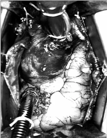
All patients underwent inverted-L mini-sternotomy, the skin incision extending from the manubrium of the ster-num to the 3rd right intercostal space, for 10 to 11cm (fig. 1).
Opening of the right pleural cavity or lesion of the right in-ternal thoracic artery, or both, did not occur when the trans-versal segment of the sternotomy was completed.
Trendelemburg’s position was adopted in all cases, which facilitated the marsupialization of the heart with fixation and traction of the pericardium to the edges of the mini-incision.
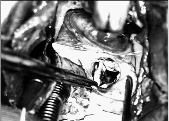
Arterial cannulation was easily performed with a pur-se-string suture in the ascending aorta, close to the inno-minate vein. Venous cannulation was performed through a single ¼-inch wire cannula introduced into the right atrial auricle through a purse-string suture. The use of negative pressure in the venous line made perfect venous drainage possible, facilitating the exposure of the structures (fig. 2). Aspiration of the left cavities was performed with the aid of an aspirator introduced into the left atrium through a purse-string suture in the right superior pulmonary vein in most cases. In 5 patients, very good aspiration was obtained by introducing the aspirator into the pulmonary artery.
All patients underwent surgery with hypothermia at 28ºC and cardioplegia with St. Thomas solution.
Removal of calcifications on the leaflets of the aortic valve and section of the fusions between these leaflets were pos-sible in 3 patients, with good results on direct visualization and intraoperative echocardiographic assessment (fig. 3).
The remaining patients underwent valvar replacement with removal of the annular calcifications when present. One patient received a bioprosthesis 21mm in diameter, 7 patients each received bioprostheses of 23mm, 9 patients each received bioprostheses of 25mm, 7 patients each recei-ved bioprostheses of 27mm, and 3 patients each receirecei-ved bioprostheses of 29mm of diameter (fig. 4).
Fig. 1 – Intraoperative photography showing skin incision of approximately 10cm.
Fig. 2 – Intraoperative photography showing aorta and right atrium cannulated and ready for perfusion. Note the very good exposure of the ascending aorta.
Arq Bras Cardiol 2001; 77: 225-8.
Dias et al Mini-sternotomy 227 227 227 227 227
The management of patients with pure aortic insuffici-ency was much easier than that of patients with intensely calcified aortic stenosis.
An important point is mediastinal drainage, which should be performed immediately before the end of the ano-xic cardiac arrest, prior to reestablishment of cardiac beats. A small incision in the aponeurosis, right below the sternal furcula, really facilitates the introduction of a retrosternal clamp up to the end of the mini-sternotomy to provide fixa-tion of the drain and its exteriorizafixa-tion below the sternal fur-cula. The still empty cardiac cavities make this procedure easy and safe.
Sternal closing was performed with stitches of steel thread only on the manubrium, no stitch being required for the transversal segment of the sternotomy. The patients were referred to the postoperative unit at the end of the procedure.
Results
The duration of the anesthesia ranged from 225 to 480min (mean of 327.22min), the duration of extracorporeal circulation ranged from 53 to 140min (mean of 90.38min), and the duration of the anoxic cardiac arrest ranged from 37 to 103min (mean of 63.11min).
No significant bleeding occurred in this group of patients. It is worth noting that, after regaining consciousness, the patients had greater mobility and greater ability to per-form their physiotherapeutic exercises proper to this phase of the postoperative period.
The length of stay in the intensive care unit ranged from 24 to 48h, and the time in the ward before hospital discharge was around 6 to 7 days.
One patient died due to renal and pulmonary pro-blems not related to the incision.
In our series, ambulation occurred much sooner and was performed more easily and with more willingness compared with that in patients undergoing conventional sternotomy.
After hospital discharge, the patients have been pe-riodically followed up in the cardiac valve outpatient care unit, all of them being in functional class I.
Discussion
Our initial experience shows that the mini-sternotomy technique is safe, perfectly feasible, and relatively easy to perform. When comparing inverted-L mini-sternotomy with right paramedian vertical thoracotomy, proposed by some authors 1,4, we can see that the incision above the
manubri-um of the sternmanubri-um is more benign and freer from pleuropul-monary complications.
We do not consider it necessary to extend the incision as far as the 4th intercostal space 5, because the 3rd
intercos-tal space provides enough room for the creation of the pou-ch in the right atrial auricle, as well as for drainage of the left chambers through a catheter introduced into the superior pulmonary vein.
Caution, however, is required because this access via should not be considered of unlimited application. We em-phasize that in short and obese patients, the use of this tech-nique may not be recommended.
In addition to the cosmetic benefit of the small skin in-cision (10cm), the greatest benefit is the comfort experienced by the patients as soon as they recover from anesthesia. The firmness provided by the integrity of the inferior 2/3 of the sternum makes it possible for the patients to cough more easily, to perform their physiotherapeutic exercises more comfortably, and to ambulate sooner with minimum pain in the incision. InCor is an institution affiliated with a university, has well-defined postoperative protocols for conventional procedures, and we did not find it necessary to make any change in these protocols in this initial phase of the procedure. Therefore, no substantial reduction existed in the periods of stay in the intensive care unit or in the period of hospitalization as a whole.
The durations of anesthesia, of perfusion, and of ano-xic cardiac arrest also did not differ from those of conven-tional surgery.
We think that greater experience with and wider appli-cation of this type of surgical approach may certainly redu-ce the duration of hospital stay and the stay in the intensive care unit, with obvious advantages to the patients.
Simple maneuvers, such as those reported, make the procedure easier, provide greater safety, and facilitate its application. Among these maneuvers, we highlight the wide marsupialization of the heart, the venous drainage with a thinner wire cannula, Trendelemburg’s position, and aspiration through the pulmonary artery, when necessary.
In our environment 2,3, this incision and its variants
have been used with good results, as has been the case at other international services 6.
In conclusion, we consider inverted-L mini-sternoto-my a safe and efficient approach that enhances the recovery of patients in the early postoperative period.
Late results still require further assessment. However, it is very likely that they will be linked to a lower incidence of problems related to the incision, with no direct interference with heart disease.
228 228 228 228 228
Dias et al Mini-sternotomy
Arq Bras Cardiol 2001; 77: 225-8.
1. Cosgrove DM, Sabik JF. Minimally invasive approach for aortic valve opera-tions. Ann. Thorac Surg 1996; 62: 596-7.
2. Mulinari LA, Tyszka AL, Costa F, et al. Miniesternotomia. Um acesso seguro para cirurgia cardíaca. Anais do 24º Congresso Nacional de Cirurgia Cardíaca. Campo Grande, MS 1997: 95.
3. Pereira WM, Frota Filho JD, Jung LA, et al. Acesso restrito para cirurgia da valva aórtica (Minimamente invasiva) Anais do 24º Congresso Nacional de Cirurgia Cardíaca, Campo Grande, MS, 1997: 115.
References
4. Cohn L, Gundry SR. Adult cardiac symposium. 77th Annual Meeting of the
American Association for Thoracic Surgery, p.6-8. Washington, 4 to 7 may, 1997. 5. Dias RR, Avelar Júnior SF, Lima SGG, et al. Miniesternotomia para a cirurgia da valva aórtica: experiência inicial. Rev Bras Cir Cardiovasc 1998; 14: 317-20. 6. Kouchoukos NT, Gundry SR, Smith CR. Controversies in Cardio Thoracic
Sur-gery. Acquired cardiac controversies. Aortic valve replacement should be perfor-med through a mini- sternotomy 80th annual Meeting. American Association for