Hospital das Clínicas da Universidade Federal do Rio Grande do Sul - Porto Alegre Mailing address: Paulo R. A. Caramori – Rua Ramiro Barcelos, 2350 – S/2059 -90035-003 - Porto Alegre, RS - Brazil
Paulo R. A. Caramori, Alcides J. Zago
Porto Alegre, RS - Brazil
Endothelial Dysfunction and Coronary Artery Disease
For several decades, the vascular endothelium was considered a unicellular layer acting as a semipermeable membrane between the blood and the interstitium. Recently, it has been demonstrated that the endothelium performs a large range of important biological functions, participating in several metabolic and regulatory pathways. Along with long-known specialized functions like gaseous exchange in the pulmonary circulation and phagocytosis in the hepatic and splenic circulation, the vascular endothelium performs universal roles in the circulation that include participation in thrombosis and thrombolytic control, vascular growth, platelet and leukocyte interactions with the vascular wall, and vasomotor tone.
The study of endothelium-dependent vasomotor reactivity has produced over the years, scientific evidence fundamental for the understanding of the endothelium’s role in physiological and pathological situations. In 1977, Moncada et al, published the first report indicating that the endothelium plays a central role in the control of vascular tone via the production of vasoactive substances 1. In 1980,
Furchgott and Zawadzki 2 demonstrated in an experimental
preparation of the rabbit aorta, the obligatory role played by endothelial cells in vascular relaxation in response to effec-tors like acetylcholine, and postulated the existence of a vascular relaxing factor derived from the endothelium. In 1987, two research groups, lead by Ignarro et al 3, and by
Palmer et al 4, demonstrated that the relaxing factor derived
from the endothelium was nitric oxide, an odorless gas until then considered as a mere pollutant.
Endothelial dysfunction was first characterized in hu-mans in 1986 by Ludmer et al, 5 who demonstrated that
athe-rosclerotic coronary arteries contracted in response to in-tracoronary infusion of acetylcholine, while normal corona-ries showed dilatation. In 1992, endothelial dysfunction was documented by Celermajer et al 6 in children and otherwise
healthy young adults with risk factors for atherosclerosis. Under physiological conditions, the endothelium keeps a reduced vasomotor tone, prevents leukocyte and platelet adhesion, and inhibits the proliferation of vascular
smooth muscle cells. In contrast, endothelial dysfunction ap-pears to play a pathogenic role in the initial development of atherosclerosis 7-9 and of unstable coronary syndromes 10,
being associated with atherosclerotic disease risk factors 11-18,
and being present even before vascular involvement be-comes evident 6,19-21.
Recent clinical studies have demonstrated that some drugs well known to reduce the incidence of cardiovascular events, improve endothelial function 22-25. On the other
hand, clinical interventions like the continuous administra-tion of organic nitrates and percutaneous coronary inter-ventions may be associated with adverse effects on the vascular endothelium. In the present article, we will discuss vascular endothelial function versus dysfunction, and their impact on cardiovascular disease, in particular atheros-clerosis.
The endothelium in cardiovascular homeostasis
-Vascular endothelium may be considered a dynamic, hetero-geneous organ, having secretory, synthesizing, metabolic, and immunological functions, vital to human beings 26. The
endothelium regulates the flow of nutrient substances, of various biologically active molecules, and of blood cells through the entire human body. It is selectively permeable, possessing various cell membrane receptors for molecules that include proteins (growth factors, coagulation, and anticoagulation proteins), lipid-transporting particles (LDL), metabolites (nitric oxide, serotonin), and hormones (endothelin-1). The endothelium plays a central role in the regulation of vascular tone and blood flow by the secretion and capture of paracrine vasoactive substances, contrac-ting or dilacontrac-ting specific vascular beds in response to various stimuli.
The endothelium also possesses important anticoa-gulant, antiplatelet, and fibrinolytic actions. Endothelial cells are the largest sites of reactions involving thrombin 27.
Some of the stimuli that activate platelets (adenosine di-phosphate and thrombin) also stimulate the release of prostacyclin by the endothelium, inhibiting platelet aggre-gation 1,28. In response to stimuli like noradrenaline,
vaso-pressin, thrombin, and vascular stasis, endothelial cells se-crete tissue plasminogen activator 29, a potent thrombolytic
factor, and thrombomodulin. When stimulated by certain physical or chemical factors, the endothelial cell undergoes phenotypical modifications that determine its transforma-tion into a thrombogenic surface. The dynamic equilibrium existing between these two states often permits the endo-thelial cell to return to its basal state, once the thrombogenic stimulus has ceased.
Injury or activation in response to various pathologi-cal factors leads to modifications of the endothelial cell’s re-gulatory functions. The endothelium becomes incapable of maintaining vascular homeostasis. This characterizes a condition of endothelial dysfunction, which can be defined as an imbalance between relaxing and contracting factors, between procoagulant and anticoagulant mediators, or between stimulants and inhibitors of cell growth and proli-feration, respectively 30.
Endothelial vasomotor function - The endothelium
plays a fundamental role in the regulation of vasomotor tone via the synthesis and release of vasodilator substancesni-tric oxide, prostacyclin, and the endothelium-derived hyper-polarizing factor, as well as by the liberation of vasocons-trictor substances like endothelin-1 and platelet-activating factor. Nitric oxide is probably the main mediator of va-somotor tone in physiological situations, small amounts being continuously secreted by the endothelial cells 4,31 to
maintain a reduced arterial tone in the systemic and pulmo-nary circulations 32. The vasodilator activity of nitric oxide is
due to its interaction with the iron atom of the heme prosthetic group of guanylate cyclase, causing its activa-tion and increasing the intracellular levels of cyclic guanidi-ne monophosphate (cGMP), 33. In smooth muscle cells, this
decreases intracellular calcium concentration, causing vas-cular relaxation 34.
Nitric oxide is a free radical produced by the oxidation of L-arginine to L-citrulline, via nitric oxide synthetase, an enzyme that has at least three isoforms 35. Nitric oxide
synthetase type III is a constitutive enzyme of the endothe-lial cell, which continuously produces small amounts of nitric oxide. In contrast to other vasomotor agents (prosta-cyclin, endothelin-1, and the platelet activating factor), which are synthesized primarily in response to local factors, the production of nitric oxide is regulated by various chemi-cal and physichemi-cal stimuli.
Endothelial cell constitutive nitric oxide synthetase can be activated by stimuli that include thrombin, adeno-sine diphosphate, bradykinin, substance P, muscarinic ago-nists, catecholamines, and shear stress 12. Estrogens and
shear stress stimulate the expression of this synthase’s gene. Two other forms of nitric oxide synthetase are presently known: the neuronal constitutive form (type I) and the inducible form (type II). The latter has been obser-ved in various cell types, including vascular smooth mus-cle, the endothelium, and macrophages. Inducible nitric oxi-de synthetase is activated by cytokines like interleukin-1β and the tumor-necrosing factor, being capable of producing large amounts of nitric oxide in inflammatory processes.
In the presence of a normal endothelium, the release of nitric oxide in response to catecholamines counteracts alpha-adrenergic vasoconstrictor effects. In contrast, when the endothelium is dysfunctional, an increase in coronary va-soconstriction in response to adrenergic stimuli occurs 36,37.
The increased synthesis of nitric oxide consequent to shear stress, contributes to the flow-mediated phenomenon of vasodilatation that is an important auto-regulatory physio-logical mechanism 38. The production of nitric oxide can be
blocked in vivo by analogues of L-arginine, like NG–
monomethyl-L-arginine (L-NMMA). Such blockade has been considerably useful for the study of the role of nitric oxide in physiological and pathological situations. The infusion of L-NMMA in the brachial human circulation leads to an increase in vascular peripheral resistance, and intravenous infusion causes an increase in systemic arterial pressure. These findings indicate that the vasculature is in a constant state of vasodilatation due to the continuous release of nitric oxide (fig. 1).
In addition to the modulation of vasomotor tone, endothelial cell-derived nitric oxide has several important vascular effects. Nitric oxide inhibits adhesion, activation, and platelet aggregation 39 and promotes platelet
deaggre-gation, in part by a cGMP-dependent mechanism. Nitric oxide produced in response to thrombin inhibits platelets and mo-dulates blood coagulation. Nitric oxide derived from the endo-thelium also inhibits leukocyte adhesion to the endoendo-thelium
40,41, migration 42, and proliferation 43 of vascular smooth
muscle cells and stimulates the migration and proliferation of endothelial cells 44.
The contribution of endothelial cells to the regulation of vasomotor tone involves the production of other vasodi-lator compounds like prostacyclin and the endothelium-de-rived hyperpolarizing factor. Prostacyclin is synthesized from arachidonic acid by cyclo-oxygenase 1, being rapidly
produced and released from endothelial cells 45 in response
to humoral and hemodynamic factors. It interacts synergi-cally with nitric oxide, causing vasodilatation and inhibition of platelet adhesion and aggregation 46. The stimulation of
adenyl cyclase and increased intracellular concentration of cyclic adenosine monophosphate in smooth muscle cells and platelets mediate its actions. Prostacyclin does not appear to be continuously produced by endothelial cells 47,
but to be synthesized in response to specific stimuli like bradykinin, adenosine diphosphate, hypoxia, and increa-sed shear stress.
Endothelium-derived hyperpolarizing factor, another vasodilator substance produced by the endothelium, pro-motes vascular smooth muscle cell relaxation by increasing cell membrane conductance of potassium 48. This factor is
also secreted in response to acetylcholine and blocked by ouabain, an inhibitor of sodium/potassium ATPase. The en-dothelial-derived hyperpolarizing factor has not yet been isolated, and its physiological role remains uncertain.
vasoconstrictor known, endothelin-1 49. Endothelins
constitute a family of polypeptides produced by various cell types. Of the three isoforms known, endothelial cells appear to produce only endothelin-1. This is a 21 amino-acid peptide formed from its inactive precursor pre-endo-thelin-1, which seems to exert a role as an arterial blood flow regulator in both normal and pathologic conditions 50. In
res-ponse to stimuli like thrombin, adrenalin, angiotensin II, hypoxia, and increased shear stress, endothelin-1 is relea-sed from endothelial cells, binding to specific receptors in vascular smooth muscle cells causing increased intracellu-lar concentration of calcium leading to vasoconstriction 51.
Intramyocardial vessels are more sensitive to endothelin, suggesting that this peptide plays a major role in blood flow control. It is interesting that in functionally intact endothe-lia, endothelin stimulates the production of nitric oxide and of prostacyclin, which, therefore, modulates vasoconstric-tor action and reduces the synthesis of endothelin itself. Two types of vascular receptors for endothelin have been identified. Receptor ETB is observed in endothelial cells, being responsible for the stimulation of nitric oxide and prostacyclin formation. Receptors ETA and ETB, observed in smooth muscle cells, mediate contraction and prolifera-tion of these cells. A large number of endothelin receptor antagonists developed in recent years, are being tested ex-perimentally and clinically.
Thromboxane A2 and prostaglandin H2 are cons-trictor factors also secreted by the endothelium. They acti-vate the thromboxane receptor in smooth muscle cells and platelets, in opposition to the effects of nitric oxide and prostacyclin. However, the role of these substances in coro-nary circulation has not been clearly established. Platelet activation factor is another vasoconstrictor synthesized and released by endothelial cells in response to humoral and hemodynamic stimuli, which probably participates in the regulation of vasomotor tone. Finally, the endothelium also expresses the angiotensin-converting enzyme, which is identical to kinase II, which metabolizes bradykinin. The-refore, the angiotensin-converting enzyme also determines local levels of bradykinin, which stimulates nitric oxide and prostaglandin production. In addition, the angiotensin-converting enzyme synthesizes angiotensin, which directly stimulates the production of endothelin.
Pathophysiology of endothelial dysfunction –
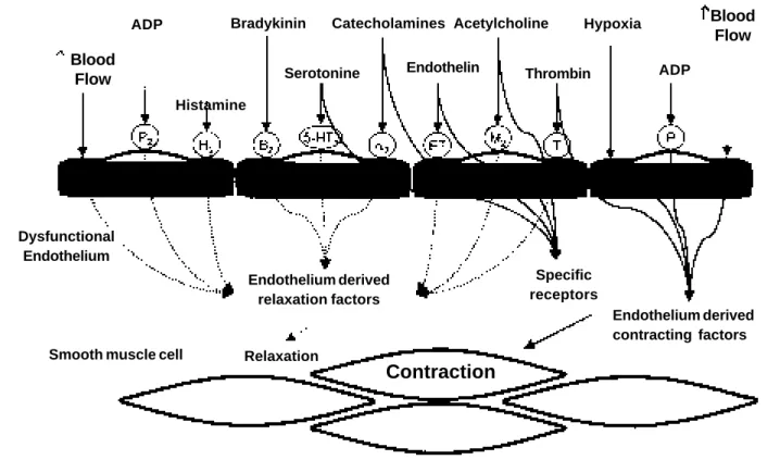
Endo-thelial dysfunction can be determined by the reduction of the endothelium-derived vasodilators, by local increases in antagonists to these substances, or by an association of these two factors (fig. 2). Reduction in the synthesis or local availability of nitric oxide have been frequently considered the major causes of endothelial dysfunction in various cli-nical conditions. Nitric oxide release from the endothelium is Fig. 1 – Diagram describing the action of various effectors on functionally intact endothelium. Receptor stimulation or direct action of these agents led to the liberation of endothelium-derived relaxation factors (nitric oxide, prostacyclin) that cause vascular smooth muscle cells to dilate. In contrast, serotonin, catecholamines, endothelin, acetylcholine, thrombin, hypoxia, adenosine diphosphate (ADP), and the stress of shearing (blood flow) may cause contraction of vascular smooth muscle cells. In functionally intact endothelium, vasodilatation predominates (H2- histamine receptor, a2- a-adrenergic receptor; 5-HT- serotoninergic receptor; B- bradykinin receptor; M- muscarinic recep-tor; P- purinergic receprecep-tor; ET- endothelin receprecep-tor; T- thrombin receptor).
Blood Flow
ADP
Histamine
Bradykinin
Serotonine Catecholamines
Endothelin Acetylcholine Thrombin
Hypoxia ADP
Blood Flow
Endothelium
Smooth muscle cell
Contraction
Relaxation
Endothelium derived relaxation factors
Specific
decreased in patients with established coronary atheroscle-rosis 5,52. A reduction in vascular availability of nitric oxide
determines damage to endothelium-dependent vasodilata-tion, an increased tendency for platelet aggregation and adhesion of monocytes to the endothelium, and influences the proliferation of vascular smooth muscle cells, probably contributing to the onset and progression of atheroscle-rosis. In animal models of hypercholesterolemia, pharma-cological inhibition of nitric oxide synthesis accelerates atherosclerosis 53, but increased availability of nitric oxide
decreases and may even lead to the regression of the disease 54,55.
The inactivation of nitric oxide by oxygen-derived free radicals can be an important factor in the development of en-dothelial dysfunction 56. Experimental studies suggest that
antioxidant agents may reestablish endothelial function 57,58.
Vitamin C, a potent antioxidant in vivo and in vitro 59 that
inhibits superoxide-mediated lipid peroxidation 60, improves
endothelial function in the brachial artery of coronary arte-ry disease patients 61, in patients with diabetes mellitus 62
and in smokers 63.
An increase in endogenous inhibitors of nitric oxide synthesis may also be involved in the genesis of endothelial dysfunction. In particular in renal insufficiency, plasma levels of methylated analogues of arginine (asymmetric dimethylarginine) are significantly increased and may com-pete with L-arginine in the synthesis of nitric oxide 64. More
recently, it was demonstrated that asymmetric
dimethylar-ginine levels are increased in young individuals with hyper-cholesterolemia and that this increase is associated with en-dothelium-dependent vasomotor dysfunction 65.
Another frequently observed mechanism of vasomo-tor endothelial dysfunction is the increase of endothelin-1. High plasma concentrations of endothelin-1 have been re-ported in myocardial infarction, cardiogenic shock, unsta-ble angina pectoris, coronary artery disease in general, car-diac failure, and essential hypertension 66,67. Endothelin-1
action, unopposed by nitric oxide, tends to promote vaso-constriction and proliferation of vascular smooth muscle cells in states of endothelial dysfunction 68.
Evaluation of endothelial function – The most
fre-quently employed method in clinical studies of endothelial function has been the evaluation of endothelium-depen-dent vasomotor responses to pharmacological stimuli or modifications of blood flow in conduction arteries and resis-tance vessels. In humans, the study of endothelial control of vascular tone is limited by various factors that need to be considered for adequate interpretation of results obtained. These limitations are related to the pharmacological and physical interventions used to stimulate endothelium-de-pendent mechanisms of vasodilatation and to the methods used to measure vascular response secondary to such inter-ventions.
The majority of clinical studies have evaluated endo-thelial function in regional circulatory beds, in particular
Fig. 2 – Diagram describing the actions of various effectors on dysfunctional endothelium. In the presence of endothelial dysfunction, a reduction in the action of endothelium-derived relaxation factors occurs, with predominance of vasoconstriction (H2- histamine receptor, a2- a-adrenergic receptor; 5-HT- serotoninergic receptor; B- bradykinin recep-tor; M- muscarinic receprecep-tor; P- purinergic receprecep-tor; ET- endothelin receprecep-tor; T- thrombin receptor).
Blood Flow
ADP
Histamine
Bradykinin
Serotonine
Catecholamines
Endothelin
Acetylcholine
Thrombin Hypoxia
ADP
Blood Flow
Dysfunctional Endothelium
Smooth muscle cell Relaxation
Contraction
Endothelium derived relaxation factors
Specific receptors
the forearm or the coronary circulation. The administration of endothelium-dependent agents to regional circulatory compartments allows the use of relatively low doses. It is expected that this precaution will prevent the adminis-tered agent from setting off systemic reflex responses. The absence of modifications of blood pressure and of heart rate is normally used as evidence of a purely local effect. Although, undetected, small systemic effects with conse-quent reflex activation may occur. Also, the standardiza-tion of the concentrastandardiza-tions administered is difficult to obtain, due to the variability of blood flow at basal con-ditions and in response to the administration of endothe-lium-dependent vasodilators 69. Regarding acetylcholine,
its in vivo concentration is also affected by the action of circulating pseudocholinesterase. Furthermore, the phy-siological role of these various agents has not yet been clearly defined.
Acetylcholine is the most frequently used agent in cli-nical studies of endothelial function. When infused into the coronary or brachial circulation of normal individuals, acetylcholine causes dose-dependent vasodilatation and increased blood flow. The vasodilatation is partly mediated by this increased blood flow, which in turn is caused by arteriolar dilatation with reduction of peripheral resistance. The direct action on endothelial cells of acetylcholine, asso-ciated with the increased flow of blood, leads to the produc-tion and release of nitric oxide, causing tone reducproduc-tion and vasodilatation. In opposition, acetylcholine also causes vasoconstriction by its direct effect on muscarinic recep-tors of the vascular smooth muscle cells 70,71. In the presence
of endothelial dysfunction, inbalance occurs between the dilator (endothelium-mediated) and constrictor (smooth muscle cell-mediated) actions of acetylcholine, with predo-minance of vasoconstriction.
Other endothelium-dependent vasodilator agents used for the evaluation of endothelial function, include se-rotonin, bradykinin, and substance P. While bradykinin and substance P do not possess vasoconstrictor actions, cau-sing solely endothelium-dependent vasodilatation, seroto-nin has a double effect, similar to that of acetylcholine, de-termining vasoconstriction by direct stimulation of vascu-lar smooth muscle. Mental activity and exposure to cold can also be used for the study of endothelial vasomotor func-tion. These stimuli are associated with the release of cate-cholamines, which have their vasoconstrictor action accen-tuated in the presence of endothelial dysfunction 36,37.
The vasomotor response to endothelium-dependent agents is frequently compared to the response to vasodila-tors that act independently of the endothelium, like sodium nitroprusside or nitroglycerin. These substances act by a common pathway that is the intracellular production or liberation of nitric oxide, leading to the activation of guany-late cyclase and relaxation of smooth muscle cells 72,73.
Dilatation of conducting arteries in response to increa-sed blood flow has also been uincrea-sed as an indicator of endo-thelial function. One of the stimuli most commonly used to increase blood flow is reactive hyperemia determined by
ischemia induced by temporary interruption of arterial blood flow, causing metabolic vasodilatation of the microcir-culation and arterioles. Similar flow increases can be obtained by the administration of adenosine or dipyridamole, which cause arteriolar vasodilatation. Physical exercise and pace-maker-induced tachycardia can also be used to obtain in-creased blood flow. Pacemaker-induced tachycardia produ-ces a lesser increase in flow, associated with metabolic vaso– dilatation. Physical exercise causes a complex physiological response, involving metabolic vasodilatation and systemic release of catecholamines. The use of the response to an in-creased blood flow as an index of endothelial function is vali-dated by the experimental demonstration that flow-dependent vasodilatation of conductance arteries is determined by the release of nitric oxide from the endothelium 74-77.
The quantification of vasodilatation or vasoconstric-tion of the arterial conducts in response to a stimulus can be made by radiographic or ultrasonographic techniques or by plethysmography. The determination of the response of coronary conducting arteries to endothelium-dependent agents is obtained by the injection of radiological contrast media and measurement of coronary diameter by quantita-tive analysis of angiograms, preferably using computer as-sisted systems. The study of variations in coronary blood flow secondary to endothelium-dependent responses of the microcirculation demands the utilization of invasive methods like intracoronary Doppler. Vascular spasm in res-ponse to coronary catheter or Doppler guide wire may ren-der the interpretation of these measurements difficult.
In the peripheral circulation, endothelial function can be evaluated in a noninvasive manner from the vasomotor response of the brachial artery or the forearm’s microcircula-tion, using respectively, ultrasound or plethysmography. Responses of the peripheral microcirculation can also be evaluated noninvasively, by measuring blood flow by vas-cular Doppler. Few authors have used other vasvas-cular beds like the lower limb/femoral artery for the study of endothelial function.
Besides endothelium-dependent relaxation, other en-dothelial functions that may be investigated in human beings include the condition of the vascular renin-angioten-sin system 78,79, adhesive endothelial properties related to
leukocytes and platelets 80,81 and factors involved in
throm-botic and fibrinolytic homeostasis 82. In this connection,
circulatory levels of endothelin, bradykinin, prostaglandins, von Willebrand factor, tissue plasminogen activator, and soluble forms of cell surface adhesion molecules (like E-se-lectin, ICAM-1, and VCAM-1) are potentially useful indi-cators of endothelial function. However, the functional role of some of these substances in human beings has not been clarified. Furthermore, the impact of different clinical ditions on the levels of these substances and on the con-centration of the soluble forms of adhesion molecules, remains undetermined.
Clinical implications - The implications of endothelial
unders-tood. Nevertheless, there is convincing evidence that injury and dysfunction of the endothelium play a pathogenic role in the initial development of atherosclerosis 7-9 and, in a more
de-layed way, in unstable coronary syndromes 10. Endothelial
dysfunction has been associated with diverse risk factors for atherosclerotic disease 11, including the presence of
hy-percholesterolemia 12, smoking 13, arterial hypertension 14,
diabetes mellitus 15, family history of premature coronary
di-sease 16, hyperhomocysteinemia 17, and aging 18, even
befo-re vascular damage becomes evident.
Like atherosclerosis, endothelial dysfunction is evidenced earlier in the bifurcations of human coronary arteries 19. In the presence of coronary atherosclerosis, the
intensity of endothelial dysfunction is directly related to the atherosclerotic damage 5. In primates, diet-induced
de-velopment of atherosclerosis is preceded by endothelial dysfunction, and the regression of the atherosclerotic pla-que is associated with the normalization of responses to acetylcholine 20. It has also been demonstrated that
endo-thelial dysfunction precedes the development of obstruc-tive coronary disease in cardiac transplant patients 21. To
date, no studies are available that demonstrate whether other groups of patients with endothelial dysfunction will develop atherosclerosis.
A fundamental physiological function of the endothe-lium is to facilitate blood flow by providing an antithrombotic surface, which inhibits platelet adhesion and thrombi formation. As we have discussed, the injured or activated en-dothelial cell may either loose this anticoagulant activity or acquire pro-coagulant properties, or both. Although the role of the endothelium in pathogenesis of thrombosis in vivo has not been clearly documented, available evidence indica-tes that endothelial dysfunction is fundamental for the de-velopment of various thrombotic disturbances, in particular in acute ischemic syndromes.
It is probable that endothelial dysfunction in addition to involvement in the development of atherosclerosis and acute ischemic events, potentiates the development of myocardial ischemia even in the absence of obstructive atherosclerotic lesions by hindering an appropriate increa-se in blood flow in situations of stress. Up to 40% of the total coronary resistance resides in small diameter arteries (110-400mm) that are not under metabolic control 83. These small
arteries may importantly influence coronary resistance 84
and, consequently, maximal velocity of blood flow. Under physiological conditions, vasomotor tone of these small ar-teries is indirectly coupled with metabolic necessities by flow-mediated vasodilatation. This means that when arteriolar vasodilatation causes increased blood flow, the re-sulting increase in shear stress will increase nitric oxide pro-duction and dilate the small arteries 83-85, leading to an
ad-ditional reduction in peripheral resistance and increased blood flow. When endothelial dysfunction is present, flow-mediated dilatation may be reduced or lost in small diameter arteries, causing subtotal increases in blood flow.
Several clinical studies have associated intracoronary infusion of endothelium-dependent vasodilators, with the development of angina pectoris in some patients with endothelial dysfunction. Recently, Hasdai et al. 86
demons-trated the presence of perfusion defects detected by 99mTc
sestambi in patients with reduced coronary flow in response to intracoronary acetylcholine. However, the clinical rele-vance of these findings remains arguable, because in this study the radioactive drug was administered together with the infusion of acetylcholine. In another study, where we compared the vasomotor response to acetylcholine with results of effort myocardial perfusion scintillography or with dobutamine stress echography in patients free of significant coronary stenosis, we failed to find an asso-ciation between the development of coronary vaso-constriction and the presence of reversible ischemia 87.
This incapacity of adequately increasing blood flow as-sociated with endothelial dysfunction has been conside-red as one of the possible mechanisms of development of angina in patients with microvascular angina, or syn-drome X. In this group of patients, we demonstrated that endothelium-dependent vasomotor dysfunction is pre-sent in more that 50% of cases, becoming progressively more severe with aging, but not being related to other risk factors for coronary artery disease 88.
In the same way, endothelial dysfunction appears to play a pathogenic role in various clinical situations, inclu-ding systemic and pulmonary arterial hypertension, conges-tive heart failure, and septic shock.
Clinical interventions on endothelial function - Recent
clinical studies have demonstrated improved endothelial function following the use of drugs like angiotensin-con-verting enzyme inhibitors 22, oral hypolipemic agents 23,24,
and acetylsalicylic acid 25, known to reduce the incidence of
cardiovascular events. At least part of the clinical benefits due to these therapeutic interventions are probably related to the reversal of endothelial dysfunction. These studies vouch for the role of endothelial function in the maintenan-ce of vascular homeostasis.
The beneficial effects of acetylsalicylic acid in the evolution of atherosclerosis are well substantiated, being attributed to its antiplatelet action. Recently, the effects of acetylsalicylic acid on endothelial function were clinically evaluated in 19 patients with atherosclerosis or with risk factors for cardiovascular disease 25. Acetylsalicylic acid
improved endothelium-mediated vasodilatation in respon-se to acetylcholine in atherosclerotic patients. This sugges-ted that the drug might improve endothelial function by re-ducing a tendency towards vasoconstriction and thrombo-sis inhibiting in this way as well, the progress of atheroscle-rosis. Inhibition of angiotensin-converting enzyme with quinapril 22 and inhibition of HMG-CoA reductase with
lo-vastatin 23,24 improved endothelial function in coronary
for the reduction of adverse coronary events caused by the use of these drugs.
Reversal of endothelial dysfunction has also been ob-tained by the administration of antioxidant vitamins C and E in various clinical situations 61-63,89-92, estrogen replacement
therapy 93, and the administration of folic acid to
hyper-homocysteinemic 94 or hypercholesterolemic 95 patients. It
remains open for discussion whether a relevant clinical be-nefit has been achieved by these interventions.
In contrast, other clinical interventions may be asso-ciated with adverse effects on the vascular endothelium. In two recently published clinical studies 96,97, we have
evaluated the effects of potentially deleterious interven-tions on endothelium-dependent vasomotor function in coronary arteries. One of these studies demonstrated that the prolonged use of nitroglycerin leads to the develop-ment of endothelial dysfunction 96. Fifteen patients were
randomized to receive 0.6mg/hour of transdermal nitrogly-cerin for five days or to a control group. In comparison to the controls, greater coronary constriction in response to acetylcholine was observed in the patients who had received nitroglycerin; this response persisted for at least three hours following discontinuation of the nitroglycerin treatment (fig. 3). These findings are in agreement with those of animal experiments demonstrating that the continuous administration of organic nitrates leads to biochemical changes in the vascular wall such as increa-sed oxidative stress 98 and increased production of
endo-thelin-1 99, which may evoke endothelial dysfunction.
These results have clinical implications related to the de-velopment of nitrate tolerance and the potential for re-bound following prolonged nitroglycerin therapy.
Percutaneous coronary angioplasty is another clinical intervention that might intensify endothelial dysfunction in atherosclerotic patients. Angioplasty of coronary stenosis determines a severe mechanical lesion of the vascular wall 100.
Although the injured endothelium appears to regenerate, endothelium-dependent vasodilatation remains altered for a long time, even following re-endothelialization 101,102.
These alterations in endothelial vasomotor function are as-sociated with increased oxidative stress 103, which may be
reversed by the administration of antioxidant vitamins 104. In
agreement with these phenomena, studies in humans have shown abnormal endothelium-dependent vasomotor func-tion in arteries several months following coronary balloon angioplasty 105-107.
The long-term effects on endothelial function of diffe-rent percutaneous coronary interventions are not known. Following a coronary intervention, the severety of the en-dothelial dysfunction may depend on the intensity of the injury, as well as on the specific type of the percutaneous intervention performed. The implantation of coronary en-doprostheses, or stents, may cause more severe arterial injury 108,109, and a more intense inflammatory response in
the vascular wall than other percutaneous coronary in-terventions 110,111, and be associated with incomplete
en-dothelial regeneration 112. Recent experimental evidence
indicates that stent implantation may be associated with both more severe and prolonged endothelial dysfunc-tion 113.
To evaluate endothelial function following a percuta-neous coronary intervention, we studied vasomotor res-ponses to acetylcholine of the coronary arteries of 39 pati-ents who had undergone more than six months earlier a per-cutaneous intervention for stenosis in the anterior descen-ding artery and did not have a recurrence of the stenosis 97.
Twelve of these patients had received stents, 15 had had angioplasty by balloon catheter, and 12 had had directional atherectomy. Patients who received stents had significantly more endothelial dysfunction in comparison with those treated with balloon catheter angioplasty or directional atherectomy (fig. 4). These findings may have implications regarding the progress of atherosclerosis in coronary arteries treated with percutaneous interventions, in particu-lar stent implantation; these findings require confirmation by additional studies.
Conclusion
The endothelium plays a central role in vascular home-ostasis: endothelial dysfunction contributes to pathological conditions characterized by vasospasm, vasoconstriction, excessive thrombosis, and abnormal vascular proliferation. In fact, deterioration of endothelium–dependent vascular Fig. 3 – Percent modification of the average luminal diameter of the anterior descending coronary artery (LAD) from the baseline, in response to an intracoronary infusion of acetylcholine (10 – 4 molar) in patients who had received nitroglycerin versus patients in
a control group. * P<0.01 versus controls during nitroglycerin therapy; † P<0.01 versus controls following withdrawal of nitroglycerin; ‡ P<0.05 in nitroglycerin group versus controls. = nitroglycerin group. = control group.
LAD Change in diameter (%)
During nitroglycerin infusion
References
relaxation has been documented in practically all forms of cardiovascular disturbances, including hypercholes-terolemia, diabetes mellitus, hypertension, cardiac failure, and atherosclerosis. The vasomotor dysfunction is a reflection of a global endothelial alteration associated with the deterioration of other endothelial functions like the regula-tion of anti-thrombotic, profibrinolytic, leukocyte adhesive, and vascular proliferative activities. Endothelial de-terioration precedes the development of atherosclerosis, becoming evident in normal individuals with risk factors for coronary artery disease. By preventing appropriately increased blood flow in stressful situations, endothelial dysfunction probably potentiates the unfolding of myocar-dial ischemia.
Clinical interventions using angiotensin-converting enzyme inhibitors 22, HMG-CoA reductase inhibitors 23,24,
and acetylsalicylic acid 25 improve endothelial function and
decrease cardiovascular events. Other interventions like the continued use of nitroglycerin and the implantation of stents appear to be associated with an abnormal response of the coronary arteries. The possibility that such therapeu-tic modalities cause unfavorable development of atheros-clerosis and acute coronary syndromes is, at present, a spe-culation that requires further clinical investigation.
1. Moncada S, Herman AG, Higgs EA, Vane JR. Differential formation of prostacy-clin (PGX or PGI2) by layers of the arterial wall. An explanation for the anti-thrombotic properties of vascular endothelium. Thromb Res 1977;11: 323-44. 2. Furchgott RF, Zawadzki JV. The obligatory role of endothelial cells in the
relaxa-tion of arterial smooth muscle by acetylcholine. Nature 1980; 288: 373-6. 3. Ignarro LJ, Byrns RE, Buga GM, Wood KS. Endothelium-derived relaxing factor
from pulmonary artery and vein possesses pharmacologic and chemical proper-ties identical to those of nitric oxide radical. Circ Res 1987; 61: 866-79. 4. Palmer RM, Ferrige AG, Moncada S. Nitric oxide release accounts for the
biolo-gical activity of endothelium-derived relaxing factor. Nature 1987; 327: 524-6. 5. Ludmer PL, Selwyn AP, Shook TL, et al. Paradoxical vasoconstriction induced by acetylcholine in atherosclerotic coronary arteries. N Engl J Med 1986; 315: 1046-51.
6. Celermajer DS, Sorensen KE, Gooch VM, et at. Non-invasive detection of endo-thelial dysfunction in children and adults at risk of atherosclerosis. Lancet 1992; 340: 1111-5.
7. Ross R. The pathogenesis of atherosclerosis: a perspective for the 1990s. Nature 1993; 362: 801-9.
8. Choen R. The role of nitric oxide and other endothelium-derived vasoactive substances in vascular disease. Prog Cardiovasc Dis 1995; 38: 105-28. 9. Schwartz SM, Gajdusek EM, Selden SC. Vascular wall growth control: the role of
endothelium. Arteriosclerosis 1981; 1: 107-61.
10. Okumura K, Yasue H, Matsuyama K, et al. Effect of acetylcholine on the highly stenotic coronary artery: difference between the constrictor response of the in-farct-related coronary artery and that of the noninin-farct-related artery. J Am Coll Cardiol 1992; 19: 752-8.
11. Vita JA, Treasure CB, Nabel EG, et al. Coronary vasomotor response to acetyl-choline relates to risk factors for coronary artery disease. Circulation 1990; 81: 491-7.
12. Sorensen KE, Celermajer DS, Georgakopoulos D, Hatcher G, Betteridge DJ, De-anfield JE. Impairment of endothelium-dependent dilation is an early event in children with familial hypercholesterolemia and is related to the lipoprotein(a) level. J Clin Invest 1994; 93: 50-5.
13. Celermajer DS, Sorensen KE, Georgakopoulos D, et al. Cigarette smoking is asso-ciated with dose-related and potentially reversible impairment of endothelium-dependent dilatation in healthy young adults. Circulation 1993; 88: 2149-55.
14. Panza JA, Quyyumi AA, Brush JE Jr, Epstein SE. Abnormal endothelium depen-dent vascular relaxation in patients with essential hypertension. N Engl J Med 1990; 323: 22-7
15. McVeigh GE, Brennan GM, Johnston GD, et al. Impaired endothelium-depen-dent and indepenendothelium-depen-dent vasodilation in patients with type 2 (non-insulin-depen-dent) diabetes mellitus. Diabetologia 1992; 35: 771-6.
16. Clarkson P, Celermajer DS, Powe AJ, Donald AE, Henry RM, Deanfield JE. Endo-thelium-dependent dilatation is impaired in young healthy subjects with a fami-ly history of premature coronary disease. Circulation 1997; 96: 3378-83. 17. Woo KS, Chook P, Lollin YI, et al. Hyperhomocyst(e)inemia is a risk factor for
anterial endothelial dysfunction in humans. Circulation 1997; 96: 2542-4. 18. Egashira K, Inou T, Hirooka Y, et al. Effects of age on endothelium-dependent
va-sodilation of resistance coronary artery by acetylcholine in humans. Circulation 1993; 88: 77-81.
19. McLenachan JM, Vita JA, Fish RD, et al. Early evidence of endothelial vasodila-tor dysfunction at coronary branch points. Circulation 1990; 82: 1169-73 20. Harrison DG, Armstrong ML, Freiman PC, Heistad DD. Restoration of
endothe-lium-dependent relaxation by dietary treatment of atherosclerosis. J Clin Invest 1987; 80: 1808-11.
21. Fish RD, Nabel EG, Selwyn AP, et al. Response of coronary arteries of cardiac transplant patients to acetylcholine. J Clin Invest 1988; 81: 21-31. 22. Mancini GB, Henry GC, Macaya C, et al. Angiotensin-converting enzyme
inhi-bition with quinapril improves endothelial vasomotor dysfunction in patients with coronary artery disease. The TREND (Trial on Reversing Endothelial Dys-function) Study. Circulation 1996; 94: 258-65.
23. Anderson TJ, Meredith IT, Yeung AC, Frei B, Selwyn AP, Ganz P. The effect of cho-lesterol-lowering and antioxidant therapy on endothelium-dependent coronary vasomotion. N Engl J Med 1995; 332: 488-93.
24. Treasure CB, Klein JL, Weintraub WS, et al. Beneficial effects of cholesterol-lo-wering therapy on the coronary endothelium in patients with coronary artery di-sease. N Engl J Med 1995; 332: 481-7.
25. Husain S, Andrews NP, Mulcahy D, Panza JA, Quyyumi AA. Aspirin improves endothelial dysfunction in atherosclerosis. Circulation 1998; 97: 716-20. 26. Fishman AP. Endothelium: a distributed organ of diverse capabilities. Ann N Y
Acad Sci 1982; 401: 1-8.
Fig. 4 - Percent modification of the average luminal diameter of the anterior descen-ding coronary artery (LAD) from the baseline onwards, in response to an intracoro-nary infusion of acetylcholine (10–4 molar) in patients who underwent a
percuta-neous coronary intervention. C, Ach-6 , Ach -5 , Ach -4 indicate respectively,
intraco-ronary control, and 10-6, 10-5, 10-4 molar acetylcholine (Ach) infusions. * P = 0.02
versus angioplasty by balloon catheter and directed atherectomy. = stent group. = angioplasty by balloon catheter group. = directional atherectomy group.
Intracoronary Infusions
27. Machovich R. Choices among the possible reaction routes catalyzed by throm-bin. Ann N Y Acad Sci 1986; 485: 170-83.
28. Jaffe EA. Cell biology of endothelial cells. Hum Pathol 1987; 18: 234-9. 29. Hekman CM, Loskutoff DJ. Fibrinolytic pathways and the endothelium. Semin
Thromb Hemost 1987; 13: 514-27.
30. Rubanyi GM. The role of endothelium in cardiovascular homeostasis and disea-ses. J Cardiovasc Pharmacol 1993; 22(suppl 4): S1-S4.
31. Vanhoutte PM, Rubanyi GM, Miller M, Houstin DS. Modulation of vascular smooth muscle cell contraction by the endothelium. Ann Rev Physiol 1986; 48: 349-80.
32. Stamler JS, Loh E, Roddy MA, Currie KE, Creager MA. Nitric oxide regulates ba-sal systemic and pulmonary vascular resistance in healthy humans. Circulation 1994; 89: 2035-40
33. Arnold WP, Mittal CK, Katsuki S, Murad F. Nitric oxide activates guanylate cy-clase and increases guanosine 3': 5'-cyclic monophosphate levels in various tis-sue preparations. Proc Natl Acad Sci USA 1977; 74: 3203-7.
34. Loscalzo J, Welch G. Nitric oxide and its role in the cardiovascular system. Prog Cardiovasc Dis 1995; 38: 87-104.
35. Stamler JS, Singel DJ, Loscalzo J. Biochemistry of nitric oxide and its redox-acti-vated forms. Science 1992; 258: 1898-902.
36. Yeung AC, Vekshtein VI, Krantz DS, et al. The effect of atherosclerosis on the va-somotor response of coronary arteries to mental stress. N Engl J Med 1991; 325: 1551-6.
37. Zeiher AM, Drexler H, Wollschlager H, et al. Coronary vasomotion in response to sympathetic stimulation in humans: Importance of the functional integrity of the endothelium. J Am Coll Cardiol 1989; 14: 1181-90.
38. Loscalzo J, Vita JA. Ischemia, hyperemia, exercise, and nitric oxide. Complex phy-siology and complex molecular adaptations. Circulation 1994; 90: 2556-9. 39. Mendelsohn ME, O’Neill S, George D, Loscalzo J. Inhibition of fibrinogen
bin-ding to human platelets by S-nitroso-N-acetylcysteine. J Biol Chem 1990; 265: 19028-34.
40. Kubes P, Suzuki M, Granger DN. Nitric oxide: an endogenous modulator of leu-kocyte adhesion. Proc Natl Acad Sci USA 1991; 88: 4651-5.
41. De Caterina R, Libby P, Peng HB, et al. Nitric oxide decreases cytokine-induced endothelial activation. Nitric oxide selectively reduces endothelial expression of adhesion molecules and proinflammatory cytokines. J Clin Invest 1995; 96: 60-8.
42. Marks DS, Vita JA, Folts JD, Keaney JF Jr, Welch GN, Loscalzo J. Inhibition of neointimal proliferation in rabbits after vascular injury by a single treatment with a protein adduct of nitric oxide. J Clin Invest 1995; 96: 2630-8. 43. Grag UC, Hassid A. Nitric oxide (NO) and 8-bromo-cyclic GMP inhibits
mito-genesis and proliferation of culturated rat vascular smooth muscle cells. J Clin Invest 1989; 83: 1974-8.
44. Fukuo K, Inoue T, Morimoto S, et al. Nitric oxide mediates cytoxicity and basic fi-broblast growth factor release in cultured vascular smooth muscle cells. J Clin Invest 1995; 95: 669-72
45. Weksler BB, Marcus AJ, Jaffe EA. Synthesis of prostaglandin I2 (prostacyclin) by cultured human and bovine endothelial cells. Proc Natl Acad Sci USA 1977; 74: 3922-6.
46. Stamler JS, Vaughan DE, Loscalzo J. Synergistic disaggregation of platelets by tissue-type plasminogen activator, prostaglandin E1, and nitroglycerin. Circ Res 1989; 65: 796-804.
47. FitzGerald GA, Pedersen AK, Patrono C. Analysis of prostacyclin and thrombo-xane biosynthesis in cardiovascular disease. Circulation 1983; 67: 1174-7. 48. Garland CJ, Plane F, Kemp BK, Cocks TM. Endothelium-dependent
hyperpola-rization: a role in the control of vascular tone. Trends Pharmacol Sci 1995; 16: 23-30.
49. Levin ER. Endothelins. N Engl J Med 1995; 333: 356-63.
50. Haynes WG, Webb DJ. Contribution of endogenous generation of endothelin-1 to basal vascular tone. Lancet 1994; 344: 852-4.
51. Pigazzi A, Heydrick S, Folli F, Benoit S, Michelson A, Loscalzo J. Nitric oxide inhibits thrombin receptor-activating peptide-induced phosphoinositide 3-kinase activity in human platelets. J Biol Chem 1999; 274: 14368-75. 52. Vita JA, Treasure CB, Ganz P, Cox DA, Fish RD, Selwyn AP. Control of shear
stress in the epicardial coronary arteries of humans: impairment by atherosclero-sis. J Am Coll Cardiol 1989; 14: 1193-9.
53. Cayatte AJ, Palacino JJ, Horten K, et al. Chronic inhibition of nitric oxide pro-duction accelerates neointima formation and impairs endothelial function in hy-percholesterolemic rabbits. Atheroscler Thromb 1994; 14: 753-9.
54. Cooke JP, Singer AH, Tsao P, et al. Anti-atherogenic affects of L-arginine in the hypercholesterolemic rabbit. J Clin Invest 1992; 90: 1168-72.
55. De Meyer GRY, Bult H, Üstünes L, et al. Effect of nitric oxide donors on neointima formation and vascular reactivity in the collered carotid artery rabbits. J Cardio-vasc Pharmacol 1995; 26: 272-9.
56. Gryglewski RJ, Palmer RM, Moncada S. Superoxide anion is involved in the
breakdown of endothelium-derived vascular relaxing factor. Nature 1986; 320: 454-6.
57. Keaney JF Jr, Gaziano JM, Xu A, et al. Dietary antioxidants preserve endothe-lium-dependent vessel relaxation in cholesterol-fed rabbits. Proc Natl Acad Sci USA 1993; 90: 11880-4.
58. Keaney JF Jr, Xu A, Cunningham DC, Jackson T, Frei B, Vita JA. Dietary probucol preserves endothelial function in cholesterol-fed rabbits by limiting vascular oxidative stress and superoxide production. J Clin Invest 1995; 95: 2520-9. 59. Frei B. Reactive oxygen species and antioxidant vitamins: mechanisms of action.
Am J Med 1994; 97: 5s-13s.
60. Frei B, England L, Ames, BN. Ascorbate is an outstanding antioxidant in human blood plasma. Proc Natl Acad Sci USA 1989; 86: 6377-81.
61. Levine GN, Frei B, Koulouris SN, Gerhard MD, Keaney JF Jr, Vita JA. Ascorbic acid reverses endothelial vasomotor dysfunction in patients with coronary arte-ry disease. Circulation 1996; 93: 1107-13.
62. Ting H, Timimi F, Haley E, Roddy M, Ganz P, Creager M. Vitamin C improves en-dothelium-dependent vasodilation in forearm resistance vessels of humans with hypercholesterolemia. Circulation 1997; 95: 2617-22.
63. Heirtzer T, Hanjörg J, Münzel T. Antioxidant Vitamin C improves endothelial dysfunction in chronic smokers. Circulation 1996; 94: 6-9.
64. Vallance P, Leone A, Calver A, Collier J, Moncada S. Accumulation of an endoge-nous inhibitor of nitric oxide synthesis in chronic renal failure. Lancet 1992; 339: 572-5.
65. Boger RH, Bode-Boger SM, Szuba A, et al. Asymmetric dimethylarginine (ADMA): A novel risk factor for endothelial dysfunction: its role in hypercho-lesterolemia. Circulation 1998; 98: 1842-7.
66. Cernacek P, Stewart DJ. Immunoreactive endothelin in human plasma: marked elevations in patients in cardiogenic shock. Biochem Biophys Res Commun 1989; 161: 562-7.
67. Shichiri M, Hirata Y, Ando K, et al. Plasma endothelin levels in hypertension and chronic renal failure. Hypertension 1990; 15: 493-6.
68. Lopez JA, Armstrong ML, Piegors DJ, Heistad DD. Vascular responses to endo-thelin-1 in atherosclerotic primates. Arteriosclerosis 1990; 10: 1113-8. 69. Chowienczyk PJ, Cockcroft JR, Ritter JM. Blood flow responses to intra-arterial
acetylcholine in man: effects of basal flow and conduit vessel length. Clin Sci (Colch) 1994; 87: 45-51.
70. Lefroy DC, Crake T, Uren NG, Davies GJ, Maseri A. Effect of inhibition of nitric oxide synthesis on epicardial coronary artery caliber and coronary blood flow in humans. Circulation 1993; 88: 43-54.
71. Vallance P, Collier J, Moncada S. Effects of endothelium-derived nitric oxide on peripheral arteriolar tone in man. Lancet 1989; 2: 997-1000.
72. Sinoway LI, Hendrickson C, Davidson WR Jr, Prophet S, Zelis R. Characteristics of flow-mediated brachial artery vasodilation in human subjects. Circ Res 1989; 64: 32-42.
73. Drexler H, Zeiher AM, Wollschlager H, Meinertz T, Just H, Bonzel T. Flow-de-pendent coronary artery dilatation in humans. Circulation 1989; 80: 466-74. 74. Holtz J, Forstermann U, Pohl U, Giesler M, Bassenge E. Flow-dependent,
endo-thelium-mediated dilation of epicardial coronary arteries in conscious dogs: effects of cyclooxygenase inhibition. J Cardiovasc Pharmacol 1984; 6: 1161-9 75. Cooke JP, Stamler J, Andon N, Davies PF, McKinley G, Loscalzo J. Flow stimula-tes endothelial cells to release a nitrovasodilator that is potentiated by reduced thiol. Am J Physiol 1990; 259(3 Pt 2): H804-12.
76. Rubanyi GM, Romero JC, Vanhoutte PM. Flow-induced release of endothelium-derived relaxing factor. Am J Physiol 1986; 250(6 Pt 2): H1145-9.
77. Hornig B, Maier V, Drexler H. Physical training improves endothelial function in patients with chronic heart failure. Circulation 1996; 93: 210-4.
78. Bank AJ, Kubo SH, Rector TS, Heifetz SM, Williams RE. Local forearm vasodila-tion with intra-arterial administravasodila-tion of enalaprilat in humans. Clin Pharmacol Ther 1991; 50: 314-21.
79. Ridker PM, Gaboury CL, Conlin PR, Seely EW, Williams GH, Vaughan DE. Sti-mulation of plasminogen activator inhibitor in vivo by infusion of angiotensin II. Evidence of a potential interaction between the renin-angiotensin system and fibrinolytic function. Circulation 1993; 87: 1969-73.
80. Bevilacqua MP, Nelson RM, Mannori G, Cecconi O. Endothelial-leukocyte adhesion molecules in human disease. Annu Rev Med 1994; 45: 361-78. 81. Krejcy K, Schwarzacher S, Wolfgand F, Plesch C, Cybulsky MI, Weidinger FF.
Expression of VCAM-1 in rabbit iliac arteries is associated with vasodilator dysfunction of regenerated endothelium following balloon injury. Atheroscle-rosis 1996; 122: 59-67.
82. Meidell RS. Southwestern Internal Medicine Conference: endothelial dysfunc-tion and vascular disease. Am J Med Sci 1994; 307: 378-89.
83. Komaru T, Lamping KG, Eastham CL, Dellsperger KC. Role of ATP-sensitive potassium channels in coronary microvascular autoregulatory responses. Circ Res 1991; 69: 1146-51.
vascular resistance in beating left ventricle. Am J Physiol 1986; 251(4 Pt 2): H779-88.
85. Ishibashi Y, Duncker DJ, Zhang J, Bache RJ. ATP-sensitive K+ channels, adeno-sine, and nitric oxide-mediated mechanisms account for coronary vasodilation during exercise. Circ Res 1998; 82: 346-59.
86. Hasdai D, Gibbons RJ, Holmes DR, Higano ST, Lerman A. Coronary endothelial dysfunction in humans is associated with myocardial perfusion defects. Circula-tion 1997; 96: 3390-5.
87. Glied M, Caramori PRA, Sasson Z, Adelman AG. Coronary artery endothelial dysfunction is not associated with reversible myocardial ischemia. J Am Coll Cardiol 1998; 31(5suppl C): 100C.
88. Seidelin PH, Caramori PRA, Schampaert E, Gilbert BW, Adelman AG. Endothe-lial dysfunction in patients with angina-like chest pain and normal coronary ar-teries is a function of age. Canadian J Cardiol 1997; 13(suppl C): 73C. 89. Nappo F, De Rosa N, Marfella R, et al. Impairment of endothelial functions by
acu-te hyperhomocysacu-teinemia and reversal by antioxidant vitamins. JAMA 1999; 281: 2113-8.
90. Chambers JC, McGregor A, Jean-Marie J, Obeid OA, Kooner JS. Demonstration of rapid onset vascular endothelial dysfunction after hyperhomocysteinemia: an effect reversible with vitamin C therapy. Circulation 1999; 99: 1156-60 91. Kugiyama K, Motoyama T, Doi H, et al. Improvement of endothelial vasomotor
dysfunction by treatment with alpha-tocopherol in patients with high remnant lipoprotein levels. J Am Coll Cardiol 1999; 33: 1512-8.
92. Heitzer T, Yla Herttuala S, Wild E, Luoma J, Drexler H. Effect of vitamin E on endo-thelial vasodilator function in patients with hypercholesterolemia, chronic smo-king or both. J Am Coll Cardiol 1999; 33: 499-505.
93. Gerhard M, Walsh BW, Tawakol A, et al. Estradiol therapy combined with pro-gesterone and endothelium-dependent vasodilation in postmenopausal women. Circulation 1998; 98: 1158-63.
94. Bellamy MF, W McDowell IF, Ramsey MW, Brownlee M, Newcombe RG, Lewis MJ. Oral folate enhances endothelial function in hyperhomocysteinaemic sub-jects. Eur J Clin Invest 1999; 29: 659-62.
95. Verhaar MC, Wever RM, Kastelein JJ, et al. Effects of oral folic acid supplementa-tion on endothelial funcsupplementa-tion in familial hypercholesterolemia. A randomized placebo-controlled trial. Circulation 1999; 100: 335-8.
96. Caramori PRA, Adelman AG, Azevedo ER, Newton GE, Parker AB, Parker JD. Therapy with nitroglycerin increases coronary vasoconstriction in response to acetylcholine. J Am Coll Cardiol 1998; 32: 1969-74.
97. Caramori PRA, Lima VE, Seidelin PH, Newton GE, Adelman AG. Endothelial dysfunction long term after stent deployment. J Am Coll Cardiol. (in press). 98. Münzel T, Sayegh H, Freeman BA, Tarpey MM, Harrison DG. Evidence for
enhan-ced vascular superoxide anion production in nitrate tolerance. A novel mecha-nism underlying tolerance and cross-tolerance. J Clin Invest 1995; 95: 187-94.
99. Münzel T, Giaid A, Kurz S, Stewart DJ, Harrison DG. Evidence for a role of endo-thelin 1 and protein kinase C in nitroglycerin tolerance. Proc Natl Acad Sci USA 1995; 92: 5244-8.
100. Waller BF. Pathology of transluminal balloon angioplasty used in the treatment of coronary heart disease. Hum Pathol 1987; 18: 476-84.
101. Shimokawa H, Flavahan NA, Shepherd JT, Vanhoutte PM. Endothelium-depen-dent inhibition of ergonovine-induced contraction is impaired in porcine coro-nary arteries with regenerated endothelium. Circulation 1989; 80: 643-50. 102. Cox RH, Haas KS, Moisey DM, Tulenko TN. Effects of endothelium regeneration
on canine coronary artery function. Am J Physiol 1989; 257(5 Pt 2): H1681-H1692.
103. Laurindo FR, da Luz PL, Uint L, Rocha TF, Jaeger RG, Lopes EA. Evidence for su-peroxide radical-dependent coronary vasospasm after angioplasty in intact dogs. Circulation 1991; 83: 1705-15.
104. Nunes GL, Robinson K, Kalynych A, King SB 3rd, Sgoutas DS, Berk BC. Vita-mins C and E inhibit O2- production in the pig coronary artery. Circulation 1997; 96: 3593-601.
105. Suter TM, Buechi M, Hess OM, Haemmerli-Saner C, Gaglione A, Krayenbuehl HP. Normalization of coronary vasomotion after percutaneous transluminal co-ronary angioplasty? Circulation 1992; 85: 86-92.
106. Mc Fadden EP, Bauters C, Lablanche JM, Quandalle P, Leroy F, Bertrand ME. Response of human coronary arteries to serotonin after injury by coronary angio-plasty. Circulation 1993; 88: 2076-85.
107. Vassanelli C, Menegatti G, Zanolla L, Molinari J, Zanotto G, Zardini P. Coronary vasoconstriction in response to acetylcholine after balloon angioplasty: possi-ble role of endothelial dysfunction. Coronary Artery Disease 1994; 5: 979-86. 108. Hanke H, Kamenz J, Hassenstein S, et al. Prolonged proliferative response of smooth muscle cells after experimental intravascular stenting. Eur Heart J 1995; 6: 785-93. 109. Hoffmann R, Mintz GS, Dussaillant GR, et al. Chronic arterial response to stent
implantation: a serial intravascular ultrasound analysis of Palmaz-Schatz stents in native coronary arteries. J Am Coll Cardiol 1996; 28: 1134-9.
110. Kollum M, Kaiser S, Kinscherf R, Metz J, Kubler W, Hehrlein C. Apoptosis after stent implantation compared with balloon angioplasty in rabbits. Role of macro-phages. Arterioscler Thromb Vasc Biol 1997; 17: 2383-8.
111. Hofma SH, Whelan DM, van Beusekom HM, Verdouw PD, van der Giessen WJ. Increasing arterial wall injury after long-term implantation of two types of stent in a porcine coronary model. Eur Heart J 1998; 19: 601-9.
112. Grewe PH, Deneke T, Machraoui A, Barmeyer J, Muller KM. Pathogenesis of neo-intima-generation after coronary stent-implantation. Morphological findings in 18 stents. Circulation 1997; 96: I-403.