4 4 8
Albano et al Friedreich's ataxia
Arq Bras Cardiol 2002; 78: 448-51.
Child’s Institute of Hospital das Clínicas - FMUSP
Mailing address: Lilian M. J. Albano, Instituto da Criança, Hospital das Clínicas da FMUSP - Av. Dr. Enéas C. Aguiar, 647 - 05403-900 - São Paulo, SP - E-mail: [email protected]
Received for publication on 12/8/00 Accepted on 4/18/01
Objective - Cardiac evaluation (clinical, electrocar-diographic and echocarelectrocar-diographic) of 25 Brazilian patients with clinical diagnosis of Friedreich’s ataxia (FA) related to the frequency and the size of GAA repeats (unstable expansion of trinucleotide repeats that results in the disease).
Methods - Clinical and cardiac study including electrocardiogram and echocardiogram of all patients and molecular analysis to detect the frequency and the size of GAA expansion, by polymerase chain reaction analysis.
Results - Homozygous GAA expansion was detected in 17 patients (68%) – all typical cases. In 8 (32%) cases (6 atypical and 2 typical), no GAA expansion was obser-ved, therefore it was not considered Friedreich’s ataxia. All patients with GAA expansion (100%) had electrocar-diographic abnormalities, and only 25% of the cases without GAA expansion had some abnormality on this exam. However, only 6% of all patients revealed some signals/symptoms suggestive of cardiac involvement.
Conclusion - A molecular analysis is essential to confirm the diagnosis of Friedreich’s ataxia; however, an adequate cardiac evaluation, including an electrocar-diogram, was extremely useful to better screening the pa-tients which should perform these molecular analysis.
Key words: Friedreich’s ataxia, cardiac abnormalities, hypertrophic cardiomyopathy.
Arq Bras Cardiol, volume 78 (nº 5), 448-51, 2002
Lilian Maria José Albano, Silvana Angelina Dório Nishioka, Regina Lucia Moysés, Jaqueline Wagenführ, Débora Bertola, Sofia Mizuho Miura Sugayama, Chong A. Kim
São Paulo, SP - Brazil
Friedreich’s Ataxia. Cardiac Evaluation of 25 Patients with
Clinical Diagnosis and Literature Review
Original Article
Friedreich’s ataxia1 is a progressive neurodegenerative
disease and the commonest of all inherited ataxia, affecting the central and peripheral nervous system, bone and heart. It is characterized by a recessively inherited spinocerebellar degeneration with selective loss of large myelinated fibers in the dorsal root ganglia. Its incidence is approximately 1:50.000 and the carrier frequency is 1:70-110. The gene was mapped in 19882 and, in 1996, it was cloned by Campuzano et al 3.
Patients present a progressive gait ataxia and neurologic signals and symptoms occur in reason of the degeneration of dorsal root ganglia with loss of large sensory neurons, followed by thinning of the spinal cord due to atrophy of the posterior columns and spinocerebellar tracts. The onset of disease usually occur, in almost cases, before 25 years of age with a progressive ataxia, sensory loss and muscle weakness, often associated with scoliosis, pes cavus, hypertrophic cardiomyopathy. There is an increased risk of diabetes mellitus. It may also be present a neurosensorial deafness, nystagmus and optic atrophy. The disease leads to a progressive physical handicap at a young age and the most part of the patients are wheelchair bound at about 20 years of age. Currently, no treatment can delay the progression of the disease and, in ge-neral, survival is about 15 to 20 years after disease onset 4.
The presence of hypertrophic cardiomyopthy, scolio-sis and pes cavus is characteristic and it may be also asso-ciated to diabetes mellitus, in about 20% of cases 5. Cardiac
involvement is frequent, being over 90% in the Quebec’s collaborative study and in other reports 6-10.
Methods
We evaluate 25 patients in a prospective study bet-ween January 1997 and May 1999. They were selected from the reference charts or referred to the Genetics Unit of the Child Institute of the “Hospital das Clínicas”, University of São Paulo as having Friedreich’s ataxia, by one or more neurologists from the original service. Inclusion clinical cri-teria were those of Geoffroy et al 11 and Harding 4; however,
Arq Bras Cardiol 2002; 78: 448-51.
Albano et al Friedreich's ataxia
4 4 9
Patients and their family members were studied by means of a questionnaire (submitted to the Ethics Committee of the Department of Pediatrics and of the Hospital das Clíni-cas – FMUSP). Thereafter, a complete clinical investigation was performed and based on the pedigree analysis, other suspected relatives were identified and examined, as were unaffected first-degree relatives. The neurological evalua-tion of the selected patients was made together with the cer-tified neurologists from de origin service.
Fast glucose blood test, oral glucose tolerance test, electrocardiogram, echocardiogram and cranial and/or spine computadorized tomography and the following eva-luations were requested: cardiac, otolaryngologic and oph-thalmologic, even without symptoms. The magnetic nuclear resonance and other subsidiary exams, relevant to each case and depending on its necessity, were also requested. The results of clinical, neurological, laboratory, and molecular (frequency and size of GAA expansion – unsta-ble expansion of trinucleotide repeats that results in the di-sease) evaluations were studied relate to the: age of onset and of wheelchair bound; presence of signals of cardiac ab-normalities and/or abab-normalities observed on the electrocar-diogram, the echocarelectrocar-diogram, or both; presence of consan-guinity and duration of the disease.
DNA extraction were performed at the Child’s Institute of HC-FMUSP, from 10 ml of whole blood leukocytes, as previously described by Miller, Dykes, Polesky 12. The
poly-merase chain reaction was performed in the Laboratorio do Dipartimento de Patologia Cellulare e Molecolare – Univer-sitá Federico II, Naples, Italy, according Filla et al 13.
Results
Fifteen families were evaluated, involving 25 patients. Mean age of onset of the disease was 9,8 ± 4,14 years (vary-ing from 2–18 years) and, after a mean length of 5,40 years (from the date of need for support to walking), the patients were wheelchair bound.
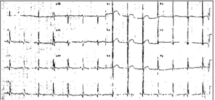
All cases with FRDA mutation who underwent elec-trocardiography (14/18) showed some abnormality and the commonest were those related to ventricular repolarization (50%) (fig. 1). We also have observed: left ventricular hy-pertrophy, signals of ischemia, atrial tachycardia, auriculo-ventricular block, sinusal bradycardia and sinusal
tachycar-dia. On the other hand, only 25% of the cases without GAA expansion, presented some abnormality in the electro-cardiogram.
Out of 17, only two patients of FRDA mutation pre-sented some abnormality in echocardiogram (2/9 – 22%), both with concentric ventricular hypertrophy.
The molecular analysis of 25 patients with a clinical diagnosis of Friedreich’s ataxia revealed the FRDA muta-tion constituted by GAA homozygous expansion in 17 ca-ses, confirming the diagnosis of Friedreich’s ataxia in these patients. All of them presented abnormalities in ECG, but only 25% of patients without GAA expansion (not-Friedreich) presented some cardiac abnormality. These data attest the importance of performing a complete cardiac evaluation in Friedreich’s ataxia, especially if we consider that only 6% (1/17) of the patients presented signals and/or symptoms of a cardiac involvement: auriculo flutter with variable block.
Discussion
In Friedreich’s original report of 1863, one patient had died of typhoid fever with a fatty infiltration in the heart. Five of the 6 patients originally described by Friedreich had cardiac involvement and, in spite of Pitt (1887) and Saury (1905) had emphasized these features, they were unnoticed until Mollaret, in 1929, reported the electrocardiographic ab-normalities present in the disease. Few studies were subse-quently published about the cardiac abnormalities obser-ved in Friedreich’s ataxia and only in 1938 did Loiseau raise the possibility that this association was not merely casual. Thereafter, many reports were published in the European literature, especially by the French, who were more interest in the cardiac aspects of Friedreich’s ataxia than were the Americans and British. Afterwards, it was realized that car-diac disturbances were, in fact, much more frequent than thought 14,15.
Cardiomyopathy, usually in its hypertrophic form, is a cardinal feature of FA. It is variable in its clinical and electric presentation and apparently, does not parallel the severity of other aspects of the disease (such as ataxia or muscle weakness). Although cardiomyopathy is typical of Friedrei-ch’s ataxia it is not exclusive of disease and maybe both, ata-xia and miocardiopathy are manifestations of a common pa-thogenic defect, most probably inherited and biochemical in nature 16.
Therefore, the cardiac disturbs in Friedreich’s ataxia ap-pear to be much more frequent than previously believed 17-19
and it is accepted that the prominence of electrocardiogra-phic defect parallels that of the neurologic deficit, in which the absent tendon reflexes in the lower extremities, extensor toe signs, scoliosis and pes cavus are most often correlated with electrocardiographic alterations 14.
The main and more frequent clinical manifestations in-dicative of myocardial involvement are rhythm disturban-ces and myocardial insufficiency terminating in a congesti-ve heart failure. These cardiac arrhythmias occur alone or in various combinations, including a complete heart block
4 5 0
Albano et al Friedreich's ataxia
Arq Bras Cardiol 2002; 78: 448-51.
with Stokes-Adams syndrome. In general, patients do not exhibit any symptomatology. Breathlessness, paroxysmal tachycardia, palpitations and thoracic pain or discomfort, and respiratory movements with short amplitude have also been reported.
Cardiac enlargement when present, occurs late in the course of the illness. Anginal pain or decubitus angina has already been reported (12%) and no correlation was found between this symptom and the coronarial changes someti-mes described in some patients 19-21. About 73% of the
patients either died of heart failure or showed some clinical evidence of cardiac dysfunction during life. Of those who died with heart failure, it was demonstrated that 25% develo-ped heart failure six months or more before death; and in 75% of them, the heart failure occurred only during the last six months of life 22. Electrocardiographic abnormalities,
ob-served in 75-100% of patients 10,22-26, were more common in
patients with absent tendon reflexes and extensor respon-ses and, in general, these electrocardiographic alterations may precede neurological signs by many years. Often, se-vere electrocardiogram abnormalities may be found in asymptomatic patients 6,20. In the present report, all typical
patients with FRDA mutation presented electrocardio-graphic abnormalities in electrocardiogram, but only 25% of patients without GAA expansion and therefore, without Friedreich’s ataxia, showed alterations in electrocardio-gram (table I).
Therefore, the best indicator of myocardial involve-ment appeared to be the presence of electrocardiographic abnormalities. It may also help to establish the diagnosis of Friedreich’s ataxia when the neurological manifestations are not altogether typical of the condition: an abnormal tracing lens support to the diagnosis, but a normal curve does not exclude it 6,10.
Child et al 27 found a normal electrocardiogram in only
8% of the cases and nonspecific ST-T wave changes were
the most common abnormality detected (75%). They also detected: abnormally broad inferolateral Q waves (13%), left ventricular hypertrophy (16%), short PR interval (24%), QRS axis with right axis deviation, tall right precordial R wa-ves. Within a family some authors observed similar patterns of electrocardiographic changes 6,19. Cardiac arrhythmias
occurred in 50% of the fatal cases (Hewer) 22 and atrial
fibri-lation occurred in 42% of them. ST-T wave changes were the most common abnormality observed (56-75%) 14,25-27.
Some authors have found severe electrocardiogram al-terations in asymptomatic patients 10. Our data were similar
to the literature and only one typical patient with the FRDA mutation presented signals of cardiac involvement. He was hospitalized because of a palpitation and lately it was iden-tified an atrial flutter with variable blockade.
The abnormalities in the echocardiogram were less fre-quent (50-73%) 10,24 and they are: asymmetric septal
hyper-trophy (9%), concentric left ventricular hyperhyper-trophy (11%), “dilated” cardiomyopathy, mitral valve prolapse, mitral val-ve motion for superior systolic displacement of the bellies of the leaflets and the coaptation point relative to the plane of the mitral anulus and for systolic anterior motion of the anterior mitral leaflet 27. In the present study, two patients
with FRDA mutation showed concentric ventricular hypertrophy (table I).
The cardiomyopathy in Friedreich’s ataxia is almost ever represented by the concentric form. Asymmetric septal hypertrophy constitute the minor part, being the most part represented by the concentric hypertrophy, which can occur in mild forms or during the beginning of the disease. In the classical hypertrophic cardiomyopathy concentric hypertrophy is a rare event, presenting frequently an asym-metric septal hypertrophy. Hypertrophic cardiomyopathy shows familial transmission and it has also been observed dominant autosomal pattern of inheritance, whereas the cardiac involvement of Friedreich’s ataxia does not have an independent transmission, being attached with neurologic abnormality 10,25,28-30.
The combination of electrocardiography and echocar-diography detected one or more abnormalities in 95% of the Child et al patients and it appears there are two fundamental-ly indifferent types of cardiac disease in Friedreich’s ataxia: a) a common dystrophic form manifested by electrocardio-graphic initial force deformities without detectable echogra-phic wall motion abnormalities, but occasionally by exten-sion throughout the left ventricle with global hypokinesia and reduced QRS voltage; and b) a hypertrophic form represented by symmetric or asymmetric left ventricular hypertrophy with normal cavity size and ventricular function 27,28.
In the present study, the definitive diagnosis of Frie-dreich’s ataxia was established in 68% of the patients with a previous clinical diagnosis of Friedreich’s ataxia (17/25) and in 89,5% of the typical cases (17/19) that fulfilled the disea-ses’s criteria 4,11 – all with electrocardiographic
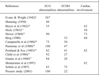
abnormaliti-es. On the other hand, only 25% of the patients without GAA expansion (not Friedreich) presented electrocardio-Table I - Abnormalities in electrocardiogram (ECG),
echocardio-gram (ECHO) and/or evidences of cardiac involvement in Friedreich’s ataxia ( %), according to the literature
References ECG ECHO Cardiac
abnormalities abnormalities involvement
Evans & Wright (1942)6 56* Manning (1950) 80
Boyer et al (1962)18 55 62
Heck (1963)19 50
Hewer (1968)22 90 73
Berg (1980) 33 60
Campanella et al (1980)24 75 50 Pasternac et al (1980)10 100 47
Pentland & Fox (1983)30 82 41 82 Child et al (1986)21 92 27
Giunta et al (1988)25 84 28
Montermini et al (1997) 82
Schöls et al (1997) 83 75 Present study (2001) 100 22
Arq Bras Cardiol 2002; 78: 448-51.
Albano et al Friedreich's ataxia
4 5 1
gram abnormalities. Therefore, it can be said that the definitive diagnosis of Friedreich’s ataxia is only likely with molecular analysis. Nevertheless, a good clinical evaluation, including a complete cardiac evaluation or, at least an electrocardiogram, helps physicians to better screening the cases which should perform a molecular analysis.
Currently, one of the biggest advantages of a clear diagnosis of Friedreich’s ataxia lies in the evidence of impro-vement in cardiac function and the therapeutic possibilities of acknowledging the neuroprotective effects of some substances with antioxidant properties 31,32.
Acknowledgments
To the members of Laboratorio do Dipartimento de Patologia Cellulare e Molecolare - Universitá Federico II, Naples, Italy, specially to Dra. Antonella Monticelli and Dr Sergio Cocozza, to perform our molecular analysis. To Dra Telma Okay and Dr. Roberto Raíz Júnior of the Laboratory of Instituto da Criança, for performing the DNA extraction. To Dra. Maria Joaquina Marques-Dias, Dr. Fernando Kok, Dr. Sérgio Rosemberg, Dr. Lúcio Coelho Miranda, Dra. Maria Bernardete Dutra Resende, Dr. Ivan Ferrareto e Dr. Jorge David Aivazoglow Carneiro, for sending their patients.
1. OMIM - Online Mendelian Inheritance in Man, OMIM ™. Center for Medical Genetics, Johns Hopkins University (Baltimore, MD) and National Center for Biotechnology Information, National Library of Medicine (Bethesda, MD), 1996. Available at: http://www3.ncbi.nlm.nih.gov:80/htbin-post/Omim/ dispmim?229300.
2. Chamberlain S, Shaw J, Rowland A, et al. Mapping of mutation causing Friedreich’s ataxia to human chromosome 9. Nat Genet 1988; 334: 248-50. 3. Campuzano V, Montermini L, Moltò MD, et al. Friedreich’s ataxia: autosomal
recessive disease caused by an intronic GAA triplet repeat expansion. Science 1996; 271: 1423-7.
4. Harding AE. Friedreich’s ataxia: a clinical and genetic study of 90 families with an analysis of early diagnostic criteria and intrafamilial clustering of clinical features. Brain 1981; 104: 589-620.
5. Finocchiaro G, Baio G, Micossi P, Pozza G, Di Donato S. Glucose metabolism alterations in Friedreich’s ataxia. Neurology 1988; 38: 1292-6.
6. Evans W, Wright G. The electrocardiogram in Friedreich disease. Br Heart J 1942; 4: 91-9.
7. Thorén C. Diabetes mellitus in Friedreich’s ataxia. Acta Paediatr (Stockholm) 1962; 135(suppl): 239-47.
8. Hewer RL. The heart in Friedreich’s ataxia. Br Heart J 1969; 31: 5-14. 9. Malo S, Latour Y, Cote M, Geoffroy G, Lemieux B, Barbeau A.
Electrocardio-graphic and vectocardioElectrocardio-graphic findings in Friedreich’s ataxia. Can J Neurol Sci 1976; 3: 323-8.
10. Pasternac A, Król, R, Petitclerc R, Harvey C, Andermann E, Barbeau A. Hy-pertrophic cardiomyopathy in Friedreich’s ataxia: symmetric or ou asymmetric? Can J Neurol Sci 1980; 7: 379-82.
11. Geoffroy G, Barbeau A, Breton G, et al. Clinical description and roentge-nologic evaluation of patients with Friedreich’s ataxia. Can J Neurol Sci 1976; 3: 279-86.
12. Miller SA, Dykes DD, Polesky HF. A simple salting out procedure for extracting DNA from human nucleated cells. Nucleic Acids Res 1988; 6: 1215. 13. Filla A, De Michele G, Cavalcanti F, Pianese L, Monticelli A, Campanella G. The
relationship between trinucleotide (GAA) repeat length and clinical features in Friedreich ataxia. Am J Hum Genet 1996; 59: 554-60.
14. Albano LMJ. Estudo molecular de portadores de ataxia de Friedreich. Tese de Doutorado Faculdade de Medicina da Universidade de São Paulo. São Paulo, 2000. 15. Albano LMJ. Genética clínica e molecular das doenças neurológicas com
mutações dinâmicas. São Paulo: Manole, 2000: 185p.
16. Barbeau A. The Quebec cooperative study of Friedreich’s ataxia: 1974-1984 - 10 years of reasearch. Can J Neurol Sci 1984b; 11(suppl): 646-60.
17. Hartman JM, Booth RW. Friedreich’s ataxia: a neurocardiac disease. Am Heart J 1960; 60: 716-20.
18. Boyer SH, Chisholm AW, McKusick VA. Cardiac aspects of Friedreich’s ataxia. Circulation 1962; 25: 493-505.
19. Heck AF. Heart disease in Friedreich’s ataxia: clinical studies and review of the literature (part I). Neurology 1963; 13: 587-95.
20. Flipse ME, Dry TJ, Woltman HW. The heart in Friedreich’s ataxia. Minnesota Med 1950; 33: 1000-03.
21. Salisachs P. La historia natural de la enfermedad de Friedreich: a proposito de 13 casos. Med Clin 1974; 63: 1-10.
22. Hewer RL. Study of fatal cases of Friedreich’s ataxia. Br Med J 1968b; 3: 649-52. 23. Andermann E, Remillard GM, Goyer C, Blitzer L, Andermann F, Barbeau A. Genetic
and family studies in Friedreich’s ataxia. Can J Neurol Sci 1976; 3: 287-301. 24. Campanella G, Filla A, De Falco F, Mansi D, Durivage A, Barbeau A. Friedreich’s
ataxia in the south of Italy: a clinical and biochemical survey of 23 patients. Can J Neurol Sci 1980; 7: 351-7.
25. Giunta A, Maione S, Biagini R, Filla A, De Michele G, Campanella G. Noninva-sive assesment of systolic and diastolic function in 50 patients with Friedrei-ch’s ataxia. Cardiology 1988; 75: 321-7.
26. Filla A, De Michele G, Caruso G, Marconi R, Campanella G. Genetic data and natural history of Friedreich’s disease: a study of 80 Italian patients. J Neurol 1990; 237: 345-51.
27. Child JS, Perloff JK, Bach PM, Wolfe AD, Perlman S, Kark P. Cardiac involvement in Friedreich’s ataxia: a clinical study of 75 patients. J Am Coll Cardiol 1986; 7: 1370-8.
28. Pellicelli AM, Borgia C, Ferranti E, et al. Caratteristiche anatomo-cliniche ed ecocardiografiche della cardiopatia in corso di atassia di Friedreich: descrizione di un caso clinico. G Ital Cardiol 1994; 24: 47-51.
29. D’Angelo A, Di Donato S, Negri G, et al. Friedreich’s ataxia in northern Italy: I. Clinical, neurophysiological and biochemical studies. Can J Neurol Sci1980; 7: 359-65.
30. Pentland B, Fox KAA. The heart in Friedreich’s ataxia. J Neurol Neurosurg Psychiatr 1983; 46: 1138-42.
31. Shulz JB, Dehmer T, Schöls L, et al. Oxidative stress in oxidative stress in patients with Friedreich’s ataxia. Neurology 2000; 55: 1719-21.
32. Sherer T, Greenamyre JT. A therapeutic target and biomarker in Friedreich’s ataxia. Neurology 2000; 55: 1600-1.
33. Manning GW. Cardiac manifestations in Friedreich's ataxia. Am Heart J 1950; 39: 799-816.
34. Berg RA, Kaplan AM, Jarrett PB, Molthan ME. Friedreich's ataxia with acute cardiomyopathy. Am J Dis Child 1980; 134: 390-3.
35. Montermini L, Richter A, Morgan K, et al. Phenotypic variability in Friedreich ataxia role of the associated GAA triplet repeat expansion. Ann Neurol 1997; 41: 675-82.
36. Schöls L, Amoiridis G, Przuntek H, Frank G, Epplen JT, Epplen C. Friedreich's ataxia: revision of the phenotype according to molecular genetics. Brain 1997; 120: 2131-40.