Arq Bras Oftalmol. 2009;72(2):257-60
Vitreorretinopatia exsudativa familiar (FEVR) associada à
osteoporose infantil: relato de caso
Trabalho realizado na Clínica Particular Ocular Oftal-mologia - Vitória (ES) - Brasil.
1Doctor in Medicine. Universidade Federal de Minas
Gerais - UFMG - Belo Horizonte (MG) - Brazil; Profes-sor of Ophthalmology - Escola Superior de Ciências da Santa Casa de Misericórdia de Vitória EMESCAM -Vitória (ES) - Brazil.
2Medical Student da Escola Superior de Ciências da
EMESCAM, Vitória (ES) - Brazil.
3Professor da Universidade Federal do Espírito Santo
-UFES - Vitória (ES) - Brazil. Vice-Director - CCHN (Cen-tro de Ciências Humanas e Naturais). PhD in Bioche-mistry and Molecular Genetics, USA. Posdoc in Molecu-lar Biology of Cancer - USA.
Endereço para correspondência: Laurentino Biccas Neto. Rua Fortunato Ramos , 411 - Vitória (ES) CEP 29055-290
E-mail: [email protected]
Recebido para publicação em 20.03.2008 Última versão recebida em 22.10.2008 Aprovação em 21.11.2008
Nota Editorial: Depois de concluída a análise do arti-go sob sigilo editorial e com a anuência da Dra. Rosane Cruz Ferreira sobre a divulgação de seu nome como revisora, agradecemos sua participação neste processo. Laurentino Biccas Neto1
Arthur Silva de Mesquita2
Iuri Drumond Louro3
Familial exudative vitreoretinopathy (FEVR)
associated with infantile osteoporosis: case report
Keywords: Osteoporosis/complications; Vitreoretinopathy, proliferative; Retinopathy of prematurity; Eye diseases, hereditary/etiology; Fluorescein angiography; Fundus oculi; Visual acuity; Vitreous body/surgery; Retinal detachment; Human; Female; Child; Case reports [Publication type]
Familial exudative vitreoretinopathy (FEVR) is an inherited blinding condition characterized by abnormal development of the retinal vascu-lature. The authors describe a rare case of the disease associated with severe infantile osteoporosis in a young female patient. The patient was submitted to multiple vitreoretinal procedures in both eyes due to tractional macular detachments. The case was complicated by diffuse uveitis of difficult control in one eye, which stimulated proliferative vitreoretinopathy and retinal redetachment. The inflammatory potential of drugs used in the control of the osteoporosis, in contrast with the inherent inflammatory activity the disease, are discussed.
ABSTRACT
RELATOS DE CASOS
INTRODUCTION
Familial exudative vitreoretinopathy (FEVR or Criswick-Schepens syndrome) is an inherited disease characterized by aberrant retinal deve-lopment in non-premature infants(1). Hallmark features include peripheral
retina non-perfusion, retinal neovascularization, subretinal exudation, formation of an abnormal vitreoretinal interface and retinal detachment(2-6).
As in retinopathy of prematurity (ROP), FEVR is a retinal vascular disease in which the peripheral retinal vessels fail to grow into the far peripheral retina. The clinical course is variable, but the most severe forms may be active for life, with variable periods of quiescence(2).
CASE REPORT
In 2001 an eight-year-old Caucasian girl born at full term was referred to one of the authors (LBN) due to visual loss in her eye and esotropia. Her parents were non-consanguineous and there was no familial history of relevant ocular problems. Since early childhood, the patient suffered from severe osteoporosis and showed reduced bone mineral density (BMD) on sequential densitometries, sustaining spontaneous fractures of the lower limbs. BMD was found to be reduced by 59% and volumetric Z scores as low as -2.55 were observed during her follow-up.
258Familial exudative vitreoretinopathy (FEVR) associated with infantile osteoporosis: case report
Arq Bras Oftalmol. 2009;72(2):257-60
normal in OU. The retina was attached in OD and temporal macular dragging could be seen bilaterally. There was trac-tional retinal detachment in OS involving the dystrophic macula temporally under thick epiretinal membranes (Figure 1). A broad linear macular fold due to posterior hyaloidal con-tracture was also present. Retinal vessels were straightened as they reached the equator and a fluorescent angiography (FA) disclosed important non-perfusion in the retinal periphery in OU (Figure 2).
She underwent retinopexy and pars plana vitrectomy in OS two months after the initial diagnosis, with delamination of the posterior hyaloid and complete peeling of the epiretinal membranes followed by silicone oil tamponade. Cryotherapy was applied to the avascular areas in the peripheral retina. Four months later, cryotherapy was also applied to the peripheral retina in OD, also targeting ischemic areas that had been seen on FA. After six months, silicone oil was removed and phacoemulsification was performed in OS. In the most recent evaluation, the retina remained reattached, with dystrophic
alterations, externally, in the macula (BCVA of 20/150 after an YAG laser capsulotomy).
As new areas of retinal non-perfusion and vessel proli-feration were seen in FA temporally in OD, new cryotherapy was undertaken in Nov/2002. After the procedure, vision wor-sened to 20/100 in this eye due to cystoid macular edema associated with epiretinal membranes. An intraocular injec-tion of triamcinolone acetonide (8 mg) was performed, resul-ting in improvement of the macular edema on optical cohe-rence tomography (OCT) and a BCVA of 20/40.
In May/2005 she complained of severe visual loss and a retinal detachment was first seen in OD. There were atrophic holes peripherally but also dense epiretinal membranes, with moderate flare (++/4), granular keratic precipitates and vitreitis (++/4). The diffuse uveitis was addressed with local and systemic steroids and a pars plana vitrectomy associated with pha-coemulsification was performed, leaving a silicone oil tampona-de. Special care was taken to dissect the posterior hyaloid. After surgery, both local and systemic steroids had to be maintained to
Figure 1 - Fundus photography of OD (A) and of OS (B and C) at presentation
C A
259 Familial exudative vitreoretinopathy (FEVR) associated with infantile osteoporosis: case report
Arq Bras Oftalmol. 2009;72(2):257-60
control local inflammation, with intense fibrin formation in the anterior chamber. Postoperatively, vision was 20/100 in OD and cystoid macular edema was seen on OCT.
Four months after the procedure, thick epiretinal membra-nes recurred around the optic disk and over the macular region, causing recurrent tractional retinal detachment. A new vitrec-tomy was undertaken in Oct/2005, with membrane peeling and a new silicone oil tamponade. Once more, uveitis flared up and was controlled by systemic and local (periocular) ste-roids, which worsened the osteoporosis. This led to the use of alendronate sodium (Fosamax; Merck & Co Inc, Whitehouse Station, NJ) systemically.
In Jan/2006 epimacular membranes returned, leading again to macular tractional detachment in OD and a new pars plana vitrectomy and membrane peeling was done under silicone oil. As she evolved favorably, on May/2006 the silicone oil was removed along with delicate epimacular membrane peeling and a posterior capsulotomy, leaving the eye with a gas tamponade (C3F8). The retina remained reattached for one year in OD, with an IOP of 15 mmHg and no recurrence of epiretinal membranes. The BCVA was 20/80 but every attempt to taper the steroids was followed by a flare up of uveitis. A year after the last procedure, there was severe recurrence of inflammation in this eye - despite sustained systemic steroids (0.5 mg/kg/daily) - leading to a total retinal redetachment associated with exuberant vitreoretino-pathy. This time, no further surgery was indicated and the case was considered to be unsolvable.
DISCUSSION
There are very few reports of FEVR in Brazilian patients(7-8),
but this disease may be more common than is currently appre-ciated(9).
We have described a young girl with osteoporosis, redu-ced bone mass and a retinal phenotype typical of FEVR, including avascularity of a large portion of the peripheral
retina, reduced visual acuity, temporal dragging of the vessels emanating from the optic disc, epiretinal membranes and, eventually, tractional retinal detachment.
Diffuse and rebel uveitis occurred in one eye and may be related to poor visual outcome. Inflammatory elements are found histologically in eyes with FEVR, supporting the pro-liferative process(10) and may account for the uveitic process.
The association of juvenile osteoporosis with FEVR-associated uveitis posed a therapeutic dilemma: steroids had to be used systemically to control inflammation, worsening the balance in bone mass gain. On the other hand, bisphosphonates as alen-dronate - used as potent inhibitors of osteoclast-mediated nor-mal and abnornor-mal bone resorption - have been associated with ocular inflammation including iritis, nonspecific conjunc-tivitis, anterior uveitis, episcleritis, and scleritis(11-14). Every
at-tempt to taper systemic steroids - including periocular injec-tions of triamcinolone acetonide - was followed by a flare up of the uveitis, but aggravation of osteoporosis - complicated by spontaneous fractures of the legs - did not allow discontinuation of bisphosphonates. It is possible that cryotherapy may have triggered the inflammatory response in OD, as some patients with FEVR evolve unfavorably with this approach(7).
The association of FEVR with osteoporosis, although rare, should be remembered to avoid early complications such as spontaneous fractures and bone malformation in childhood.
ACKNOWLEDGMENTS
We thank Dr. Sergio Ragi Eis for his most valuable help in the control of the osteoporosis.
RESUMO
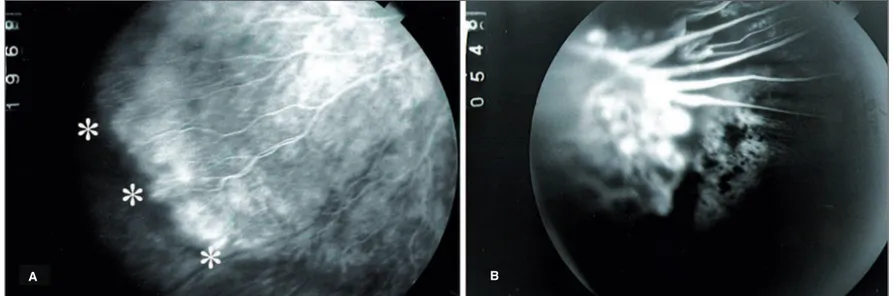
Relato de um caso incomum de vitreorretinopatia exsudativa familiar (FEVR) bilateral em criança do sexo feminino porta-dora de grave osteoporose infantil. A paciente foi submetida a Figure 2 - Peripheral retinal non-perfusion on the FA in OD (A) and in OE (B)
260Familial exudative vitreoretinopathy (FEVR) associated with infantile osteoporosis: case report
Arq Bras Oftalmol. 2009;72(2):257-60
vitrectomias sucessivas para correção de descolamentos tra-cionais maculares em ambos os olhos. O quadro foi complica-do em um olho por uveíte difusa de difícil controle, que esti-mulava o aparecimento de proliferação vitreorretiniana de di-fícil abordagem, responsável por redescolamentos sucessivos da retina. Discute-se sobre o potencial pró-inflamatório de drogas usadas no controle da osteoporose, em contraste com a atividade inflamatória inerente à doença.
Descritores: Osteoporose/complicações; Vitreorretinopatia
proliferativa; Retinopatia da prematuridade; Oftalmopatias hereditárias/etiologia; Angiofluoresceinografia; Fundo de olho; Acuidade visual; Corpo vítreo/cirurgia; Descolamento reti-niano; Humano; Feminino; Criança; Relatos de casos [Tipo de publicação]
REFERENCES
1. Criswick VG, Schepens CL. Familial exudative vitreoretinopathy. Am J Ophthal-mol. 1969;68(4):578-94.
2. Benson WE. Familial exudative vitreoretinopathy. Trans Am Ophthalmol Soc. 1995;93:473-521.
3. Ebert EM, Mukai S. Familial exudative vitreoretinopathy. Int Ophthalmol Clin. 1993;33(2):237-47.
4. Gole GA, Goodall K, James MJ. Familial exudative vitreoretinopathy. Br J Ophthalmol. 1985;69(1):76.
5. Gow J, Oliver GL. Familial exudative vitreoretinopathy. An expanded view. Arch Ophthalmol. 1971;86(2):150-5.
6. Tasman W, Augsburger J, Shields J, Caputo A, Annesley WH Jr. Familial exudative vitreoretinopathy. Trans Am Ophthalmol Soc. 1981;79:211-26. 7. Portella E, Ramos ARB, Moreira JCA, Moreira ATR. Manejo da
vitreoreti-nopatia exsudativa familial. Rev Bras Oftalmol. 1995;54(8):565-8. 8. Abreu GBM, Abreu M. Vitreo-retinopatia familial exsudativa: relato de uma
família. Arq Inst Penido Burnier. 1992;34(1):11-6.
9. Vianna RNG, Serop S, Meire F. Familial exudative vitreoretinopathy. Rev Bras Oftalmol. 1994;53(1):19-23.
10. Boldrey EE, Eg, Gass JD, Friberg T. The histopathology of familial exuda-tive vitreoretinopathy. A report of two cases. Arch Ophthalmol. 1985;103(2): 238-41.
11. Siris ES. Bisphosphonates and iritis. Lancet. 1993;341(8842):436-7. 12. Mbekeani JN, Slamovits TL, Schwartz BH, Sauer HL. Ocular inflammation
associated with alendronate therapy. Arch Ophthalmol. 1999;117(6):837-8. 13. Leung S, Ashar BH. Miller RG. Bisphosphonate-associated scleritis: a case
report and review. South Med J. 2005;98(7):733-5.