Arq Bras Oftalmol. 2009;72(5):622-5
Comparação entre as medidas da espessura da camada de fibras nervosas da
retina e da mácula pela tomografia de coerência óptica na detecção da
perda axonal progressiva decorrente à neuropatia óptica traumática
From the Division of Ophthalmology, University of São Paulo Medical School, São Paulo, Brazil. 1Pós-graduando (Doutorado) da divisão de Clínica
Oftal-mológica do Hospital das Clínicas da Faculdade de Medicina da Universidade de São Paulo - USP - São Paulo (SP) - Brazil.
2Pós-graduanda (Doutorado) da divisão de Clínica Oftal-mológica do Hospital das Clínicas da Faculdade de Medicina da USP - São Paulo (SP) - Brazil. 3Professor Livre Docente da Clínica Oftalmológica do
Hospital das Clínicas da USP - São Paulo (SP) - Brazil. 4Livre-docente, Professor Adjunto da divisão de Clínica OftalmológiClínica da Faculdade de Medicina da USP -São Paulo (SP) - Brazil
Address for correspondence: Leonardo Provetti Cu-nha. Av. Barão do Rio Branco, 4.051 - Juiz de Fora (MG) CEP 36021-630
E-mail: [email protected] Recebido para publicação em 28.10.2008 Última versão recebida em 03.08.2009 Aprovação em 04.08.2009
Leonardo Provetti Cunha1
Luciana Virginia Ferreira Costa-Cunha2
Roberto Freire Santiago Malta3
Mário Luiz Ribeiro Monteiro4
Comparison between retinal nerve fiber layer
and macular thickness measured with OCT
detecting progressive axonal loss following
traumatic optic neuropathy
Keywords: Optic nerve injuries; Tomography, optical coherence; Retina; Retinal ganglion cells; Nerve fibers; Macula; Visual acuity
Purpose: To compare the optical coherence tomography retinal nerve fiber layer and macular thickness measurements for detection of pro-gressive axonal loss following acute traumatic optic neuropathy in a longitudinal study. Methods: Three patients with unilateral traumatic optic neuropathy were evaluated sequentially after trauma. Macular and retinal nerve fiber layer thickness measurements were obtained using optical coherence tomography weekly for five weeks and around the twelfth week after trauma. Results: All patients showed progressive macular and retinal nerve fiber layer thickness reduction. The mean retinal nerve fiber layer thickness on the first week was 114 µm and reduced sequentially over the first five weeks and was 46 µm on the
twelfth week. For macular parameters, the mean average thickness on the first week was 248 µm and also reduced over the first five weeks and was
218 µm on the twelfth week. When compared to the initial measurement,
macular thickness average reduction rate at the 12th week was 14% while peripapillary retinal nerve fiber layer thickness average reduction rate was 59%. Conclusions: Although both measurements reduce signifi-cantly after trauma, retinal nerve fiber layer thickness measurements show greater and faster retinal neural reduction if compared to macular thickness measurements in traumatic optic neuropathy.
ABSTRACT
INTRODUCTION
623
Comparison between retinal nerve fiber layer and macular thickness measured with OCT detecting progressive axonal loss following traumatic optic neuropathy
Arq Bras Oftalmol. 2009;72(5):622-5
used for such purpose particularly in eyes with optic disc anomalies or edema. Although several studies have investi-gated the changes in RNFL and macular thickness measu-rements in optic pathway diseases, no study has investigated longitudinally which set of measurements is altered first in the course of optic nerve diseases.
Although previous studies have documented in single ca-se-reports that OCT is able to document both RNFL(3) and macular thickness(4) measurements in traumatic optic neuro-pathy (TON), no study has performed a direct comparison between RNFL and macular thickness measurements in such patients. The purpose of this study was to evaluate longitudi-nally the RNFL and macular thickness measurements follo-wing TON in three patients using OCT and to compare RNFL and macular thickness measurements documenting progres-sive retinal axonal loss.
METHODS
Three eyes of three patients with TON were examined sequentially for three months after trauma. All patients had a history of acute indirect traumatic optic nerve injury, without any trauma to the eye (Table 1).
Systemic, neurological and neuro-imaging findings were unremarkable in all patients. Patient characteristics and visual outcomes are summarized in Table 1. All patients showed acute and severe visual loss with relative afferent pupillary defect on the affected eye, a normal looking fundus examination at the first examination, and developed severe optic disc pallor on follow-up. All patients were treated with intravenous me-thylprednisolone (1 gram/day for 3 days) followed by oral prednisone tapering in five weeks.
Patients underwent ocular imaging with dilated pupils using a commercially available Stratus-OCT (Carl Zeiss Me-ditec, Dublin, California, USA). Peripapillary RNFL and ma-cular thickness scans were obtained weekly from the first to the fifth week after trauma. Measurements were also obtained at the twelfth week after the optic nerve injury. Quality asses-sment of Stratus OCT scans was evaluated by an experienced examiner. Good-quality scans had to have focused images and signal strength equal to or higher than 7,and presence of a centered ring around the optic disc for RNFL scans. For macula scans, the radial scans had to be centered on the fovea. External fixation was used since all patients presented severe visual loss in the study eye. Approval from the Institutional Review Board Ethics Committee was obtained for the study.
The study followed the principles of the Declaration of Helsinki and informed consent was obtained from all participants.
The fast RNFL algorithm was used to obtain RNFL thi-ckness measurements with Stratus-OCT. Three images were acquired from each subject, with each image consisting of 256 A-scans along a 3.4 mm-diameter circular ring around the optic disc. Peripapillary (360º measure) RNFL thickness average was automatically calculated by existing Stratus-OCT soft-ware (version 4.0.1). The fast macular thickness protocol was used to obtain macular thickness measurements. Average ma-cular thickness was calculated as the weighted average of the sectoral macular thickness measurements excluding the fovea. In order to improve the reproducibility, for each session at least three scans protocols for both RNFL and macular mea-surements in each patient were performed.
RESULTS
Three patients showed progressive reduction of both ma-cular and peripapillary RNFL average thickness measure-ments after TON documented sequentially by OCT (Figures 1 and 2). Table 2 shows macular and RNFL thickness measure-ments along the first five weeks after trauma with remarkable reduction in the twelfth week. Based on the values from a large series of normal individuals recently examined with the same equipment (Stratus-OCT) at our institution(5) and using 2 standard deviation as the lower limit of normal, values below 223 µm and 88 µm were considered as the lower limit
of normal for macular and RNFL thickness measurements, respectively. When these values were used, average RNFL thickness reduction became more apparent 4 weeks after trau-ma although some reduction was observed 2 weeks after trauma. Using the same reasoning, macular thickness reduc-tion became apparent only at the 12th week after trauma al-though some reduction could be seen at least at the 5th week after trauma (Table 2).
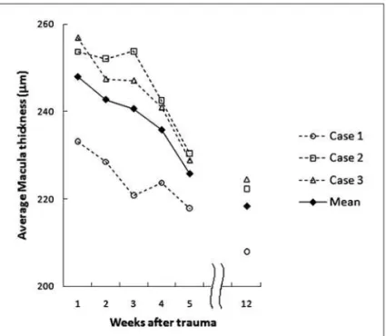
When compared to the values obtained at the first measure-ment after trauma, macular thickness at the 12th week was reduced in 11%, 14% and 14% in cases one, two and three, respecti-vely. Average macular reduction rate in the three patients was 14% (Table 2, Figure 1). Accordingly, based on the values from the first measurement after trauma, RNFL thickness measure ment reduction rate at the 12th week was 56%, 6 4% and 58% in cases one, two and three, respectively. Average RNFL thickness reduction rate in the three cases was 59% (Table 2, Figure 2).
Table 1. Characteristics and visual outcomes of three patients with traumatic optic neuropathy
Case Age Gender Eye Initial VA Type of injury Final VA
1 24 Female OD LP Stab injury to the orbit HM
2 20 Male OD LP Head trauma after fall HM
3 34 Female OS HM Head trauma after bicycle fall 20/400
624Comparison between retinal nerve fiber layer and macular thickness measured with OCT detecting progressive axonal loss following traumatic optic neuropathy
Arq Bras Oftalmol. 2009;72(5):622-5
DISCUSSION
Most studies have employed RNFL thickness to evaluate axonal damage, mainly for glaucoma patients but also in other anterior pathway optic nerve diseases, such as demyelinating and compressive optic neuropathies(5). More recently, several studies have shown that macular thickness measurements can also be used as indicator of neural damage(6-9). As the ganglion cell layer accounts for up to 40% of the thickness in the macular
area, estimation of macular thickness can be used to investigate possible ganglion cell loss(6,8). Although RNFL measurements are usually considered the most reliable way to quantify axonal loss, the use of macular thickness measurements as an estimator of neural loss may be of interest in some cases, particularly if paripapillary RNFL thickness assessment is impaired in con-ditions such as optic disc edema, optic disc anomalies and in cases of large peripapillary scarring or atrophies. In fact, pre-vious studies have shown that OCT macular thickness measu-rements are significantly thinner in many optic nerve diseases, like glaucoma(6,8) and band atrophy of the optic nerve from chiasmal compression(2) and could be a useful tool for clinical assessment of ganglion cell loss in many optic nerve diseases.
Of interest, RNFL thickness represent the measurement of all retinal nerve fibers entering the optic disc, while macular thickness represents the measurement of the retina in macular area(10). Studies comparing macular and RNFL thickness mea-surements are important for understanding how they can be used for diagnosis and during follow-up in conditions with nerve phase loss, or optic nerve diseases. Furthermore, it might be possible that both sets of measurements can be used in association to increase diagnostic sensitivity and specificity.
Recently, four reports(11-14) documented RNFL loss after TON using scanning laser polarimetry (SLP) and showed that RNFL thickness measurements first decreased 4 to 9 weeks after trauma. On the other hand some authors(3), using OCT after TON, found that RNFL thickness measurements started to decrease 20 days after the trauma, suggesting that OCT can show RNFL loss earlier when compared SLP. Recently, a case of progressive macular thinning after severe indirect TON was documented using OCT(4). Progressive reduction of macular thickness parameters was observed in conjunction with thin-ning of RNFL thickness. At time 77 days after trauma, severe thinning of the macular thickness parameters was observed.
In the present study, all three patients showed progressive macular and RNFL thickness reduction documented sequen-tially by OCT after trauma. RNFL thickness measurements showed greater reduction than macular thickness measure-ments. The reduction rate of RNFL thickness was greater than 55% in all cases, while for the macular thickness parameters, the reduction rate was approximately 13%. Furthermore, RNFL thickness measurements reduced at least 2 weeks earlier than macular thickness measurement.
CONCLUSION
The current study indicates that both macular and RNFL thickness measurements may be used to assess and monitor axonal loss after acute optic nerve diseases such as traumatic optic neuropathy. Although both sets of measurements can be used to monitor retinal neural damage, clinicians should be aware that macular thickness measurements show a smaller and later reduction in size when compared to RNFL thickness measurements.
Figure 1 - Macular thickness measurements (µµµµµm) over the first five
weeks and in the twelfth week in three patients after acute traumatic optic neuropathy
Figure 2 - Retinal nerve fiber layer thickness measurements (µµµµµm) over
625
Comparison between retinal nerve fiber layer and macular thickness measured with OCT detecting progressive axonal loss following traumatic optic neuropathy
Arq Bras Oftalmol. 2009;72(5):622-5
RESUMO
Objetivo: Comparar as medidas da espessura da camada de fibras nervosas da retina e macular obtidas pela tomografia de coerência óptica na detecção da perda axonal progressiva após neuropatia óptica traumática aguda e durante o segui-mento clínico. Métodos: Três pacientes com neuropatia óp-tica traumáóp-tica unilateral aguda foram avaliados sequencial-mente após o trauma. Medidas da espessura macular e da camada de fibras nervosas da retina foram obtidas pela to-mografia de coerência óptica semanalmente por 5 semanas consecutivas e ao redor da décima segunda semana após o trauma. Resultados: Todos os pacientes apresentaram redu-ção progressiva dos valores da espessura macular e da ca-mada de fibras nervosas da retina. A espessura média da camada de fibras nervosas da retina foi de 114 µm na primeira semana e reduziu sequencialmente ao longo das primeiras cinco semanas e foi de 46 µm na décima segunda semana.
Para parâmetros macular, a espessura média foi de 248 µm na primeira semana, e também reduziu ao longo das primeiras cinco semanas e foi de 218 µm na décima segunda semana.
Quando comparado às medidas iniciais, a taxa de redução das médias da espessura macular foi 14% na décima segunda semana após o trauma, enquanto que a taxa de redução das médias da espessura da camada de fibras nervosas da retina foi 59%. Conclusões: Os valores da espessura da camada de fibras nervosas da retina apresentaram uma redução maior e mais rápida se comparada às medidas da espessura macular na neuropatia óptica traumática.
Descritores: Traumatismos do nervo óptico; Tomografia de coerência óptica; Retina; Fibras nervosas; Células ganglio-nares da retina; Mácula; Acuidade visual
REFERENCES
1. Huang D, Swanson EA, Lin CP, Schuman JS, Stinson WG, Chang W, et al. Optical coherence tomography. Science. 1991;254(5035):1178-81.
2. Moura FC, Medeiros FA, Monteiro ML. Evaluation of macular thickness measurements for detection of band atrophy of the optic nerve using optical coherence tomography. Ophthalmology. 2007;114(1):175-81.
3. Medeiros FA, Moura FC, Vessani RM, Susanna R Jr. Axonal loss after traumatic optic neuropathy documented by optical coherence tomography. Am J Ophthalmol. 2003;135(3):406-8.
4. Vessani RM, Cunha LP, Monteiro ML. Progressive macular thinning after indirect traumatic optic neuropathy documented by optical coherence tomogra-phy. Br J Ophthalmol. 2007;91(5):697-8.
5. Costa-Cunha LV, Cunha LP, Malta RF, Monteiro ML. Comparison of Fourier-domain and time-Fourier-domain optical coherence tomography in the detection of band atrophy of the optic nerve. Am J Ophthalmol. 2009;147(1):56-63.
6. Greenfield DS, Bagga H, Knighton RW. Macular thickness changes in glauco-matous optic neuropathy detected using optical coherence tomography. Arch Ophthalmol. 2003;121(1):41-6.
7. Guedes V, Schuman JS, Hertzmark E, Wollstein G, Correnti A, Mancini R, et al. Optical coherence tomography measurement of macular and nerve fiber layer thickness in normal and glaucomatous human eyes. Ophthalmology. 2003; 110(1):177-89.
8. Lederer DE, Schuman JS, Hertzmark E, Heltzer J, Velasquez LJ, Fujimoto JG, et al. Analysis of macular volume in normal and glaucomatous eyes using optical coherence tomography. Am J Ophthalmol. 2003;135(6):838-43.
9. Medeiros FA, Zangwill LM, Bowd C, Vessani RM, Susanna R Jr, Weinreb RN. Evaluation of retinal nerve fiber layer, optic nerve head, and macular thickness measurements for glaucoma detection using optical coherence tomo-graphy. Am J Ophthalmol. 2005;139(1):44-55.
10. Garway-Heath DF, Holder GE, Fitzke FW, Hitchings RA. Relationship bet-ween electrophysiological, psychophysical, and anatomical measurements in glaucoma. Invest Ophthalmol Vis Sci. 2002;43(7):2213-20.
11. Kuo MT, Lai IC, Teng MC. Serial follow-up in traumatic optic neuropathy using scanning laser polarimetry and visual field testing. Chang Gung Med J. 2005; 28(8):581-6.
12. Miyahara T, Kurimoto Y, Kurokawa T, Kuroda T, Yoshimura N. Alterations in retinal nerve fiber layer thickness following indirect traumatic optic neuropathy detected by nerve fiber analyzer, GDx-N. Am J Ophthalmol. 2003;136(2):361-4. 13. Medeiros FA, Susanna R Jr. Retinal nerve fiber layer loss after traumatic optic neuropathy detected by scanning laser polarimetry. Arch Ophthalmol. 2001; 119(6):920-1.
14. Meier FM, Bernasconi P, Stürmer J, Caubergh MJ, Landau K. Axonal loss from acute optic neuropathy documented by scanning laser polarimetry. Br J Ophthalmol. 2002;86(3):285-7.
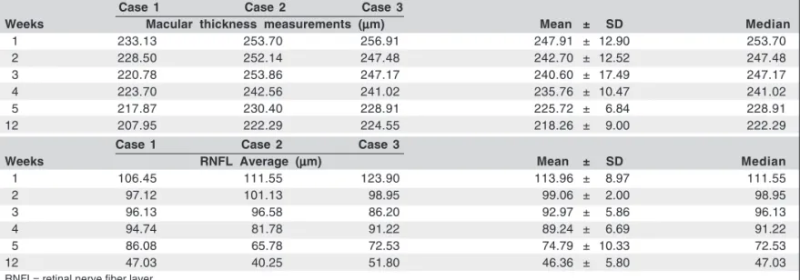
Table 2. Macular and RNFL thickness measurements along the first five weeks and later in twelfth week, in three eyes with traumatic optic neuropathy
Case 1 Case 2 Case 3
Weeks Macular thickness measurements (µµµµµm) Mean ± SD Median
1 233.13 253.70 256.91 247.91 ± 12.90 253.70
2 228.50 252.14 247.48 242.70 ± 12.52 247.48
3 220.78 253.86 247.17 240.60 ± 17.49 247.17
4 223.70 242.56 241.02 235.76 ± 10.47 241.02
5 217.87 230.40 228.91 225.72 ± 6.84 228.91
12 207.95 222.29 224.55 218.26 ± 9.00 222.29
Case 1 Case 2 Case 3
Weeks RNFL Average (µµµµµm) Mean ± SD Median
1 106.45 111.55 123.90 113.96 ± 8.97 111.55
2 97.12 101.13 98.95 99.06 ± 2.00 98.95
3 96.13 96.58 86.20 92.97 ± 5.86 96.13
4 94.74 81.78 91.22 89.24 ± 6.69 91.22
5 86.08 65.78 72.53 74.79 ± 10.33 72.53
12 47.03 40.25 51.80 46.36 ± 5.80 47.03