INTRODUCTION
In 1904, Teleky described the irst case of esophageal cancer (EC) after corrosive ingestion. It is known in this patient population the incidence of EC is 1%-4% and the duration between caustic ingestion and development of EC is 15-30 years(11,18,21). Alkalis corrosive agents combine with tissue proteins and cause liquefac-tive necrosis and saponiication, and penetrate deeper into tissues, helped by a higher viscosity and a longer contact time through the esophagus. Additionally, alkali absorption leads to thrombosis in blood vessels, impeding blood low to already damaged tissue(6).
Esophageal neoplasms (both adenocarcinoma and squamous cell carcinoma) may develop as a late complication of caustic injury at a rate 1000-3000 times higher than expected in patients of a similar age(22). When squamous cell carcinoma (SCC) occurs in these patients it appears in the stenotic ring and may be due to repeated trauma of dilation, slow passage of food (stasis), relux
Lugol’s iodine chromoendoscopy versus Narrow
Band Image enhanced endoscopy for the detection
of esophageal cancer in patients with stenosis
secondary to caustic/corrosive agent ingestion
Caterina Maria Pia Simoni
PENNACHI
1, Diogo Turiani Hourneaux de
MOURA
1,
Renato Bastos Pimenta
AMORIM
2, Hugo Gonçalo
GUEDES
1, Vivek
KUMBHARI
3and
Eduardo Guimarães Hourneaux de
MOURA
1Received 27/10/2016 Accepted 30/1/2017
ABSTRACT – Background – The diagnosis of corrosion cancer should be suspected in patients with corrosive ingestion if after a latent period of negligible symptoms there is development of dysphagia, or poor response to dilatation, or if respiratory symptoms develop in an otherwise stable patient of esophageal stenosis. Narrow Band Imaging detects supericial squamous cell carcinoma more frequently than white-light imaging, and has signiicantly higher sensitivity and accuracy compared with white-light. Objective – To determinate the clinical applicability of Narrow Band Imaging versus Lugol´s
solution chromendoscopy for detection of early esophageal cancer in patients with caustic/corrosive agent stenosis. Methods – Thirty-eight patients,
aged between 28-84 were enrolled and examined by both Narrow Band Imaging and Lugol´s solution chromendoscopy. A 4.9mm diameter endoscope was used facilitating examination of a stenotic area without dilation. Narrow Band Imaging was performed and any lesion detected was marked for later biopsy. Then, Lugol´s solution chromoendoscopy was performed and biopsies were taken at suspicious areas. Patients who had abnormal indings
at the routine, Narrow Band Imaging or Lugol´s solution chromoscopy exam had their stenotic ring biopsied. Results – We detected nine suspicious
lesions with Narrow Band Imaging and 14 with Lugol´s solution chromendoscopy. The sensitivity and speciicity of the Narrow Band Imaging was 100% and 80.6%, and with Lugol´s chromoscopy 100% and 66.67%, respectively. Five (13%) suspicious lesions were detected both with Narrow Band
Imaging and Lugol’s chromoscopy, two (40%) of these lesions were conirmed carcinoma on histopathological examination. Conclusion – Narrow Band
Imaging is an applicable option to detect and evaluate cancer in patients with caustic /corrosive stenosis compared to the Lugol´s solution chromoscopy.
HEADINGS – Esophageal neoplasms. Squamous cell carcinoma. Caustics, adverse effects. Iodides, therapeutic use.
Declared conflict of interest of all authors: none Disclosure of funding: no funding received
1 Serviço de Endoscopia Gastrointestinal do Departamento de Gastroenterologia da Faculdade de Medicina da Universidade de São Paulo (FM-USP), SP, Brasil; 2 Departamento de Gastro-enterologia, Irmandade da Santa Casa de Misericórdia de São Paulo, SP, Brasil; 3 Bariatric Endoscopy, Division of GI & Hepatology, Johns Hopkins Bayview, The Johns Hopkins School of Medicine, Baltimore, Maryland, USA.
Correspondence: Caterina Maria Pia Simoni Pennacchi. Departamento de Endoscopia Gastrointestinal, Faculdade de Medicina da Universidade de São Paulo. Rua Carvalho Aguiar, 255, 6º andar – CEP: 05422-090 – São Paulo, SP, Brasil. E-mail: [email protected]
of the food due to the shortening of the esophagus and of the scarring process itself(1,9,18,19).
One-stage resection and reconstruction is the best way to treat the radically operable patients. In patients with esophageal cor-rosive stricture in need of operation, both a bypass procedure and resection can be performed, but it should be pointed out that malignancy may develop even years after the operation in the remaining part of the gullet. Total esophagectomy is therefore suggested instead of by-pass(17).
The diagnosis of corrosion cancer should be suspected in pa-tients with corrosive ingestion if after a latent period of negligible symptoms there is development of dysphagia, or poor response to dilatation, or if respiratory symptoms develop in an otherwise stable patient of esophageal stenosis(25).
Currently, there are many diagnostic methods available to facili-tate the early detection of EC, among which upper GI endoscopy, is the most important. Upper endoscopy when combined with imaging magniication and chromoscopy may be the key to improv-ing diagnosis and may subsequently improve survival. The need to develop techniques to improve the early diagnosis of esophageal SSC without side effects has spawned research into new endoscopic optical techniques. For example, NBI which was demonstrated irst in 1994, which uses spectroscopy(2,14).
The technique known as “Narrow Band Imaging” (NBI), uses narrow light spectrum, which penetrates in the supericial tissues, allowing the identiication of small blood vessels and details of the mucosal pattern(2). With the NBI technique the normal mucosa appears light blue/green and the vessels brown, lighting up suspi-cious lesions, similarly to Lugol’s chromoscopy(5,16). This allows for demarcation of suspicious areas and hence targeted biopsy(2,18). NBI detects supericial SCC more frequently than white-light imaging, and has signiicantly higher sensitivity and accuracy compared with white-light. In addition, narrowband imaging magnified endoscopy (NBI-ME) is useful for differentiating cancerous from non-cancerous lesions and assessing tumor depth and invasion by analysis of the microvascular patterns. Generally, the presence of irregular loop-shaped microvessels suggests noninvasive cancer in the mucosal layer, and thick non-looped vessels suggest invasive cancer in typical SCC(13).
The aim of this study is to determinate the clinical applicability of Narrow Band Imaging versus Lugol’s solution chromendoscopy for detection of early esophageal cancer in patients with caustic/ corrosive agent stenosis.
METHODS
This is a single center, observational transversal trial, cross-over study, that enrolled 38 patients with esophageal stenosis secondary to corrosive ingestion who underwent upper GI endoscopy with NBI and Lugol’s iodine chromoendoscopy at the Gastrointestinal Endoscopy Unit of Hospital das Clínicas da Universidade de São Paulo. This project was granted approval by the Ethics Committee, and was registered in the Sao Paulo University Ethics Committee was number 1087/06.
Patients were included if they had their ingestion >20 years ago and had esophageal stenosis secondary to caustic lesion caused by ingestion. Informed consent was obtained in all patients. Patients were excluded if they had their caustic injury in the last 20 years, refused to consent, had an iodine allergy, had under 21 years age or refused to undergo upper endoscopy.
The gold standard were histopathological, surgery and follow-up.
Endoscopic procedure
Upper gastrointestinal endoscopy was performed with the vid-eogastroscope (Olympus 180 – GIF Type N180 “slimsight”) with 4.9mm diameter by only one endoscopist (E.G.H.M., with more than 25 years’ experience in experience of conventional endoscopy). He had experienced more than 70,000 esophagogastroduodenosco-pies and had more than 6 years of experienced with NBI.
The exam sequence was:
Monitorization with pulse oximetry and oxygen catheter, intravenous access, sedation with midazolam (0.025 – 0.1 mg/kg), associated to fentanyl (0.7 - 2µg/kg) and propofol (0.5 – 1mg/kg).
Conventional endoscopic exam and removal of luids and secre-tion of the esophagus with saline and N-Acetylcysteine.
Exam with image enhanced endoscopy (NBI) and when the eventually lesions were found, were taken notes about their locali-zation in relation to incisors and esophageal walls.
Then, Lugol’s iodine solution 1.25% chromoscopy was per-formed and eventual found lesion were taken notes similarly to NBI exam.
Previously marked suspicious lesions were biopsied with dispos-able forceps. If no lesion was identiied, then the stenotic ring was biopsied and sent for histopathological examination.
The lesion was considered suspicious (positive indings) when presents Lugol voiding area >5 mm in Lugol’s chromoendoscopy and brownish and dark brown spots in NBI chromoendoscopy (Figures 1 and 2).
FIGURE 1. Narrow Band Imaging (1A) and Lugol (1B) showing suspicious lesion that was negative to carcinoma.
FIGURE 2. Narrow Band Imaging (1A) and Lugol (1B) showing a suspicious lesion with irregular surface and positive to carcinoma.
No suspicious areas were considered negative indings. The chromoendoscopy exam with Lugol’s iodine and NBI have had the same execution time.
Statistical analysis
RESULTS
Thirty-eight patients (22 female – 57.8%) with a median age of 56 years (28-84) were enrolled. In this study, abnormal (pre-malignant or (pre-malignant) lesions no were detected during regular endoscopy. The median length of follow-up in the entire cohort was 4 months. Of these, 29 (76.3%) patients had negative indings and 9 (23.7%) positive indings with NBI. Lugol identiied 24 (63.2%) negative indings and 14 (36.8%) positive indings identiied. All the suspected lesions were located adjacent to stenosis. Histo-pathologic analysis showed 36 (94.7%) no cancer biopsies versus 02 (5.3%) SCC anatomopathological diagnosis. All the patients are followed annually, and additional controls exams were performed on patients with negative indings or dysplasia, that not conirmed positive cancer samples.
Lugol iodine chromoendoscopy identiied 14 suspicious areas (Lugol voiding area) and two (14.3%) were conirmed as SCC. There was no case of neoplastic disease not identiied by Lugol iodine chromoendoscopy resulting in a sensitivity was 100% (exact Fisher’s Test 0.1294). The speciicity was 66.67%. Therefore, there were 12 (85.7%) false positives. The positive predicted value was of 14.3% and the negative predictive value was 100%. The likelihood ratio of a positive test was of 2.9, and its negative correspondent doesn’t exist. The accuracy of this diagnostic test was of 73% (Table 1).
When assessing the conformity between the NBI and the Lu-gol’s chromoendoscopy in diagnosis of EC, both methods revealed ive (55.6%) positive indings, two of those were cases of neoplasia. Nine (31%) negative indings with the NBI technique and positive with Lugol’s solution; four (44.4%) negative indings with Lugol’s solution and positive with NBI technique. Twenty (69%) were negative indings in both methods (Table 3A and 3B).
TABLE 1. Evaluation of conformity beetwen chromoscopy with Lugol’s solution and the result of the histopathological exam in the detection of early esophageal cancer in patients with esophagus stenosis secondary to caustic ingestion. According to this, there is a week conformity between the tests (Kappa = 0.174).
Kappa 0.174 [-0.213 – 0.561]
Sensitivity 100.00% [34.2 –100.0]
Speciicity 66.67% [50.3 – 79.8]
Positive predictive value 14.30% [4.0 – 39.9] Negative predictive value 100.00% [86.2 – 100.0] Likelihood ratio of a positive test 2.9
Likelihood ratiof of a negative test Inexistent
Accuracy 0.73
TABLE 2. Evaluation of conformity beetwen the NBI technique and the histopathological exam. According to this, there is a week conformity between the tests (Kappa = 0.239).
Kappa 0.304 [-0.162 – 0.77]
Sensitivity 100% [34.2 –100.0]
Speciicity 80.56% [65.0 – 790.2]
Positive predictive value 22.20% [6.3 – 54.7]
Negative predictive value 100% [74.1 – 100.0]
Likelihood ratio of a positive test 5 Likelihood ratio of a negative test 0.0
Accuracy 0.81
NBI identiied nine suspicious areas (brownish and dark brown spots) considered positive indings which 2 (22.2%) were conirmed as SCC in the histopathological examination. Seven of nine were false positives, since brownish areas were negative for SCC in the histopathological examination. According to the table there was no case of neoplasia that wasn’t identiied by the NBI technique, 29 were true negatives (exact Fisher’s Test 0.0512) (Table 2).
TABLE 3A. Evaluation of conformity between the NBI technique and the Lugol’s chromoendoscopy for detection of early esophageal cancer in patients with esophageal stenosis secondary to caustic lesion.
Esophagus neoplasia
Lugol Exact Fisher’s test Positive Negative
NBI 0.245
Positive 5 (55.6%) 4 (44.4%)
Negative 9 (31.0%) 20 (69.0%)
TABLE 3B. Evaluation of conformity between the NBI technique and the Lugol’s chromoendoscopy for detection of early esophageal cancer in patients with esophagus stenosis secondary to caustic lesion. According to this, there is a weak conformity beetwen the techniques (Kappa = 0.206).
Kappa 0.206 [-0.144 – 0.556]
Sensitivity 35.70% [16.3 – 61.2]
Speciicity 83.30% [64.1 – 93.3]
Positive predictive test 55.60% [26.7 – 81.1]
Negative predictive test 69.00% [50.8 – 82.7]
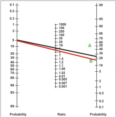
To improve the data analysis the Fagan’s Nomogram was used with the likelihood ratio of a positive test for both methods. There-fore, the likelihood ratio of a positive test was 2.9 for the Lugol and 5 for the NBI (Figure 3).
Figure 3 shows that the pre test probability of EC in these patients was 5%. Using the likelihood ratio of a positive test for the NBI (A, Black line) of 5 and for the Lugol (B, red line) of 2.9, the post test probability obtained is 22% for the NBI and for the Lugol. The clinical use of these values indicates that a patient with esophageal stenosis secondary of caustic ingestion has 18% and 22% higher chance of having SCC diagnosed by Lugol and NBI, respectively, than a healthy patient.
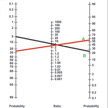
According to Figure 4, we notice that the pre test probability for the SCC in patients with stenosis secondary to caustic lesion was 5%. Using the likelihood ratio of a positive test for the NBI (A, Black line) and for Lugol (B, red line), we have achieved a post test prob-ability of 22% for the NBI and 18% for the Lugol. Trying to improve the current data analysis, considering the pre test probability of 5% and applying a likelihood ratio of a positive test of 5 for the NBI, there is a post test probability of 22% Following the same process, considering the pre test probability now of 22% and the likelihood ratio of a positive test for the Lugol, there is a post test probability of 52%, highly improving the odds of diagnosis using both methods.
FIGURE 4. Fagan’s Nomogram. “A” (Black line) is NBI and “B” (red line) is Lugol.
DISCUSSION
This is an observational study, assessing 38 patients with es-ophageal stenosis secondary to caustic lesion. We compared the use of NBI and Lugol chromoscopy for its ability to diagnose EC. The study was performed by one proceduralist and both methods were performed during the same endoscopic exam. We identiied two cases of SCC among the 38 selected patients, which gave the prevalence of EA of 5%. This is consistent with a study Goldman presents a prevalence of 0.8%-4%(4).
In this study we used an ultra slim gastroscope, with 4.9 mm diameter, which allowed the evaluation of the stenotic ring’s mucosa, without needing dilatation, excluding some distortion factors caused by the dilation. A majority suspected lesions and esophageal
neo-plasms are diagnosed at oral side of stenosis or adjacent to stenosis. So, about early cancer diagnosis, no difference existing between standard adult gastroscope or ultrathin gastroscope. A large review of lye corrosion carcinoma of the esophagus with 63 cases showed that majority of corrosion carcinomas occur at the site of tracheal bifurcation, possibly due to stasis and more severe injury at this site because of anatomical narrowing. Fifty-three of the 63 patients in the series had cancer at the site of tracheal bifurcation(3).
Goda(26), described the use of optical chromoscopy with NBI in the detection of esophageal carcinoma, concluding that the method may improve the identiication of lat lesions that weren’t seen in conventional endoscopy Reported a case of a 65-year-old man who had a lat and small squamous cell carcinoma in the esophagus and the endoscopic observation with the NBI system was useful for detecting the lesion.
Yoshida T, et al.(23), studied patients without history of caustic ingestion, observed 41 patients with esophageal SCC had a stronger color contrast in the NBI obtained images. The NBI system im-proved the accuracy of magnifying endoscopy for evaluation of esophageal lesion. The accuracy was 85% in the evaluation of depth of invasion, compared to histologic exam(7,10,23).
Another comparative study between NBI technique and Lugol’s chromoscopy in the diagnosis of SCC in patients with history of head and neck neoplasia showed that the NBI and Lugol’s chromo-scopy were equally sensitive and easy to perform(28). We also found these results in our study, which conirm the useful of both methods.
The accuracy of NBI endoscopy in screening for esophageal cancer has been reported to be comparable to that of Lugol chro-moendoscopy, with low rates of adverse events. Especially, the speciicity of NBI endoscopy with or without magnifying imaging was higher than that of Lugol chromoendoscopy. Although Lugol chromoendoscopy is the current gold standard for screening for esophageal cancer, NBI endoscopy might be the irst-choice en-doscopy for screening in the future(15).
In the current study the sensitivity and speciicity of the Lugol’s chromoscopy was 100% and 66.67%, respectively. The sensitivity was high since all the cases of carcinoma were identiied. The probable explanation for the lower speciicity, at least with regards to Lugol’s iodine is that due to the healing process caused by the caustic injury, areas of vascular neoformation and ibrosis, present-ing low glycogen and therefore do not color by iodine dye. The positive predictive value was low, 14.29%, since it relates indirectly to the speciicity; and the negative predictive value was high, 100%, since all the cases of cancer were diagnosed. The indings were consistent to the literature, what makes NBI suitable for screening, once the chance of false negatives is minimal. The test’s accuracy was of 73%(12,20,24,27).
CONCLUSION
NBI is an applicable option to detect and evaluate esophageal carcinomain patients with caustic lesion and corrosive stenosis compared to the Lugol´s solution chromoscopy.
Authors’ contributions
REFERENCES
1. Aggarwal R, Kochhar R, Nagi B, Mehta SK. Carcinoma developing in a
pa-tient with longstanding lye stricture of oesophagus. J Assoc Physicians India. 1989;37:233-4.
2. Appelqvist P, Salmo M. Lye corrosion carcinoma of the esophagus: a review of 63 cases. Cancer. 1980;45:2655-8.
3. Bergman JJ. The endoscopic diagnosis and staging of oesophageal
adenocarci-noma. Best Pract Res Clin Gastroenterol. 2006;20:843-66.
4. BIGELOW NH. Carcinoma of the esophagus developing at the site of lye
stric-ture. Cancer. 1953 Nov;6(6):1159-64. PubMed PMID: 13106830. eng. 5. Castillo E, Lawler LP. Diagnostic radiology and nuclear medicine. J Surg Oncol.
2005;92:191-202.
6. Csíkos M, Horváth O, Petri A, Petri I, Imre J. Late malignant transformation of chronic corrosive oesophageal strictures. Langenbecks Arch Chir. 1985;365:231-8. 7. Csíkos M, Horváth OP, Petri A, Petri I. [Late malignant transformation of chronic
corrosive oesophageal strictures]. Magy Seb. 2005;58:357-62.
8. Goda K-I, Tajiri H, Kaise M, Kato M, Takubo K. Flat and small squamous
cell carcinoma of the esophagus detected and diagnosed by endoscopy with narrow-band imaging siystem. Digestive Endoscopy. 2006;18(Suppl S1):S9-S12.
9. Goldman LP, Weigert JM. Corrosive substance ingestion: a review. Am J
Gas-troenterol. 1984;79:85-90.
10. Gschossmann JM, Schroeder R, Wyler F, Scheurer U, Schiemann U. [Whether or not to perform an early endoscopy following ingestion of potentially caustic agents - a retrospective longterm analysis in a tertiary referral institution]. Z Gastroenterol. 2016;54:548-55.
11. Hashimoto CL, Iriya K, Baba ER, Navarro-Rodriguez T, Zerbini MC, Eisig JN, et al. Lugol’s dye spray chromoendoscopy establishes early diagnosis of esopha-geal cancer in patients with primary head and neck cancer. Am J Gastroenterol. 2005;100:275-82.
12. Hopkins RA, Postlethwait RW. Caustic burns and carcinoma of the esophagus. Ann Surg. 1981;194:146-8.
13. K G. An Introduction to High-Resolution endoscopy and Narrowband imaging. In: Cohen. J, editor. Advanced Digestive Endoscopy: Comprehensive Atlas of High Resolution Endoscopy and Narrowband imaging. Massachusetts; 2007. 14. Kai Y, Kato M, Hayashi Y, Akasaka T, Shinzaki S, Nishida T, et al. Esophageal
early basaloid squamous carcinoma with unusual narrowband imaging magniied endoscopy indings. World J Gastroenterol. 2014;20:12673-7.
15. Kiviranta N. Corrosive carcinoma of the esophagus. Acta Otolaryngol. 1952;102:1-9.
Pennachi CMPS, Moura DTH, Amorim RBP, Guedes HG, Kumbhari V, Moura EGH. Cromoscopia vital com Lugol versus cromoscopia óptica na detecção de câncer de esôfago em pacientes com estenoses cáusticas esofagianas. Arq Gastroenterol. 2017;54(3):250-4.
RESUMO – Contexto – A suspeita do câncer de esôfago na lesão cáustica ocorre quando os pacientes com estenoses previamente estáveis, após um período latente sem sintomas, apresentam disfagia, baixa resposta as dilatações ou sintomas respiratórios. A cromoscopia com luz de banda estreita detecta o câncer supericial de esôfago mais frequentemente que a luz branca, com alta sensibilidade e acurácia. Objetivo – Determinar a aplicabilidade clínica da luz de banda estreita versus a cromoscopia vital com Lugol na detecção do câncer precoce de esôfago em pacientes com lesões cáusticas.
Métodos – Um total de 38 pacientes, entre 28 e 84 anos, foram alocados seguidamente e submetidos à cromoscopia com luz de banda estreita e com Lugol. Um gastroscópio de 4,9 mm de diâmetro foi usado para facilitar o exame da área estenosada, sem necessidade de dilatação. A cromoscopia
com luz de banda estreita era realizada primeiro e as áreas suspeitas anotadas. Depois, a cromoscopia com Lugol era realizada e as áreas suspeitas
biopsiadas. Resultados – Detectamos nove lesões suspeitas com a luz de banda estreita e 14 com o Lugol. A sensibilidade e especiicidade da cromoscopia com luz de banda estreita foi de 100% e 80,6%, e a do Lugol foi de 100% e 66,67% respectivamente. Cinco (13%) lesões suspeitas foram detectadas
coincidentemente pelos dois métodos, sendo duas (40%) com diagnóstico anatomopatológico de câncer de esôfago. Conclusão – A cromoscopia com
luz de banda estreita é opção concreta para o diagnóstico de câncer em pacientes com estenoses esofágicas por corrosões cáusticas, comparado a cromoscopia com Lugol.
DESCRITORES – Neoplasias esofágicas. Carcinoma de células escamosas. Cáusticos, efeitos adversos. Iodetos, uso terapêutico.
16. Lecleire S, Antonietti M, Iwanicki-Caron I, Duclos A, Lemoine F, Pessot FL, et al. Lugol chromo-endoscopy versus narrow band imaging for endoscopic screening of esophageal squamous-cell carcinoma in patients with a history of cured esophageal cancer: a feasibility study. Dis Esophagus. 2011;24:418-22. 17. Mamede RC, de Mello Filho FV. Ingestion of caustic substances and its
compli-cations. Sao Paulo Med J. 2001;119:10-5.
18. Moura Ed, Maluf Filho F, Azzam R, Sakai P, Ishioka S, Iriya K, et al. Corrosive Esophagitis and Esophageal Cancer. Deinitions of predictive sings of cancer by endoscopic and pathologic evaluation. In: editore M, editor. Diseases of the esophagus. Itália.1996. p. 955-60.
19. Muto M, Hironaka S, Nakane M, Boku N, Ohtsu A, Yoshida S. Association of multiple Lugol-voiding lesions with synchronous and metachronous esophageal squamous cell carcinoma in patients with head and neck cancer. Gastrointest Endosc. 2002;56:517-21.
20. Ponchon T, Lapalus M, Saurin J, Robles-Medranda C, Chemaly M, Parmentier B, et al. Could Narrow Band Imaging (NBI) replace Lugol staining for the detec-tion of oesophageal squamous cell carcinoma? Gastrointest Endosc. 2007;65(5). 21. Rodríguez Vargas BO, Monge Salgado E, Montes Teves P, Salazar Ventura S,
Guzmán Calderón E. [Caustics injuries in the upper gastrointestinal tract: clinical and endoscopic features]. Rev Gastroenterol Peru. 2016;36:135-42.
22. Ruol A, Rampado S, Parenti A, Portale G, Giacomelli L, Battaglia G, et al. Caustic ingestion and oesophageal cancer: intra- and peri-tumoral ibrosis is associated with a better prognosis. Eur J Cardiothorac Surg. 2010;38:659-64.
23. Shiozaki H, Tahara H, Kobayashi K, Yano H, Tamura S, Imamoto H, et al. Endoscopic screening of early esophageal cancer with the Lugol dye method in patients with head and neck cancers. Cancer. 1990;66:2068-71.
24. Song LM, Adler DG, Conway JD, Diehl DL, Farraye FA, Kantsevoy SV, et al. Narrow band imaging and multiband imaging. Gastrointest Endosc. 2008;67:581-9. 25. Sreedharan A, Rembacken BJ, Rotimi O. Acute toxic gastric mucosal damage
induced by Lugol’s iodine spray during chromoendoscopy. Gut. 2005;54:886-7. 26. Yamasaki Y, Takenaka R, Hori K, Takemoto K, Kawano S, Kawahara Y, et al. Tolerability of magnifying narrow band imaging endoscopy for esophageal cancer screening. World J Gastroenterol. 2015;21:2793-9.
27. Yoshida S. Advanced Digestive Endoscopy: Comprehensive Atlas of High Resolu-tion Endoscopy and Narrow Band Imaging. Massachusetts.: Jonathan Cohen.; 2007. 28. Yoshida T, Inoue H, Usui S, Satodate H, Fukami N, Kudo SE. Narrow-band