DOI: 10.1590/0004-282X201301461
VIEWS AND REVIEWS
Prionic diseases
Doenças priônicas
Abelardo Q-C Araújo
When Stanley Prusiner coined the term “prion” in 1982, which he deined as a small infectious pathogen containing protein but apparently lacking nucleic acid, the scientiic world watched amazed a revolution in the ields of biology
and medicine1. Finally unveiling the cause of a group of mys-terious diseases known by veterinarians since 1730, when the irst cases of scrapie appear in Central Europe and England, Prusiner deservedly won the 1997 Nobel Prize for his achieve
-ment. Before him Carleton Gajdusek had won the same prize in 1976 for his discoveries in this ield.
In fact, like in other subjects, Prusiner was picking up on an idea originally proposed in the 1960s, when Tikvah Alper
and J. S. Griith from London, suggested that an infectious
agent that lacked nucleic acid could cause disease. Such an idea seemed to threaten the very foundations of molecular biology, which held that nucleic acids were the only way to transmit information from one generation to the next2.
Biology
Prions are small infectious pathogens containing pro-tein but apparently lacking nucleic acid1. he “protein only”
hypothesis proposes that the pathological prion protein (PrPSC) is a conformational isoform of a normal host protein
(PrPC), which is found predominantly on the outer surface of
Associate Professor of Neurology, the Federal University of Rio de Janeiro; Head, the Laboratory for Clinical Research in Neuroinfections, National Institute of Infectology, Evandro Chagas Research Institute, FIOCRUZ, Rio de Janeiro RJ, Brasil.
Correspondence: Abelardo Q-C Araújo; Av. das Américas 700 / bl. 3 / sala 202; 22640-100 Rio de Janeiro RJ, Brasil; E-mail: abelardo@ufrj.br ABSTRACT
Prion diseases are neurodegenerative illnesses due to the accumulation of small infectious pathogens containing protein but apparently lacking nucleic acid, which have long incubation periods and progress inexorably once clinical symptoms appear. Prions are uniquely re-sistant to a number of normal decontaminating procedures. The prionopathies [Kuru, Creutzfeldt-Jakob disease (CJD) and its variants, Gerstmann-Sträussler-Scheinker (GSS) syndrome and fatal familial insomnia (FFI)] result from accumulation of abnormal isoforms of the prion protein in the brains of normal animals on both neuronal and non-neuronal cells. The accumulation of this protein or fragments of it in neurons leads to apoptosis and cell death. There is a strong link between mutations in the gene encoding the normal prion protein in humans (PRNP) - located on the short arm of chromosome 20 – and forms of prion disease with a familial predisposition (familial CJD, GSS, FFI). Clini-cally a prionopathy should be suspected in any case of a fast progressing dementia with ataxia, myoclonus, or in individuals with pathological insomnia associated with dysautonomia. Magnetic resonance imaging, identification of the 14-3-3 protein in the cerebrospinal fluid, tonsil biopsy and genetic studies have been used for in vivo diagnosis circumventing the need of brain biopsy. Histopathology, however, remains the only conclusive method to reach a confident diagnosis. Unfortunately, despite numerous treatment efforts, prionopathies remain short-lasting and fatal diseases.
Keywords: prion, Creutzfeldt-Jakob disease, dementia, slow viruses.
RESUMO
Doenças priônicas são enfermidades neurodegenerativas devido ao acúmulo de pequenos agentes infecciosos compostos unicamente por proteína (prions), com longos períodos de incubação e de progressão inexorável para o óbito. Esses agentes são excepcionalmente resis-tentes aos processos habituais de descontaminação para germes e vírus. As prionopatias [Kuru, doença de Creutzfeldt-Jakob (CJD) e suas variantes, Síndrome de Gerstmann-Sträussler-Scheinker (GSS) e insônia familiar fatal (FFI)] resultam do acúmulo de isoformas anormais da proteína priônica no cérebro. Este acúmulo leva, em última análise, à apoptose e morte celular. Existe uma forte associação entre mutações no gene que codifica a proteína priônica normal em humanos (PRNP) - localizado no braço curto do cromossoma 20 - e formas genéticas destas doenças (CJD familiar, GSS, FFI). Clinicamente devemos suspeitar de uma prionopatia em qualquer caso de demência de rápida pro-gressão, particularmente quando associadas a ataxia, mioclonias, ou em indivíduos com insônia patológica combinada com disautonomia. Métodos diagnósticos como ressonância magnética, pesquisa da proteína 14-3-3 no líquido cefalorraquiano, biópsia de amígdalas e estu-dos genéticos têm sido utilizaestu-dos para diagnóstico in vivo, evitando-se assim a necessidade de biópsia cerebral. A despeito disso, a histopa-tologia continua a ser o único método conclusivo para se chegar a um diagnóstico definitivo. Infelizmente, apesar dos inúmeros esforços de tratamento, as prionopatias permanecem doenças de curta duração e fatais.
neurons, attached by a glycosylphosphatidyl-inositol (GPI) anchor. he abnormal conformer (PrPSC), when introduced into the organism – by external routes or by mutations – is
thought to cause the conversion of the normal PrPC into a
likeness of itself (PrPSC) (akin to the dual-personality
cha-racters described in the novel by Robert Louis Stevenson, he Strange Case of Dr Jekyll and Mr Hyde). Because the amino acid sequences of PrPC and PrPSC are often identi-cal, the cause of their functional diferences is attributed to
their variance in structure, and not to any chemical dissimi-larity. Whereas PrPC consists primarily of alpha helices and
very few beta sheets, PrPSC, though having the same primary
structure, consists in large part of beta sheets3. As yet, it is still unclear exactly what causes the lip from PrPC to PrPSC.
After the transformation of one prion, the process continues at an exponential rate, eventually infecting the entire brain of the host and causing irreparable damage. Once PrPC adopts
the beta-sheet structure of PrPSC, it becomes detached from
the cell membrane and is absorbed by vesicles within the cell.
In particular, it begins to accumulate in the cell’s lysosomes. he accumulation of PrPSC in the lysosomes causes them to
swell and eventually burst, thereby releasing the damaging proteolytic enzymes and PrPSC into the cell. In contrast to
PrPC, PrPSC accumulates within cells and does not normally
appear on the cell surface. PrPSC appears to be neurotoxic, its
accumulation leading to apoptosis and cell death4.
In addition to the mode of infection outlined above, that
is, through sporadic conformational transformation, infec-tive prions can also be transmitted from one individual to another during medical procedures such as tissue transplan-tation and injection of growth hormones, as well as
contami-nated surgical instruments. Yet a third mode of infection is
through the inheritance of mutated genes that increase the likelihood of conformational change in the prions for which they are responsible.
One characteristic feature of prions is their resistance to a
number of normal decontaminating procedures. hey are re
-sistant to processes afecting nucleic acids, such as hydroly
-sis or shearing. hus, suspected prion-contaminated medical instruments require speciic procedures5.
Prions can be transmitted between members of dife-rent species. his phenomenon is known as the violation
of the species barrier6. he determining factor seems to be
the degree of similarity between the two species of prions.
his fact became cause of a great deal of anxiety caused by
the outbreak of epidemic bovine spongiform
encephalopa-thy (BSE, or mad cow disease). If one compares the overall
evolutionary relationship of humans and cows, the general features of prions, there seems to be little cause for worry; however, it is possible that bovine prions might be similar enough to human prions to be able to cross the species bar-rier and infect human hosts with what was originally a bo-vine disease7.
Genetics
he gene encoding the normal protein in humans (”PRNP”) is located on the short arm of chromosome 20.
A strong link was established between mutations in this gene and forms of prion disease with a familial
predisposi-tion ( familial Creutzfeldt Jakob Disease ( fCJD), Gerstmann-Sträussler-Scheinker syndrome (GSS), and fatal familial in
-somnia (FFI)). A single mutation can produce diferent clinical phenotypes in diferent individuals or families. More than 50 diferent mutations have been identiied so far. his
is why some experts have advocated classifying prion disea-ses based upon the responsible mutation rather than the
tra-ditional classiications8.
he codon 129 of the PRNP gene is polymorphic; nor -mal individuals have either valine or methionine at that site.
Neither V129 nor M129 appears to be pathogenic by itself. Patients with the D178N mutation who are homozygous for valine at codon 129 appear to develop CJD, while those who are homozygous for methionine tend to have FFI9.
All GSS kindreds investigated to date have PRNP gene mutations, being the P102L mutation the most common. In fCJD, a missense mutation involving the substitution of lysine for glutamine in codon 200 is the most common gene muta
-tion worldwide. he D178N muta-tion has been the predomi
-nant mutation found in nearly all families with FFI. his mu
-tation also occurs in fCJD. It appears that patients with this mutation who are homozygous for methionine at codon 129 develop an FFI-like clinical syndrome whereas those homo
-zygous for valine develop fCJD9.
Clinical aspects
Five established human prion diseases are currently
recognized: Kuru, Creutzfeldt-Jakob disease (CJD), variant Creutzfeldt-Jakob disease (vCJD also known as new variant CJD), Gerstmann-Sträussler-Scheinker syndrome (GSS), and fatal familial insomnia (FFI)10. However, a new prion disease named “proteinase-sensitive prionopathy” was described in 200811. Patients presented at a mean of 62 years with a de
-mentia with prominent neuropsychiatric manifestations
and progressive motor decline (ataxia and/or parkinsonism). Death followed symptoms onset within a mean of 20 months.
A family history of dementia was present in many patients,
suggesting a possible genetic origin. CSF 14-3-3 was nega
-tive in all individuals, magnetic resonance imaging (MRI) demonstrated difuse atrophy without restricted difusion, and electroencephalogram (EEG) were normal or showed only difuse slowing. Neuropathologic examination revealed
spongiform degeneration in the cerebral cortex, basal gan-glia, and thalamus with relative sparing of the brainstem and
cerebellum. here was a similar anatomic distribution of PrP immunostaining but, intriguingly and diferently from all
other prion diseases, its immunoreactivity was virtually
genotype. Since its original description more cases have been reported12.
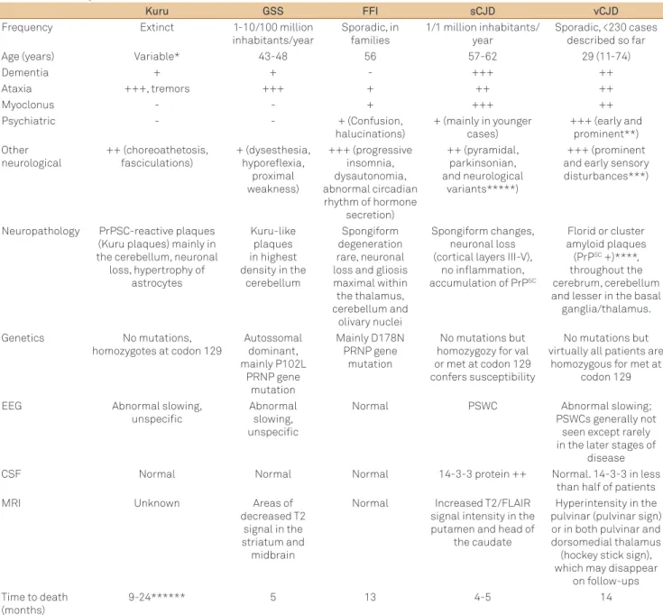
Table 1 summarizes the main aspects and diferences be -tween the classical prionopathies.
1. Kuru
Kuru (which means shivering in the Faroe language), which was endemic in Papua New Guinea among the Fore
tribes, used to be transmitted from person to person by
ritu-al cannibritu-alism. he cessation of these practices in the 1950s ended incident cases of kuru; however, 11 new cases have
been identiied between July 1996 and June 2004, with a likely incubation period of more than 50 years in some13.
No mutations in the PRNP gene have been reported in
kuru, however, like in CJD and in its variants, homozygosity at the polymorphic codon 129 of the PRNP gene has been de -tected more frequently14.
2. Gerstmann-Sträussler-Scheinker syndrome (GSS)
GSS is inherited in an autosomal dominant pattern with virtual complete penetrance. Its diagnosis cannot be made
from the usual laboratory or imaging studies. Demonstration
Table 1. Summary characteristics of the main Prion diseases.
Kuru GSS FFI sCJD vCJD
Frequency Extinct 1-10/100 million
inhabitants/year
Sporadic, in families
1/1 million inhabitants/ year
Sporadic, <230 cases described so far
Age (years) Variable* 43-48 56 57-62 29 (11-74)
Dementia + + - +++ ++
Ataxia +++, tremors +++ + ++ ++
Myoclonus - - + +++ ++
Psychiatric - - + (Confusion,
halucinations)
+ (mainly in younger cases)
+++ (early and prominent**) Other neurological ++ (choreoathetosis, fasciculations) + (dysesthesia, hyporeflexia, proximal weakness) +++ (progressive insomnia, dysautonomia, abnormal circadian rhythm of hormone
secretion) ++ (pyramidal, parkinsonian, and neurological variants*****) +++ (prominent and early sensory disturbances***)
Neuropathology PrPSC-reactive plaques (Kuru plaques) mainly in the cerebellum, neuronal
loss, hypertrophy of astrocytes
Kuru-like plaques in highest density in the
cerebellum
Spongiform degeneration rare, neuronal loss and gliosis maximal within the thalamus, cerebellum and olivary nuclei Spongiform changes, neuronal loss (cortical layers III-V),
no inflammation, accumulation of PrPSC
Florid or cluster amyloid plaques
(PrPSC +)****,
throughout the cerebrum, cerebellum and lesser in the basal ganglia/thalamus.
Genetics No mutations,
homozygotes at codon 129
Autossomal dominant, mainly P102L PRNP gene mutation Mainly D178N PRNP gene mutation
No mutations but homozygozy for val or met at codon 129 confers susceptibility
No mutations but virtually all patients are
homozygous for met at codon 129
EEG Abnormal slowing,
unspecific
Abnormal slowing, unspecific
Normal PSWC Abnormal slowing;
PSWCs generally not seen except rarely in the later stages of
disease
CSF Normal Normal Normal 14-3-3 protein ++ Normal. 14-3-3 in less
than half of patients
MRI Unknown Areas of
decreased T2 signal in the striatum and
midbrain
Normal Increased T2/FLAIR signal intensity in the putamen and head of
the caudate
Hyperintensity in the pulvinar (pulvinar sign) or in both pulvinar and dorsomedial thalamus (hockey stick sign), which may disappear
on follow-ups Time to death
(months)
9-24****** 5 13 4-5 14
of PRNP gene mutations appears to be a sensitive and highly
speciic way to diagnose GSS. Neuropathology, although less
used in practice, can also be useful.
he degree of dementia in GSS varies among afected
families and individuals within the same family. Part of the
variability of expression of this illness may be due to
dife-rences in the underlying PRNP mutation or the associated
polymorphisms in codon 12915.
3. Fatal familial insomnia (FFI)
FFI was irst identiied in Italian families, but kindreds
have now been reported throughout the world. Although rarely, sporadic cases are also described. Disease onset is ear-lier and duration is shorter in those who are homozygous for
methionine at codon 129. It is a rapidly fatal disease in which
patients characteristically develop progressive insomnia with loss of the normal circadian pattern, along with dysautono-mia and endocrine disturbances. Patients characteristically develop progressive insomnia with loss of the normal circadian sleep-activity pattern, which may manifest as a dream-like
confusional state during waking hours. Inattention, impaired
concentration and memory, confusion, and hallucinations are frequent, but overt dementia is rare16.
Methionine-ho-mozygous patients are more likely to have hallucinations and myoclonus as prominent disease features, while methionine-heterozygous patients are more likely to develop early pro-blems with ataxia, bulbar signs, and nystagmus.
Dysautonomia is characterized by hyperhidrosis, hy-perthermia, tachycardia, and hypertension. Endocrine dis-turbances include decreased ACTH secretion, increases in cortisol secretion, and loss of the normal diurnal varia-tions in levels of growth hormone, melatonin, and prolactin. Fluorodeoxyglucose PET showing decreased glucose utiliza-tion in the thalamus may be detectable even before the de-velopment of clinical symptoms. Sleep studies demonstrate a dramatic reduction in total sleep time and disruption of the
normal sleep architecture. Genetic studies are now the
diag-nostic procedure of choice for diagnosis of this condition.
Most cases are associated with the D178N PRNP gene mu -tation17. Spongiform degeneration, a characteristic feature of most of the human prion diseases, is rarely detected in FFI,
particularly in those with the methionine-homozygous gen-otype. Neuronal loss and gliosis that is maximal within the
thalamus are, however, consistent indings. hese changes
can also occur in other regions of the brain as well as in the cerebellar cortex, cerebellar nuclei, and olivary nuclei18.
4. Creutzfeldt-Jakob disease (CJD) and its variants
CJD is the most frequent of the human prion diseases. CJD most often occurs as a sporadic disorder (sCJD) although familial forms ( fCJD) have been described. It is clinically
ma-nifested by a rapidly progressive mental deterioration, often
with behavioral abnormalities, and myoclonus. Eight-ive to
ninety-ive percent of CJD cases are sporadic, while 5 to 15 percent are due to fCJD; iCJD generally accounts for less than 1 percent. Family history of CJD, a medical history of psycho -sis, history of multiple surgical procedures and residence for
more than 10 years on a farm were all signiicant risk factors for sCJD. No study so far documented an increased risk of sCJD with receipt of blood products19.
Variant CJD (vCJD) is a distinct disorder with clinical, di
-agnostic and pathological features diferent from sCJD20. It was irst reported in 1996 and since then around 230 cases have been described. he vCJD represents bovine-to-human
transmission of BSE, most patients acquiring the disorder
through ingestion of infected meat products. he removal of
organic solvents, which inactivate PrPSc, from the rendering
process for bovine ofal, and the subsequent use of the ofal
as a component of feed for cattle has been hypothesized to
be a mechanism for amplifying the epidemic in animals. he source of BSE remains unclear. he irst cases were recog
-nized in 1986. heories of its origination include the trans -mission of either sheep scrapie or another prion disease to cattle via contaminated feed. An intrinsic genetic event in cattle seems less likely21.
According to international regulations, a country may be considered a minimal-risk for BSE, if it has less than two BSE
cases/million cattle over 24 months of age during each of the
previous four consecutive years. Brazil is recognized as having
a negligible BSE risk, i.e. the most favorable category. Just one case of BSE has been described in Brazil so far (2012) but the
dead animal was destroyed and did not enter the food or feed chain. Updated information on the number of BSE cases re-ported throughout the world in imre-ported or indigenous
cat-tle is available online from the OIE website (www.oie.int).
he US Food and Drug Administration (FDA) adopted a policy that a prospective blood donor be indeinitely de -ferred if he or she had lived for more than six months in the
United Kingdom during the peak years of BSE (1981 through 1996). his policy went into efect in April, 2000, to attempt
to balance increased safety against decreased blood
availabil-ity. here is no evidence of vertical transmission (mother to child) of vCJD, although the potentially long incubation pe-riod for this mechanism makes it diicult to exclude this pos
-sibility deinitively22, 23.
Iatrogenic CJD (iCJD) has followed administration of ca -daveric human pituitary hormones, dural graft transplants, use of dural mater in radiographic embolization procedures, corneal transplants, liver transplants, and the use of contami-nated neurosurgical instruments or stereotactic depth
elec-trodes. No deinite cases of transfusion-associated CJD are
known to have occurred, although transfusion-related variant
CJD has been described. he incubation period for iCJD is
unknown and probably depends upon the mode of
acquired after administration of human growth hormone in a
population. CJD infection in health care workers is extreme
-ly rare. Physical contact with CJD patients entails no risk
of transmission and special precautions are not required in their care. However, special precautions should be employed in the handling of CSF as well as biopsy tissue; all materials and instruments used must be decontaminated according to established protocols24.
Other autoimmune, infectious, malignant, and
toxic-me-tabolic etiologies should be considered in the diferential diag nosis of CJD.
While brain biopsy is the gold standard test for diagnosis,
it is often unnecessary for CJD. A typical clinical presentation with corroborating indings on MRI, EEG, and CSF are usual-ly suicient to exclude other causes and establish a probable
diagnosis.
One feature that distinguishes vCJD from sCJD is its
prominent trophism for lymphoid organs such as the tonsils. Analysis of PrP extracted from tonsil biopsy tissue appears to
provide a sensitive and speciic method for the diagnosis of vCJD in the appropriate clinical context25. On the other hand, detection of the 14-3-3 protein in CSF is not a sensitive mar-ker for vCJD.
In sCJD MRI typically shows abnormal signal in the puta
-men and head of the caudate. Sensitivity and speciicity for typical MRI indings range between 83 to 92 percent and 87 to 95 percent respectively. A inding of periodic sharp wave complexes (PSWC) on EEG has a high speciicity for the diag-nosis of CJD, but a low sensitivity. he 14-3-3 protein test in CSF is a speciic test inding for CJD, but its sensitivity can be
low, particularly in some molecular subtypes26.
Clinical phenotypes of sporadic CJD have been associated
with molecular subtypes determined by the PRNP gene codon
129 genotype and the pathologic prion protein (PrPSC) type. he
PRNP genotype is homozygous or heterozygous for methionine
(M) or valine (V) at codon 129. Using this molecular classiica
-tion, six clinical phenotypes of sCJD have been described27: • MM1 and MV1 (myoclonic, Heidenhain variant) account
for about 70 percent of cases and correlate with the “clas
-sic CJD” phenotype of advanced age at onset, a rapidly
progressive dementia with early and prominent
myoclo-nus, and a short duration of illness (mean 3.9 months). he MM1 phenotype is the one most commonly associat -ed with periodic sharp wave complexes (PSWC) on
elec-troencephalogram (EEG).
• VV2 (ataxic variant) accounts for 15 percent or less of sCJD and presents with ataxia at onset, often as an
isola-ted feature, late dementia, and a longer duration of illness
(mean 7 to 9 months).
• MV2 (Kuru plaque variant) accounts for 9 percent and
presents with ataxia, progressive dementia with promi-nent psychiatric features, and longer duration (mean
17.1 months). he 14-3-3 protein in the CSF is a relatively
insensitive marker for the MV2 variant (about 70 per
-cent), and PSWC are only infrequently seen on EEG. • MM2 can present as either a thalamic variant or a corti
-cal variant. Some, but not all, patients have a young age at onset, and the disease course is typically long, with a
median disease duration of 14 months in one study. he 14-3-3 protein has been reported to be present in 61 to 91 percent of patients with MM2, and PSWCs on EEG are more often absent than in other MM and MV sub
-types. he clinical features of MM2 type sCJD may re
-semble those of variant CJD. he thalamic MM2 variant accounts for 2 percent of cases, and mean disease dura
-tion is 15.6 months. Insomnia, psychomotor
hyperactivi-ty, ataxia, and cognitive impairment are the predominate
manifestations, and this phenotype resembles that of FFI. he cortical MM2 variant accounts for 2 percent of cases, with a mean disease duration of 15.7 months. Dementia is
the predominate manifestation, while cerebellar and vi-sual signs are rarely described at presentation.
• VV1 accounts for 1 percent of cases and is notable for pro
-gressive dementia and longer duration (mean 15.3 months). A case series of nine patients with this subtype conirmed the slower, more prolonged course (median 21 months). All patients had elevated CSF levels of the 14-3-3 protein, but none had PSWCs on EEG, and cortical rather than basal ganglia abnormalities were more common on MRI.
PSWCs are helpful in the diferentiation of sCJD from
other prion disease. For example, PSWCs are occasionally
found in patients with fCJD, although PSWCs are found more commonly in patients with fCJD who have the codon 200 mutation; PSWCs are not found in patients with vCJD, kuru, Gerstmann-Sträussler-Scheinker syndrome, or fatal familial
insomnia; PSWCs may be more commonly absent in the
tha-lamic variant of MM2 sCJD, as well as the MV2 and VV2 sub
-types and in iatrogenic CJD.
PSWCs may not be recorded in the initial stages of the
illness. he probability of recording PSWCs corresponds to the amount of neuronal loss, and serial EEG recording may be useful in patients suspected of having sCJD when initial EEG recordings are negative. PSWCs typically disappear in later stages of sCJD, which is characterized by low voltage activity followed by electrocerebral inactivity. It must be re -membered that drugs such as barbiturates and benzodiaz-epines can mask PSWCs28, 29.
he 14-3-3 protein has been seen as a sensitive and spe
-ciic diagnostic test for sCJD. In one study, CSF 14-3-3 protein had a positive predictive value of 95 percent for patients with pathologically deinite and probable sCJD. However, subse -quent reports have found somewhat lower sensitivities and
speciicities of 53 to 88 percent. In addition, “false posi tive” elevations in CSF 14-3-3 have been noted in patients with
encephalopathies. It seems that the protein may be a marker of brain cell death rather than CJD30.
Studies have suggested that the 14-3-3 protein test may be helpful for diagnosis of the classical subtypes of sCJD but
may be falsely negative for the nonclassical subtypes. Taken
together, these studies suggest that detection of CSF 14-3-3 protein in CSF should be considered an adjunctive rather
than absolute test for the diagnosis of prion diseases30. A
ne-gative test does not exclude the diagnosis, especially in cases
of possible fCJD or nonclassical sCJD, and a positive result
can occur in nonprion diseases. However, a positive test
in-creases the probability of CJD when other clinical features are
suggestive but not diagnostic. Pathological studies of brain material to detect protease resistant PrPSc remain the gold standard for the diagnosis of prion diseases.
A variety of other CSF diagnostic tests have been
repor-ted in small series (S100 protein, neuron speciic enolase, thy -mosin β4, and tau protein). Further studies are however ne-cessary to evaluate their real usefulness for CJD diagnosis.
Prion protein diagnostic assays performed on blood samples, are in development and in one case series, elevated plasma levels
of several acute phase reactants were noted in patients with CJD.
All of these tests are of unproven diagnostic utility at present.
Routine laboratory studies are normal in CJD with the oc
-casional exception of liver function tests. he CSF contains
no cells and usually has normal glucose. An elevated CSF
protein may occur in about 40 percent of patients31.
Treatment
Prion diseases are always fatal, regardless of any
cur-rent treatment efort. Care for patients with prion disease is
therefore mainly supportive. Isolated case reports of stabi -lization or improvement following treatment with amanta-dine, acyclovir, interferons, polyanions, vidarabine, and me-thisoprinol have not been replicated32-34.
Quinacrine and chlorpromazine, which were found to in -hibit PrPSc formation in a cultured neuroblastoma cell line
(ScN2a) chronically infected with prions, also failed to show any beneit when used in humans.
Finally, Flupirtine maleate, a centrally acting nonopioid analgesic that has displayed cytoprotective activity in vitro in neurons inoculated with a prion protein fragment did not
show signiicant efect on survival time compared with pla
-cebo. However, patients performed signiicantly better on the
cognitive part of the Alzheimer Disease Assessment Scale (ADAS-Cog) and on the Mini Mental Status Examination,
but the diference did not reach statistical signiicance. Caregiver’s impressions were also signiicantly better in the lupirtine-treated group33.
Laboratory models used for studying prion diseases may
assist in the testing of new agents. he scientiic advan ces
in the understanding of the molecular pathogenesis of prion
di seases are expected to lead to the identiication of new targets for therapy [v.g., iPrP13, a peptide that can break a
beta-sheet conformation; depletion of endogenous PrPC by
molecular methods; induction of immune activation through the exposure of a prion protein epitope that is selectively ex-posed in the pathologic conformation; anti-PrP monoclonal antibodies; adenovirus vector platforms that express PrPc
single-chain fragment (scFv) antibodies]. As already men -tioned, however, at this moment, prionic diseases are incu-rable and invariably fatal.
References
1. Prusiner SB. Novel proteinaceous infectious particles cause scrapie. Science 1982; 216:136-144.
2. Alper T, Cramp WA, Haig DA, Clarke MC. Does the agent of scrapie replicate without nucleic acid? Nature 1967;214:764-766.
3. Ironside JW, Head MW. Biology and neuropathology of prion diseases. Handb Clin Neurol 2008;89:779-797.
4. Kretzschmar HA, Giese A, Brown DR, et al. Cell death in prion disease. J Neural Transm 1997;50(Suppl):S191-S210.
5. McDonnell G, Burke P. The challenge of prion decontamination. Clin Infect Dis 2003;36:1152-1154.
6. Moore RA, Vorberg I, Priola SA. Species barriers in prion diseases--brief review. Arch Virol 2005;19 (Suppl):S187-S202.
7. Biacabe AG, Jacobs JG, Bencsik A, Langeveld JP, Baron TG. H-type bovine spongiform encephalopathy: complex molecular features and similarities with human prion diseases. Prion 2007;1:61-68.
8. Lloyd SE, Mead S, Collinge J. Genetics of prion diseases. Curr Opin Genet Dev 2013;23:345-351.
9. Lloyd S, Mead S, Collinge J. Genetics of prion disease. Top Curr Chem 2011;305: 1-22.
10. Marandi Y, Farahi N, Sadeghi A, Sadeghi-Hashjin G. Prion diseases - current theories and potential therapies: a brief review. Folia Neuropathol 2012;50:46-49.
11. Gambetti P, Dong Z, Yuan J, et al. A novel human disease with abnormal prion protein sensitive to protease. Ann Neurol 2008;63:697-708.
12. Head MW, Yull HM, Ritchie DL, et al. Variably protease-sensitive prionopathy in the UK: a retrospective review 1991-2008. Brain 2013; 136:1102-1115.
13. Liberski PP, Sikorska B, Brown P. Kuru: the first prion disease. Adv Exp Med Biol 2012;724:143-153.
14. Mead S, Whitfield J, Poulter M, et al. Genetic susceptibility, evolution and the kuru epidemic. Philos Trans R Soc Lond B Biol Sci 2008;363::3741-3746.
15. Liberski PP. Gerstmann-Straussler-Scheinker disease. Adv Exp Med Biol 2012; 724:128-137.
16. Gambetti P, Petersen R, Monari L, et al. Fatal familial insomnia and the widening spectrum of prion diseases. Br Med Bull 1993;49:980-994.
18. Imran M, Mahmood S. An overview of human prion diseases. Virol J 2011;8:559.
19. de Villemeur TB. Creutzfeldt-Jakob disease. Handb Clin Neurol 2013; 112:1191-1193.
20. Keohane C. The human prion diseases. A review with special emphasis on new variant CJD and comments on surveillance. Clin Exp Pathol 1999;47:125-132.
21. Sikorska B, Liberski PP. Human prion diseases: from kuru to variant Creutzfeldt-Jakob disease. Subcell Biochem 2012;65:457-496.
22. Puopolo M, Ladogana A, Vetrugno V, Pocchiari M. Transmission of sporadic Creutzfeldt-Jakob disease by blood transfusion: risk factor or possible biases. Transfusion. 2011;51:1556-1566.
23. Lindholm PF, Annen K, Ramsey G. Approaches to minimize infection risk in blood banking and transfusion practice. Infect Disord Drug Targets 2011;11:45-56.
24. Brown P, Brandel JP, Sato T, et al. Iatrogenic Creutzfeldt-Jakob disease, final assessment. Emerg Infect Dis 2012;18:901-907.
25. Frosh A, Smith LC, Jackson CJ, et al. Analysis of 2000 consecutive UK tonsillectomy specimens for disease-related prion protein. Lancet 2004;364:1260-1262.
26. Wood H. Prion disease: New approaches to CJD diagnosis. Nat Rev Neurol 2012; 8:241.
27. Bishop MT, Will RG, Manson JC. Defining sporadic Creutzfeldt-Jakob disease strains and their transmission properties. Proc Natl Acad Sci U S A 2010;107:12005-12010.
28. Mizobuchi M, Tanaka C, Sako K, Nihira A, Abe T, Shirasawa A. Correlation between periodic sharp wave complexes and diffusion-weighted magnetic resonance images in early stage of Creutzfeldt-Jakob disease: a report of two cases. Seizure 2008; 17: 717-722.
29. Wang PS, Wu YT, Hung CI, Kwan SY, Teng S, Soong BW. Early detection of periodic sharp wave complexes on EEG by independent component analysis in patients with Creutzfeldt-Jakob disease. J Clin Neurophysiol 2008; 25: 25-31.
30. Muayqil T, Gronseth G, Camicioli R. Evidence-based guideline: diagnostic accuracy of CSF 14-3-3 protein in sporadic Creutzfeldt-Jakob disease: report of the guideline development sub committee of the American Academy of Neurology. Neurology 2012;79: 1499-1506.
31. Wang LH, Bucelli RC, Patrick E, et al. Role of magnetic resonance imaging, cerebrospinal fluid, and electroencephalogram in diagnosis of sporadic Creutzfeldt-Jakob disease. J Neurol 2013;260:498-506.