Case Report
Key words
Chagas disease; ventricular dysfunction, left; pacemaker, artificial; electrodes, implanted.
The present case reports on a patient presenting the cardiac form of Chagas disease, with left ventricular dysfunction and second-degree atrioventricular block Mobitz type II, associated with several syncope episodes. The patient underwent a double-chamber definitive artificial pacemaker implant. One year after the implant, the displacement of the atrial electrode was diagnosed and the patient was submitted to re-implantation of the atrial electrode. Two years after the first surgical procedure, the patient presented dyspnea on exertion. The physical evaluation included an echocardiogram, which detected the presence of a foreign body with metallic characteristics in the left cardiac chambers, consistent with that of an ectopic pacemaker electrode.
Ectopic Positioning of Pacemaker Electrode
Gildo Mota, Juliana Prazeres, Nelmacy Freitas, Luiz Magalhães, Francisco Reis, Roque Aras
Hospital Universitário Professor Edgard Santos - Universidade Federal da Bahia, Salvador, BA - Brazil
Mailing address: Gildo de Oliveira Mota •
Rua Conselheiro Corrêa de Menezes, 266 apt 903 - Horto Florestal - 40295-030 - Salvador, BA - Brazil
E-mail: gildomota@cardiol.br, gildomota@yahoo.com.br
Manuscript received January 19, 2009; revised manuscript received July 09, 2009; accepted October 27, 2009.
Case Report
J.T.S., a 38-year-old male individual with the cardiac form of Chagas disease, presented left ventricular dysfunction and second-degree atrioventricular block Mobitz type II, associated with several episodes of syncope. He had undergone a double-chamber definitive artificial pacemaker implant three years before the current assessment. The postoperative period after the implant was uneventful, which persisted for a year after surgery, when he started to present symptoms caused by abnormal electric diaphragmatic stimulation, followed by a diagnosis of atrial electrode displacement. At that time, he was submitted to the atrial electrode reimplantation, without complications.
Two years after the first surgical procedure, at the Outpatient Clinic of Professor Edgard Santos University Hospital (HUPES), due to dyspnea on exertion and complementary examinations, which included an echocardiogram, were performed to evaluate the left ventricular function.
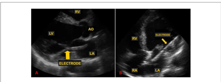
During the echocardiogram, the presence of a foreign body with typical metallic characteristics was detected in the left cardiac chambers in the initial long-axis parasternal view. Other echocardiographic projections confirmed the initial findings, demonstrating the presence of a linear metallic structure that extended from the right atrium, crossed the interatrial septum and the mitral valve and reached the basal portion of the lateral wall of the left ventricle, consistent with en ectopic pacemaker electrode (Figure 1). The electrocardiogram showed that the patient presented dual-chamber pacing with adequate capture and right bundle-branch block pattern, corroborating the artificial left ventricular stimulation (Figure 2).
The chest x-ray confirmed the abnormal position of the ventricular electrode, in the left ventricular region (Figure 2). Considering the favorable three-year evolution with the electrode located in the systemic circulation and the risks of trauma in the ventricular wall and mitral valve, in addition to the risk of thromboembolism during the electrode removal procedure, the conservative approach was chosen, associated with chronic oral anticoagulation therapy due to the increased risk for embolism determined by the presence of a foreign body in the systemic circulation.
Discussion
The location of the pacemaker electrode in the systemic circulation is a known complication; however, it is seldom reported in the literature. Most of the described cases are related to the presence of congenital anomalies of the interatrial septum, such as patent foramen ovale and interatrial communication7.
Introduction
The implant of a definitive transvenous pacemaker is a highly-effective, surgically invasive procedure with low complication rate. Among the most frequent complications are electrode fractures, surgical wound infection, large-vessel thrombosis, electrode displacement and diaphragmatic stimulation. The acute mechanical complications of the pacemaker implant are rarer, but severe and life-threatening, such as hemothorax, hemopericardium, pneumothorax and cardiac perforation1.
Electrode positioning in the left cardiac chambers occurs accidently in rare cases of pacemaker implant and can be a source of cardioembolic complications, caused by the presence of a foreign body in the systemic circulation2-5. The
echocardiogram is an accessible and important complementary assessment method to visualize intracardiac structures for the diagnosis of pacemaker electrode positioning6-7.
We describe a case of an accidental ectopic positioning of pacemaker electrode in the left ventricle, diagnosed through echocardiography.
Case Report
Mota et al Ectopic positioning of pacemaker electrode
Arq Bras Cardiol 2010; 94(5) : e59-e61
Figure 2 - (A) Twelve-lead electrocardiogram showing artiicial stimulation with right bundle branch block morphology. (B) Chest x-ray showing the pacemaker with ectopic ventricular electrode (arrow).
Figure 1 - (A) Parasternal longitudinal view of transthoracic echocardiogram showing the pacemaker electrode going past the mitral valve in the direction of the left ventricular posterior wall. (B) Apical four-chamber view of transthoracic echocardiogram showing the pacemaker electrode going through the interatrial septum, mitral valve and anchored on the LV lateral wall. RV - right ventricle; RA - right atrium; LA - left atrium; LV - left ventricle; AO - aorta.
RV
RV
RA LA
LA LV
ELECTRODE
ELECTRODE
The management of this type of complication has not been well established; however, most literature reports are favorable to the removal of the electrode in cases of an early diagnosis, in the presence of previous thromboembolic events or during cardiac surgery performed due to a different cause7-10.
For cases of which the diagnosis is a late one, as in the present case, or when the patient refuses a new surgical intervention, the indication is usually of definitive anticoagulation therapy, as chosen in the present case7.
Another important factor in patient assessment post-pacemaker implant is the attention that must be given to the analysis of complementary examinations carried out after the procedure. The presence of right-bundle branch block morphology at the electrocardiogram can be seen in some cases, even with the electrode positioned in the right ventricle, but the highest likelihood is that of an ectopic electrode implantation, and, therefore, the complementary investigation must be carried out5-7. Moreover, the location of the electrode
must be evaluated through a control chest X-ray, which is also useful when assessing the positioning of the electrode.
Finally, in cases where there are doubts about the positioning of the pacemaker electrodes, the transthoracic echocardiogram must be carried out, which, in most cases, clarifies the doubts, or the transesophageal echocardiogram, in the rare cases where the transthoracic assessment is inconclusive3,4,6.
Conclusion
The assessment of the positioning of the pacemaker electrodes must be performed early through accessible and affordable examinations, such as chest X-ray and echocardiogram, so that the electrode correction can be promptly carried out, preventing the occurrence of early or late complications.
When there has been a long time between the implant and the diagnosis or when the patient refuses to undergo a new
Case Report
Mota et al
Ectopic positioning of pacemaker electrode
Arq Bras Cardiol 2010; 94(5) : e59-e61
References
1. Geyfman V, Storm RH, Lico SC, Oren JW 4th. Cardiac tamponade as complication of active-fixation atrial lead perforations: proposed mechanism and management algorithm. Pacing Clin Electrophysiol. 2007; 30 (4): 498-501. 2. Agnelli D, Ferrari A, Saltafossi D, Falcone C. A cardiac embolic stroke due
to malposition of the pacemaker lead in the left ventricle: a case report. Ital Heart J Suppl. 2000; 1 (1): 122-5.
3. Arnar DO, Kerber RE. Cerebral embolism resulting from a transvenous pacemaker catheter inadvertently placed in the left ventricle: a report of two cases confirmed by echocardiography. Echocardiography. 2001; 18 (8): 681-4.
4. Ergun K, Tufekcioglu O, Karabal O, Ozdogan OU, Deveci B, Golbasi Z. An unusual cause of stroke in a patient with permanent transvenous pacemaker. Jpn Heart J. 2004; 45 (5): 873-5.
5. Yeh KH, Cheng CW, Kuo LT, Hung KC. Two-dimensional echocardiography for the diagnosis of interventricular septum perforation by a temporary pacing catheter. Am J Med Sci. 2006 ; 331 (2): 95-6.
6. Judson PL, Moore TB, Swank M, Ashworth HE. Two-dimensional echocardiograms of a transvenous left ventricular pacing catheter. Chest. 1981; 80 (2): 228-30.
7. Van Gelder BM, Bracke FA, Oto A, Yildirir A, Haas PC, Seger JJ, et al. Diagnosis and management of inadvertently placed pacing and ICD leads in the left ventricle: a multicenter experience and review of the literature. Pacing Clin Electrophysiol. 2000; 23 (5): 877-83.
8. Paravolidakis KE, Hamodraka ES, Kolettis TM, Psychari SN, Apostolou TS. Management of inadvertent left ventricular permanent pacing. J Interv Card Electrophysiol. 2004; 10 (3): 237-40.
9. Vanhercke D, Heytens W, Verloove H. Eight years of left ventricle pacing due to inadvertent malposition of a transvenous pacemaker lead in the left ventricle. Eur J Echocardiogr. 2008; 9: 825-7.
10. Trevisan IV, Costa R, Martinelli Filho M, Ebaid M, Jatene AD. Embolia sistêmica associada a marcapasso artificial permanente em câmaras esquerdas. Rev Bras Marcapasso e Arritmia. 1992; 5 (3): 62-4.
surgical intervention, the definitive oral anticoagulation therapy can be used as prophylaxis for thromboembolic events.
Potential Conflict of Interest
No potential conflict of interest relevant to this article was reported.
Sources of Funding
There were no external funding sources for this study.
Study Association
This study is not associated with any post-graduation program.
