422
1. Cardiac Surgeon
2. Resident Physician in Cardiac Surgery 3. PhD. Pediatric Cardiac Surgeon.
This study was carried out at Hospital Pró-Cardíaco, Rio de Janeiro, RJ, Brasil.
Andrey José MONTEIRO1, Leonardo Secchin CANALE2,Rodrigo BARBOSA1, Milton MÉIER3
Rev Bras Cir Cardiovasc 2008; 23(3): 422-424 CASE REPORT
RBCCV 44205-1011
Tamponamento cardíaco em dois recém-nascidos causado por cateter umbilical
Cardiac tamponade caused by central venous
catheter in two newborns
Abstract
Cardiac tamponade secondary to the use of central venous catheter is a rare complication; however, it is potentially reversible when it is caught in time. We report two cases of cardiac tamponade that was diagnosed using a transthoracic echocardiography, followed by urgent pericardiocentesis and surgical pericardial drainage as a complication from umbilical venous catheterization. In one case, the tip of the catheter was properly placed, and in the other case, it was not. In both cases, a hyperosmolar solution was being injected. Although it may be an uncommon situation, it should be always considered as a possibility in a newborn who develops cardiogenic shock without an apparent cause.
Descriptors: Cardiac tamponade. Infant, newborn. Umbilical veins. Catheterization, central venous/adverse effects. Catheterization/methods.
Resumo
Tamponamento cardíaco secundário ao uso de cateter venoso central é uma complicação rara, porém potencialmente tratável, quando identificada a tempo. Nós relatamos dois casos de tamponamento cardíaco, diagnosticados por ecocardiograma transtorácico, seguido de pericardiocentese de urgência e drenagem pericárdica cirúrgica como complicação de cateterização venosa umbilical. Em um caso, a ponta do cateter estava adequadamente localizada e, no outro caso, não. Em ambos os casos, solução hiperosmolar estava sendo infundida. Apesar de situação incomum, esta deve ser sempre considerada em neonato, evoluindo com choque cardiogênico sem causa aparente.
Descritores: Tamponamento cardíaco. Recém-nascido.
Veias umbilicais. Cateterismo Venoso Central/efeitos adversos. Cateterismo/métodos.
Correspondence address: Leonardo Secchin Canale
Rua General Polidoro, 192, Botafogo, Rio de Janeiro, RJ, Brasil. CEP: 22280-000.
E-mail: leonardo.canale@gmail.com
Article received on May 11st , 2008
Article accepted on July 7th, 2008
INTRODUCTION
Cardiac tamponade in newborns manifests with unspecific signs, and caution must be taken when diagnosing it. Respiratory insufficiency and circulatory shock are initial aspects that culminate in bradycardia and cardiorespiratory arrest. Upon physical examination, hypophonesis of cardiac sounds may be present. Ideally, the presence of pericardial effusion can be confirmed by echocardiogram and, in the case of signs of restriction to filling of the cavities, pericardiocentesis should be
performed as soon as possible. In the case of eventual cardiac arrest with the diagnosis of pericardial effusion, pericardiocentesis should also be promptly established.
423 MONTEIRO, AJ ET AL - Cardiac tamponade caused by central venous
catheter in two newborns
Rev Bras Cir Cardiovasc 2008; 23(3): 422-424
in the operating room when we noted the presence of about 20 ml of liquid (with the same characteristics as the previous drained liquid), and the tip of the umbilical catheter had perforated the inferior vena cava inside the pericardial sac and blood clots. The umbilical venous catheter was removed without evidence of active bleeding, and the pericardial cavity was washed with 80 ml of warm saline solution. Drain-14 was inserted. The intubated patient was referred to the neonatology unit and was monitored for 4 days. On the 9th postoperative day, the patient was discharged from the neonatology unit.
Case 2
38-week-old newborn, born by c-section, weighing 3.725g, in treatment in neonatal ICU due to streptococcal infection and respiratory distress. Two days after c-section, umbilical venous catheter was inserted in the patient (4F -Vygon polyurethane) to infuse antibiotics and dextrosis. On the same day, the patient received an echocardiography (normal examination). Chest radiography showed the tip of the catheter (which was not mobilized) in the pulmonary artery. After 48 hours of catheter implantation, the patient’s clinical condition worsened, and there was an increase of the cardiac area on chest radiography. Transthoracic echocardiogram showed large pericardial effusion with evidence of cardiac tamponade. On the same day, the patient went into cardiac arrest and needed urgent pericardiocentesis (drainage of 20ml of translucent liquid). Afterwards, an open pericardial drainage was performed in the operating room and another 40 ml of clear liquid was drained. There was no evidence of active bleeding during the procedure. Glucose level was higher than 300mg/ml, and glycemia was normal. The patient was extubated on the 10th postoperative day and was discharged from hospital after 23 days.
DISCUSSION
The pericaridal effusion physiopathology in newborns with central venous access catheter is uncertain. It can occur from a direct lesion from the tip of the catheter during its insertion, or, later, from migration or from wall necrosis caused by infusion of hyperosmolar solution. Some authors believe that the perforation is secondary to the repeated contact of the central catheter, causing lesions on cardiac walls or vessels, resulting in the formation of thrombus and adherence of the catheter to the vessel wall. Transmural lesion may be caused by the injection of hyperosmolar solution [3].
Pericardial effusion development usually occurs on the 3rd or 4th day after catheterization [2.3]. In our two cases, the central venous access [1]. Other complications reported
are: cardiac arrhythmia, intracardiac thrombosis, systemic and pulmonary embolization, endocarditis, cardiac perforation, pleural effusion, pulmonary infarction and infection related to the catheter. We aim to describe the clinical-surgical evolution of two cases of cardiac tamponade secondary to the use of central venous catheters.
CASE REPORTS
Case 1
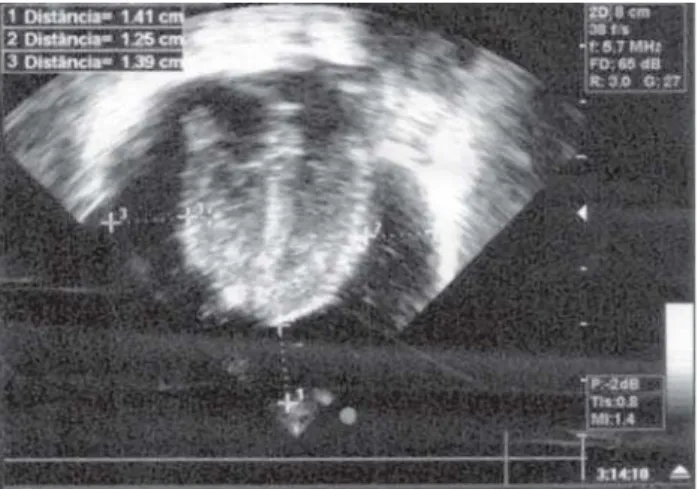
37-week-old newborn, born by c-section weighing 3.450g; pregnancy interrupted by oligohydramnios, admitted to the neonatology unit with pneumonia. On the second day of hospital stay, the infant had venous and arterial umbilical catheter inserted (Argaile 5F -polyurethane). On the seventh day of hospital stay, the patient’s clinical conditioning worsened, requiring nasal CPAP (Continuous Positive Airway Pressure). Chest radiography showed increase in the heart’s area and umbilical venous catheter in the same place previously inserted (connection between the inferior vena cava and the right atrium). The echocardiogram performed showed large pericardial effusion, of which there was no evidence in the examination performed 4 days prior (Figure 1).
Fig. 1 – Echocardiogram of Case 1 showing severe pericardial effusion
424
event was diagnosed on the 5th and 2nd day after catheterization, respectively. In both cases, the initial suspicion was sepsis.
Beardsall et al. [4] analyzed the number of central venous lines positioning and the number of pericardial effusions and/or cardiac tamponade over the course of 5 years in neonatology units of the United Kingdom. We analyzed about 46,000 central lines, and found that pericardial effusion and/or cardiac tamponade occurred in 1.8 / 1000 venous lines inserted, and found death as a result of this complication in 0.7 / 1000. Among the 82 cases of pericardial effusion and/or cardiac tamponade, 34 (41%) occurred in units that inserted less than 50 lines per year, and only 20 (24%) in units that inserted more than 100 lines per year (p = 0005). In 20 cases, the diagnosis occurred only during the autopsy.
Around the world, bedside chest radiography has been method most often used to certify the location of the umbilical catheter, and this is also the case in Brazil. Ideally, the tip of the umbilical catheter should be located at the connection of the inferior vena cava with the right atrium. Several studies have shown an incorrect positioning in patients who had correct location during the radiological examination and who underwent echographic examinations [5.6]. Only one of our patients had the tip of the catheter in the appropriate location. In the other, the tip was located in the pulmonary artery and was not mobilized. We consider such behavior inappropriate; a repositioning should have been performed.
Cardiac tamponade in newborns can occur not only as a result of the umbilical vein catheterization, but also after installing a peripherally inserted central catheter (PICC). Nadroo et al. [7] report three deaths caused by cardiac tamponade out of 390 placements of PICC. Nowlen et al. [2] reported 14 cases of pericardial effusion in newborns with deep venous access in six different units of neonatology over 2 years. This author also reported another 47 cases that were in the literature between 1970 and 1999. Among a total of 61 cases, the highest occurrence was found in cases that used the umbilical catheter (21 patients - 34.5%). The 14 patients in the series of the authors were receiving parenteral nutrition through the central venous catheter. Among our cases, one was receiving parenteral nutrition exclusively, and only one was using dextrosis.
There are insufficient data in the literature to prove the highest occurrence of pericardial effusion between different
REFERENCES
1. Onal EE, Saygili A, Koç E, Türkyilmaz C, Okumus N, Atalay Y. Cardiac tamponade in a newborn because of umbilical venous catheterization: is correct position safe? Paediatr Anaesth. 2004;14(11):953-6.
2. Nowlen TT, Rosenthal GL, Johnson GL, Tom DJ, Vargo TA. Pericardial effusion and tamponade in infants with central catheters. Pediatrics. 2002;110(1 Pt 1):137-42.
3. Rogers BB, Berns SD, Maynard EC, Hansen TW. Pericardial tamponade secondary to central venous catheterization and hyper alimentation in a very low birthweight infant. Pediatr Pathol. 1990;10(5):819-23.
4. Beardsall K, White DK, Pinto EM, Kelsall AW. Pericardial effusion and cardiac tamponade as complications of neonatal long lines: are they really a problem? Arch Dis Child Fetal Neonatal Ed. 2003;88(4):F292-5.
5. Greenberg M, Movahed H, Peterson B, Bejar R. Placement of umbilical venous catheters with use of bedside real-time ultrasonography. J Pediatr. 1995;126(4):633-5.
6. Raval NC, Gonzalez E, Bhat AM, Pearlman SA, Stefano JL. Umbilical venous catheters: evaluation of radiographs to determine position and associated complications of malpositioned umbilical venous catheters. Am J Perinatol. 1995;12(3):201-4.
7. Nadroo AM, Lin J, Green RS, Magid MS, Holzman IR. Death as a complication of peripherally inserted central catheters in neonates. J Pediatr. 2001;138(4):599-601.
types of material used for catheters. More controlled studies are needed.
CONCLUSION
Cardiac tamponade as a complication caused by the umbilical vein catheterization in newborns is rare, but serious and can occur even in cases in which the catheter is properly positioned. This complication should be considered in every newborn with central venous access whose condition suddenly worsens.
MONTEIRO, AJ ET AL - Cardiac tamponade caused by central venous catheter in two newborns