Absence of Nocturnal Dipping is Associated with Stroke and
Myocardium Infarction
Renan Oliveira Vaz de Melo
1, Juan Carlos Yugar Toledo
1, Afonso Augusto Carvalho Loureiro
1, José Paulo Cipullo
1, Heitor
Moreno Júnior
2, José Fernando Vilela Martin
1Faculdade Estadual de Medicina de São José do Rio Preto - Hospital de Base1, São José do Rio Preto, SP; Universidade Estadual de Campinas (UNICAMP)2, Campinas, SP - Brazil
Abstract
Background: The arterial hypertension varies in according to the circadian cycle, presenting physiologic fall of blood pressure (BP) during sleep (dipping). The absence of this fall or its increase associates to higher incidence of target-organ damages.
Objective: To analyze the prevalence of dipping in hypertensive individuals, to correlate dipping to the blood pressure levels, clinic, and socio-demographic factors, and biochemical characteristics and to associate it cardiovascular events (stroke and myocardial infarction).
Methods: Hypertensive individuals were submitted to the ambulatory blood pressure monitoring. Presence of dipper was defined as fall ≥10% of the systolic BP of the day for sleep.
Results: 6 evaluated patients were divided in dippers (D, n=5) and nondippers (ND, n=0). Between the groups
there was not significant difference to the age, sex, race, time of hypertension, glycemia, LDL-cholesterol, total cholesterol, triglycerides, schooling, smoking, and history of diabetes. D presented BP higher than the ND during the day and lower during sleep. ND had higher body mass index (BMI) (p=0.077), lower level of HDL-cholesterol (p=0.089), and higher pulse pressure during sleep (p=0.005). History of stroke alone (p=0,046) and combined with myocardial infarction (p=0.0) were more frequent in nondippers individuals. In the logistic regression, only ND was associated independently with stroke or myocardial infarction.
Conclusion: ND was associated in an independent way with the target-organ damages analyzed, what demonstrates its importance and strengthens the necessity of more aggressive treatment with objective to reach BP goals e, consequently, to prevent the development of new cardiologic and cerebrovascular events. (Arq Bras Cardiol 00; 94() : 74-80)
Key Words: blood pressure monitoring, ambulatory; hypertension; stroke; myocardial infarction.
Mailing address: Renan Oliveira Vaz de Melo •
Rua Professor Enjolrras Vampré, 201 / 44 Bloco 3 - Vila Santa Cândida - 15091-290 - São José do Rio Preto, SP - Brazil
E-mail: [email protected]
Manuscript received October 15, 2008; revised manuscript received Febru-ary 1st, 2009; accepted May 15, 2009.
Introduction
Blood pressure (BP) varies according to the interaction between neural-humoral, behavioral and environmental factors. One of the complementary exams capable of assessing this pressure pattern in a 24-hour period is the ambulatorial blood pressure monitoring (ABPM), which allows the indirect and intermittent register of BP, thus enabling the understanding of its variation profile at vigil and sleep periods. According to the Brazilian guidelines on ABPM, systemic arterial hypertension (SAH) is characterized by values superior to 130/80 mmHg1 during 24-hour period. Nowadays, there is
evidence that the values obtained by ABPM prognosticate better the most significant cardiovascular events, such as myocardium infarction and stroke, in comparison to the values found in consultation rooms2-6.
Among the parameters assessed by ABPM, some deserve to be emphasized. In analysis of the Syst-Eur study6, the variable
which showed better correlation to the main cardiovascular events was systolic blood pressure during sleep, followed by 24-hour and vigil systolic blood pressure. Another variable that deserves to be emphasized is the pressure drop which occurs in the vigil to sleep period, called nocturnal dipping (ND). With regard to the prognostic bound to this variable, whose normality value is a minimum 10% reduction of BP during sleep period in relation to vigil, it is known that there is an inverse correlation of BP during sleep and cardiovascular outcomes, even in the presence of normal mean values of pressure obtained by ABPM4.
Methods
A group of 163 hypertensive individuals, followed-up in a specialty ambulatory, were assessed and, later on, divided into groups with and without nocturnal dipping (dipper and non-dipper). The patients presented SAH diagnosis for at least three years. All participants signed the informed consent, which was previously approved by the Ethics Committee of the institution.
For the assessment of comorbidities, data regarding the presence of diabetes mellitus (DM), history of stroke, dislipidemia (DLP), medicines in usage, body mass index [IMC=kg/height (m2)], school degree, sex and other risk factors
or necessary information obtained from the medical record were investigated.
The patients who already presented diagnosis were considered diabetic individuals due to the presence of at least
two fast glycemia dosages ≥ 126 mg/dL or to altered glucose
oral tolerance test (OGTT)7. Patients who had OGTT in the
range of glucose intolerance, that is, glycemia of two hours after taking dextrosol with values between 140 and 199 mg/dL, were studied in the non-diabetic group, even when under treatment with metformin (three patients), which is recommended for such cases8. Analysis of stroke episodes was carried out based
on clinical history, presence of sequel and events previously identified in the medical record. MI diagnosis was based on clinical history and confirmed by analysis of medical records that showed previous enzymatic alterations (troponin and CK-MB), electrocardiographic alterations suggestive of coronary ischemia and proper treatment for this situation.
DLP was identified by total cholesterol dosages (TC), high-density lipoprotein cholesterol fraction (HDL-c) and triglycerides (TG) after 12-hour fasting9. The following reference values were
adopted: TC<200 mg/dL, HDL-c > 40 mg/dL, low-density lipoprotein cholesterol fraction (LDL-c) <130 mg/dL and TG < 150 mg/dL. LDL-c fraction was calculated by means of the formula: LDL-c = TC - HDL-c - TG/5 (for TG<400 mg/dL)10.
Individuals with alterations in the aforementioned parameters and/or those who were under treatment with HMG-CoA redutase inhibitors or other hypolipemic medicines were considered as patients with dislipidemia.
The values of systolic (SBP) and diastolic blood pressure (DBP) were obtained from 24-hour ABPM, as mean values obtained in 24 hours (_m), vigil (_v) and sleep (_s) periods were considered in the analysis. Pulse pressure (PP) was calculated with the mean values of SBP and DBP during the assessed periods (24 hours, vigil and sleep) in the formula PP
= SBP – DBP. ND was standardized as drop ≥10% of SBP in the period of vigil to sleep. The values of ≤ 130/80 within 24 hours, ≤ 135/85 during vigil and ≤ 120/70 mmHg during
sleep were considered normal1.
Spacelabs 90207 with its software for equipment programming and written reports emission was used. The installation of the equipment was made during the day, and the patient remained with it for a 24-hour period. The process of monitoring was carried out in dais that represented the patients’ daily activities. The equipment was programmed to register BP measurements every 15 minutes during vigil and every 20 minutes during sleep. All participants were instructed
as part of the exam protocol and registered their daily activities, meal, waking up and sleeping timetables, besides medications horary and presence of symptoms. Therefore, the definitions of vigil and sleep periods were based on the patients’ timetables.
The calculated size of the sample, admitting α deviation of 1% as to reject the hypothesis of nullity, was 47. For the comparison between groups, the software GraphPad Instat 3.0 was used, and the Fisher’s Exact test and Mann-Whitney test were applied for categorical and continuum variables, respectively. The multivariate logistic regression model was applied as to identify variables from the multivariate analysis that were statistically significant between groups (BMI, HDL-c and PP_s) and signifiHDL-cantly assoHDL-ciated with the studied cardiovascular events. The deviation of α at 5% with level of significance of p<0.05 was adopted.
Results
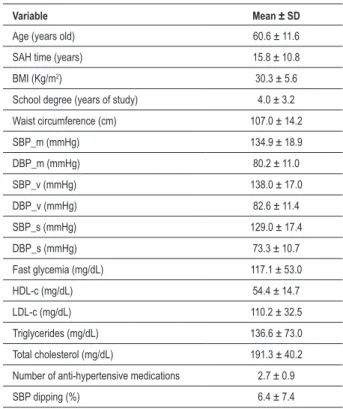
The general profile of the studied population may be observed on Table 1. The sample was composed by individuals with longtime-diagnosed SAH, whose values of BMI, waist circumference, fast glycemia and mean 24-hour BP were superior to the established limit values.
The studied casuistry consisted of 163 individuals with mean age of 60.6 ± 11.6 years old and time of SAH diagnosis of 15.8 ± 10.8 years. After ABPM analysis, the studied individuals were divided in Dipper (n=53, patients with
Table 1 - Basal proile of the studied population
Variable Mean ± SD
Age (years old) 60.6 ± 11.6
SAH time (years) 15.8 ± 10.8
BMI (Kg/m2) 30.3 ± 5.6
School degree (years of study) 4.0 ± 3.2
Waist circumference (cm) 107.0 ± 14.2
SBP_m (mmHg) 134.9 ± 18.9
DBP_m (mmHg) 80.2 ± 11.0
SBP_v (mmHg) 138.0 ± 17.0
DBP_v (mmHg) 82.6 ± 11.4
SBP_s (mmHg) 129.0 ± 17.4
DBP_s (mmHg) 73.3 ± 10.7
Fast glycemia (mg/dL) 117.1 ± 53.0
HDL-c (mg/dL) 54.4 ± 14.7
LDL-c (mg/dL) 110.2 ± 32.5
Triglycerides (mg/dL) 136.6 ± 73.0
Total cholesterol (mg/dL) 191.3 ± 40.2
Number of anti-hypertensive medications 2.7 ± 0.9
SBP dipping (%) 6.4 ± 7.4
SAH and present ND) and Non-dipper (n=110, hypertensive individuals without ND). Dipper Group was composed of 29 women and 24 men, while Non-dipper Group comprised 62 women and 48 men. Groups did not differ (p>0.05) with regard to individuals’ mean age, SAH time, school degree and waist circumference (Table 2). With regard to BMI, ND individuals presented lower mean values in comparison to hypertensive patients without ND (29.2 ± 5.0 x 30.9 ± 5.7,
p=0.0377).
Based on ABPM, it was observed that Dipper Group presented pressure levels higher than those of Non-dipper Groups during vigil period for SBP (141.6 ± 16.4 x 136.2 ± 17.0, p=0.0232) and for DBP (86.5 ± 10.4 x 80.6 ± 11.5,
p=0.0003). During sleep period, Dipper Groups presented mean values lower than those of Non-dipper Group for SBP (122.1 ± 14.1 x 132.4 ± 17.9, p=0.0001) and DBP (70.2 ± 8.6 x 74.8 ± 11.3, p=0.0113). Though pressure levels during 24-hour period followed a tendency of higher mean values in Dipper Group, they presented significant differences only for DBP (82.5 ± 9.4 x 79.0 ± 11.5, p=0.0065), while values for
Table 2 – Comparison between continuum variables of groups with and without ND by Mann-Whitney test
Variable Dipper (n=53) Non-dipper
(n=110) p-value
Age (years old) 60.4 ± 12.4 60.6 ± 11.3 NS
SAH time (years) 15.1 ± 12.1 16.1 ± 10.1 NS
BMI (Kg/m2) 29.2 ± 5.0 30.9 ± 5.7 0.0377
School degree (years
of study) 3.7 ± 3.0 4.2 ± 3.4 NS
Waist circumference
(cm) 106.9 ± 11.2 107.1 ± 15.5 NS
SBP_m (mmHg) 134.1 ± 22.6 135.4 ± 16.9 NS
DBP_m (mmHg) 82.5 ± 9.4 79.0 ± 11.5 0.0065
PP_m (mmHg) 54.2 ± 13.3 56.3 ± 12.5 NS
SBP_v (mmHg) 141.6 ± 16.4 136.2 ± 17.0 0.0232
DBP_v (mmHg) 86.5 ± 10.4 80.6 ± 11.5 0.0003
PP_v (mmHg) 55.3 ± 11.6 55.6 ± 12.4 NS
SBP_s (mmHg) 122.1 ± 14.1 132.4 ± 17.9 <0.0001
DBP_s (mmHg) 70.2 ± 8.6 74.8 ± 11.3 0.0113
PP_s (mmHg) 51.9 ± 10.9 57.6 ± 13.5 0.0025
Fast glycemia (mg/dL) 106.3 ± 32.1 122.4 ± 60.0 NS
HDL-c (mg/dL) 57.4 ± 14.2 52.9 ± 14.8 0.0189
LDL-c (mg/dL) 111.7 ± 29.1 109.4 ± 34.1 NS
Triglycerides (mg/dL) 130.9 ± 62.6 139.5 ± 77.7 NS
Total cholesterol (mg/dL) 195.3 ± 37.2 189.4 ± 41.7 NS
Number of anti-hypertensive medications
2.6 ± 0.9 2.7 ± 0.9 NS
BMI - body mass index; SBP - systolic blood pressure; DBP - diastolic blood pressure; PP - pulse pressure; _m - mean 24-hour pressure value; _v - pressure value during vigil; _s - pressure value during sleep; NS - Non-signiicant (p>0.05).
SBP were not significant (p>0.05). When PP of both groups was analyzed, we observed that the individuals from Dipper Group presented lower PP during sleep (51.9 ± 10.9 x 57.6 ± 13.5, p=0.0025) in comparison to Non-dipper Group. Such relation was not observed for PP during vigil and within the 24-hour period.
Mean HDL-c was higher in patients with ND in comparison to the mean of individuals without it (57.4 ± 14.2 x 52.9 ± 14.8, p=0.0189). Mean glycemia, TC, LDL-c and TG did not present statistically significant differences between groups.
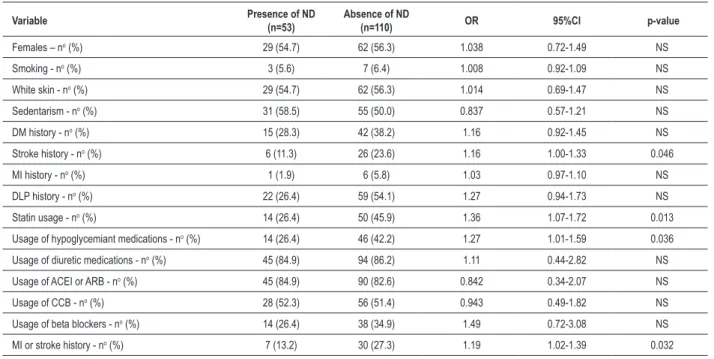
After analyzing qualitative variables (Table 3), we verified that groups did not differ (p>0.05) with regard to sex, skin color, smoking (5.6 versus 6.4%), sedentarism (58.5 versus 50.0%), MI history (1.9 versus 5.8%) in Dipper and Non-dipper Groups, respectively. Though DM and DLP histories have not presented significant p-value, both conditions were more frequent in ND individuals. Groups did not differ either regarding the main anti-hypertensive medications (diuretic, angiotensin-converting enzyme inhibitor, AT1 angiotensin II receptor blocker, calcium channel blockers and beta blockers). Stroke history (RR=1.16 95%CI 1.00-1.33), statin usage (RR=1.36 95%CI 1.07-1.72), hypoglycemiant (RR=1.27 95%CI 1.01-1.59) and associate lesions in target-organs (MI or stroke) (RR=1.19 95%CI 1.02-1.39) were related to ND absence.
As groups differed in some clinical characteristics, the logistic regression analysis was carried out as we used the presence of lesions in target-organs as dependent variable (stroke and MI) and as independent variables all clinical variables, including nocturnal dipping. BMI, HDL-c and PP_s did not constitute factors associated with stroke and MI risk. Only absence of nocturnal dipping presented independent correlation to the presence of assessed lesions in target-organs (Table 4).
Discussion
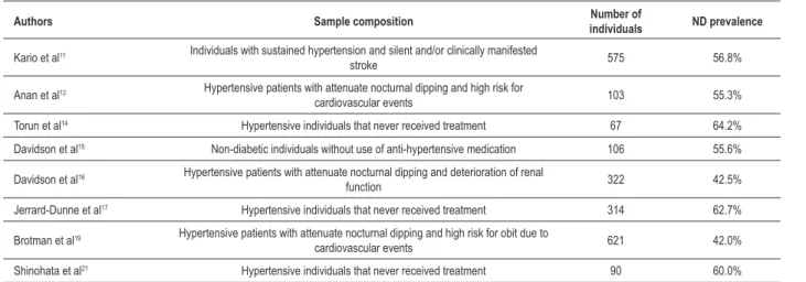
In the present study, there was a high prevalence of attenuation or absence of nocturnal dipping (67.5%). ND attenuation was associated with the presence of other cardiovascular risk factors (obesity and lower HDL-c) and also with a higher frequency of lesion in target-organ. Studies on the prevalence of cardiovascular and cerebral events varies greatly in the specific literature (Table 5), reaching a frequency of 37.5 to 64.2%11-21 under several factors’ influence, like the
definition of sleep and vigil periods22.
In our sample, ND individuals presented mean pressure superior to those presented by individuals without ND during vigil and inferior during sleep. However, when the 24-hour period was assessed, this behavior pattern was not observed. These findings, in general, differ from those found in literature14-16,18,19.
Torun et al14, after analyzing hypertensive patients with
Table 3 - Comparison between qualitative variables of groups with and without ND by Fisher’s exact test
Variable Presence of ND
(n=53)
Absence of ND
(n=110) OR 95%CI p-value
Females – no (%) 29 (54.7) 62 (56.3) 1.038 0.72-1.49 NS
Smoking - no (%) 3 (5.6) 7 (6.4) 1.008 0.92-1.09 NS
White skin - no (%) 29 (54.7) 62 (56.3) 1.014 0.69-1.47 NS
Sedentarism - no (%) 31 (58.5) 55 (50.0) 0.837 0.57-1.21 NS
DM history - no (%) 15 (28.3) 42 (38.2) 1.16 0.92-1.45 NS
Stroke history - no (%) 6 (11.3) 26 (23.6) 1.16 1.00-1.33 0.046
MI history - no (%) 1 (1.9) 6 (5.8) 1.03 0.97-1.10 NS
DLP history - no (%) 22 (26.4) 59 (54.1) 1.27 0.94-1.73 NS
Statin usage - no (%) 14 (26.4) 50 (45.9) 1.36 1.07-1.72 0.013
Usage of hypoglycemiant medications - no (%) 14 (26.4) 46 (42.2) 1.27 1.01-1.59 0.036
Usage of diuretic medications - no (%) 45 (84.9) 94 (86.2) 1.11 0.44-2.82 NS
Usage of ACEI or ARB - no (%) 45 (84.9) 90 (82.6) 0.842 0.34-2.07 NS
Usage of CCB - no (%) 28 (52.3) 56 (51.4) 0.943 0.49-1.82 NS
Usage of beta blockers - no (%) 14 (26.4) 38 (34.9) 1.49 0.72-3.08 NS
MI or stroke history - no (%) 7 (13.2) 30 (27.3) 1.19 1.02-1.39 0.032
DM - diabetes mellitus; MI - myocardium infarction; DLP - dislipidemia; ACEI - angiotensin-converting enzyme inhibitors; ARB - AT1 angiotensin II receptor blockers; CCB - calcium channel blockers; RR - relative risk; CI - conidence interval; NS - Non-signiicant (p>0.05).
is similar to those observed by Anan et al12, who emphasized
the importance of the method chosen for the investigation of hypertensive patients.
PP seems to be an independent marker of cardiovascular mortality, mainly among individuals older than 50 years old and with history of myocardium infarction, cardiac insufficiency and stroke23. The arterial rigidity and the reflection of pulse
wave are the greatest determinant factors for PP elevation due to aging. An increase in the arterial rigidity may also elevate the risk for coronary events due to rapid return of pulse wave reflected at the end of systole and the risk for cerebrovascular events due to a modification in the arteries’ walls and development of atherosclerotic plaques24. In our
sample, composed by individuals with mean age superior to 60 years old, we observed higher statistically significant PP in Non-dipper Group during sleep (57.6 mmHg), but no statistical significance in the other periods. According to the IV Brazilian directive for ABPM, the value of 53 mmHg is praised for PP as a limit which is superior to normality1. This value was
initially showed by Verdecchia et al25, who found a higher risk
for cardiovascular events when PP was superior to 53 mmHg after evaluating more than 2 thousand patients through ABPM. However, despite the fact that Non-dipper Group had higher PP, the logistic regression model did not show association with cardiovascular events in the present study.
After the analysis of sociodemographic factors, we pointed out in our sample the absence of association between ND and the remaining variables (age, SAH time, school degree, waist circumference, genre, race, smoking and sedentarism), which was confirmed by other studies12,14,15,26. Other authors
observed that individuals without ND were older16,18,19
and that this condition occurs preferentially in non-white
people19. In the current study, BMI was higher in individuals
without ND (30.9 ± 5.7 x 29.2 ± 5.0, p=0.0377), fact also observed by Kotsis et al27 in 3,216patients without previous
anti-hypertensive treatment. These authors showed a direct relation between higher BMI and higher mean pressure for ABMP, and 55% of the individuals with normal weight (normal BMI) presented ND, while only 35% of the obese individuals presented the same condition (p<0.05). Others, however, did not find such association12,14-16,19. In our sample, after
the logistic regression model, we observed that BMI was not correlated to a higher risk for stroke or MI in ND patients.
After the biochemical dosages, we found that, except for the higher levels of HDL-c in ND patients, the remaining variables did not present statistical significance, findings that are similar to those observed in other studies15,16. Despite
the lower HDL-c in non-dipper patients, there was no correlation to higher risk of lesions in target-organs. Alterations found in the levels of uric acid14,16, serum creatinine16,18,
glycemia12,18 and triglycerides15,16 in patients without ND
make their interpretation difficult due to the differences found between the studied populations as well as to the influence of medications on these biochemical parameters. Though we observed a higher prevalence of diabetic patients in the Non-dipper Group (38.2 versus 28.3%), this value was not statistically significant, which opposes to the findings of Björklund et al28, who found higher frequency of individuals
Table 5 – Prevalence of nocturnal dipping in transversal studies
Authors Sample composition individualsNumber of ND prevalence
Kario et al11 Individuals with sustained hypertension and silent and/or clinically manifested
stroke 575 56.8%
Anan et al12 Hypertensive patients with attenuate nocturnal dipping and high risk for
cardiovascular events 103 55.3%
Torun et al14 Hypertensive individuals that never received treatment 67 64.2%
Davidson et al15 Non-diabetic individuals without use of anti-hypertensive medication 106 55.6%
Davidson et al16 Hypertensive patients with attenuate nocturnal dipping and deterioration of renal
function 322 42.5%
Jerrard-Dunne et al17 Hypertensive individuals that never received treatment 314 62.7%
Brotman et al19 Hypertensive patients with attenuate nocturnal dipping and high risk for obit due to
cardiovascular events 621 42.0%
Shinohata et al21 Hypertensive individuals that never received treatment 90 60.0%
AMBP - ambulatorial monitoring of blood pressure; ND - nocturnal dipping.
Table 4 – Logistic regression model for stroke and MI in non-dipper patients
Variables β Standard error Odds Ratio (95%CI) p-value
Non-dipper 11.502 0.5764 3.16 (1.02-9.78) 0.046
BMI -0.0586 0.1022 0.94 (0.77-1.15) NS
HDL-c -0.00805 0.04166 0.99 (0.91-1.07) NS
PP_s -0.00876 0.06409 0.99 (0.87-1.12) NS
MI - myocardium infarction; BMI - body mass index; CI - conidence interval; NS - non-signiicant (p>0.05).
diabetic patients, although there was no statistically significant difference among them.
In the current paper, stroke history (p=0.046) and the association with MI or stroke (p=0.032) were statistically significant in patients without ND. Possible reasons for these findings may be found in the history of lesions in target-organs of hypertensive patients, that is, left ventricle hypertrophy, microalbuminury, renal dysfunction and cerebrovascular disease are more prevalent in individuals without ND1,14,18.
Ohkubo et al29 assessed 1,542 individuals older than 40 years
old who were followed-up for approximately 9.2 years. The authors found a linear relation between nocturnal dipping of blood pressure and mortality, and each 5% of reduction of SBP and DBP drop, the risk for cardiovascular mortality would increase in 20%. Even in the presence of normal mean
24-hour pressure levels (≤130/80 mmHg), the attenuation
of nocturnal dipping of blood pressure were associated with an increase in the risk for cardiovascular death. Kario et al11
followed-up a Japanese population of 575 individuals of ≥50
years old who were divided into groups according to nocturnal dipping of SBP. After a 41 months average, they observed that the incidence of stroke was of 12% among individuals
with accentuate nocturnal dipping (≥20%), 6.1% among
individuals with ND between 10 and 20%, 7.6% among
individuals without ND (≥0% and <10%) and 22% among
patients with nocturnal dipping of blood pressure.
The mechanisms responsible for this abnormal variation of arterial pressure remain unknown, though there is strong evidence of a possible involvement of compromising in the autonomic balance, which leads to a sympathetic hyperactivity during sleep period that may alter circadian rhythm, common occurrence among patients with sleep apnea syndrome30.
Another factor that seems to exert influence on ND phenomenon is sodium ingestion31. Some studies reported
that ND would be associated to a reduced excretion of sodium during vigil period32,33. Birkenhäger and van den Meiracker34
observed that a low sleep quality, along with daily inactivity, represented by sedentarism, might explain the phenomenon of ND absence.
Something that also deserves to be emphasized in our study is that mean pressure values during vigil, sleep and 24-hour period obtained by means of ABPM were above the limits recommended by our guideline1. In that case, measures
as adoption of a more rigid treatment and the modification of medication administration horary could contribute with the improvement of these individuals’ pressure profile and, consequently, change ND pattern35.
This previous concept that the absence of ND could be related to a worse cardiovascular and cerebral outcome has, however, exceptions and there are groups in which the influence of this variable deserves better elucidations, like in the case of individuals with previous history of cerebrovascular disease. Nakamura et al36 followed-up individuals with
previous stroke history and observed that ischemic event recurrence rate was higher among individuals with ND and under anti-hypertensive treatment. Besides, the frequency of new events was higher among treated hypertensive and ND patients than among those without nocturnal dipping. In that special case, though controlling pressure levels is a effective measure for the prevention of new events37, an
References
1. Sociedade Brasileira de Cardiologia. IV Diretriz para uso da monitorização ambulatorial da pressão arterial - II Diretriz para uso da monitorização residencial da pressão arterial IV MAPA / II MRPA. Arq Bras Cardiol. 2005; 85 (supl. 2): 1-18.
2. Clement DL, De Buvzere ML, De Bacquer DA, de Leeuw PW, Duprez DA, Fagard RH, et al. Office versus Ambulatory Pressure Study Investigators. Prognostic value of ambulatory blood-pressure recordings in patients with treated hypertension. N Eng J Med. 2003; 348 (24): 2407-15.
3. Dolan E, Stanton A, Thijs L, Hinedi K, Atkins N, McClory S, et al. Superiority of ambulatory over clinic blood pressure measurement in predicting mortality: the Dublin Outcome Study. Hypertension. 2005; 46 (1): 156-61.
4. Ben-Dov IZ, Kark JD, Ben-Ishay D, Mekler J, Ben-Arie L, Bursztyn M. Predictors of all-cause mortality in clinical ambulatory monitoring: unique aspects of blood pressure during sleep. Hypertension. 2007; 49 (6): 1235-41.
5. Giorgi DMA. Monitorização ambulatorial da pressão arterial como método de avaliação do prognóstico em hipertensão arterial. Arq Bras Cardiol. 1996; 67 (2): 145-8.
6. Staessen JA, Thijs L, Fagard R, O’Brien ET, Clement D, de Leeuw PW, et al. Predicting cardiovascular risk using conventional vs ambulatory blood pressure in older patients with systolic hypertension. Systolic Hypertension in Europe Trial Investigators. JAMA. 1999; 282 (6): 539-46.
7. American Diabetes Association. Diagnosis and classification of Diabetes Mellitus. Diabetes Care. 2004; 27 (Suppl. 1): S5-S10.
8. Knowler WC, Barrett-Connor E, Fowler SE, Hamman RF, Lachin JM, Walker EA, et al. Reduction in the incidence of type 2 diabetes with lifestyle intervention or metformin. N Eng J Med. 2002; 346 (6): 393-403.
9. Sociedade Brasileira de Cardiologia. IV Diretriz brasileira sobre dislipidemias e prevenção da doença aterosclerótica. Arq Bras Cardiol. 2007; 88 (supl. 1): 2-19.
10. Friedewald WT, Levy RI, Fredrickson DS. Estimation of the concentration of low-density lipoprotein cholesterol in plasma, without use of the preparative ultracentrifuge. Clin Chem. 1972; 18 (6): 499-502.
11. Kario K, Pickering TG, Matsuo T, Hoshide S, Schwartz JE, Shimada K. Stroke
prognosis and abnormal nocturnal blood pressure falls in older hypertensives. Hypertension. 2001; 38 (4): 852-7.
12. Anan F, Takahashi N, Ooie T, Yufu K, Saikawa T, Yoshimatsu H. Role of insulin resistance in nondipper essential hypertensive patients. Hypertens Res. 2003; 26 (9): 669-76.
13. Del Compare ME, D’Agostino D, Ferraris JR, Boldrini G, Waisman G, Krmar RT. Twenty-four hour ambulatory blood pressure profiles in liver transplant recipients. Pediatr Transplant. 2004; 8 (5): 496-501.
14. Torun D, Sezer S, Arat Z, Pelit A, Yigit F, Ozdemir FN. The frequency of combined target organ damage and the beneficial effect of ambulatory blood pressure monitoring in never treated mild-to-moderate hypertensive patients. Int Heart J. 2005; 46 (6): 1073-82.
15. Davidson MB, Vidt DG, Hoogwerf BJ, Brotman DJ. Relation of diurnal blood pressure variation and triglyceride-to-high-density lipoprotein cholesterol ratio in patients without diabetes mellitus. Am J Cardiol. 2005; 95 (1): 123-6.
16. Davidson MB, Hix JK, Vidt DG, Brotman DJ. Association of impaired diurnal blood pressure variation with a subsequent decline in glomerular filtration rate. Arch Intern Med. 2006; 166 (8): 846-52.
17. Jerrard-Dunne P, Mahmud A, Feely J. Circadian blood pressure variation: relationship between dipper status and measures of arterial stiffness. J Hypertens. 2007; 25 (6): 1233-9.
18. Muxfeldt ES, Salles GF. Pulse pressure or dipping pattern: which one is a better cardiovascular risk marker in resistant hypertension? J Hypertens. 2008; 26 (5): 878-84.
19. Brotman DJ, Davidson MB, Boumitri M, Vidt DG. Impaired diurnal blood pressure variation and all-cause mortality. Am J Hypertens. 2008; 21 (1): 92-7.
20. Ortega KC, Mion Jr D. Qual o melhor determinante do prognóstico pela monitorização ambulatorial da pressão arterial: ausência de descenso da pressão durante o sono ou elevação rápida da pressão pela manhã? Arq Bras Cardiol. 2005; 85 (3): 208-9.
21. Shinohata R, Nakatsu T, Yuki Y, Nishitani A, Mishima K, Toyonaga S, et al. Association of augmentation index of radial pressure wave form with diurnal variation pattern of blood pressure in untreated patients with essential
of the ischemic process, thus contributing to the development of cognitive debt38. It could happen due to the interaction
between excessive pressure reduction during sleep allied to the effect of anti0hypertensive drugs, which could unleash cerebral ischemia and an area’s infarction.
The main limitation of our study was the fact that the studied patients were divided into groups of people with and without ND based on only one 24-hour ABPM. Although some studies affirm that the characterization based only on one exam is not reliable, the accomplishment of the activities record by the patients allow us to exclude those who presented sleep disturbances due to the exam, thus eliminating possible trends. Another factor that is not less important is the fixation of a schedule and definition of vigil and sleep periods, fact confirmed by the standardization of these periods based on the time schedules defined by patients. Another limitation that must be mentioned was that we did not observe sleep apnea syndrome, which could directly influence the absence of ND39,40. However, patients who reported sleep disturbances
in their daily records were excluded.
Taking into account the high cardiovascular risk and the association of risk factors in patients without nocturnal
dipping, the strict control of such factors in this group of patients is justified by the potential risk reversion. Although there are no concrete data with regard to drug treatment based on ND profile, measures such as improvement of sleep quality, reduction of sodium ingestion and better pressure control are fundamental to the minimization of cardiovascular risk due to ND absence, which contributes with a better life quality and reduction of morbidity and mortality due to SAH.
Potential Conflict of Interest
No potential conflict of interest relevant to this article was reported.
Sources of Funding
There were no external funding sources for this study.
Study Association
hypertension. J Hypertens. 2008; 26 (3): 535-43.
22. Henskens LH, Kroon AA, van Oostenbrugge RJ, Haest RJ, Lodder J, de Leeuw PW. Different classifications of nocturnal blood pressure dipping affect the prevalence of dippers and nondippers and the relation with target-organ damage. J Hypertens. 2008; 26 (4): 691-8.
23. Safar ME. Systolic blood pressure, pulse pressure and arterial stiffness as cardiovascular risk factors. Curr Opin Nephrol Hypertens. 2001; 10: 257-61.
24. Laurent S, Cockcroft J, Van Bortel L, Boutouyrie P, Giannattasio C, Hayoz D, et al. Expert consensus document on arterial stiffness: methodological issues and clinical applications. Eur Heart J. 2006; 27: 2588-605.
25. Verdecchia P, Schillaci G, Borgioni C, Ciucci A, Pede S, Porcellati C. Ambulatory pulse pressure: a patent predictor of total cardiovascular risk in hypertension. Hypertension. 1998; 32: 983-8.
26. Morillo MG, Amato MC, Cendon Filha SP. Twenty-four hour blood pressure record for smokers and nonsmokers. Arq Bras Cardiol. 2006; 87 (4): 504-11.
27. Kotsis V, Stabouli S, Bouldin M, Low A, Toumanidis S, Zakopoulos N. Impact of obesity on 24-hour ambulatory blood pressure and hypertension. Hypertension. 2005; 45 (4): 602-7.
28. Björklund K, Lind L, Andrén B, Lithell H. The majority of nondipping men do not have increased cardiovascular risk: a population-based study. J Hypertens. 2002; 20 (8): 1501-6.
29. Ohkubo T, Hozawa A, Yamaguchi J, Kikuya M, Ohmori K, Michimata M, et al. Prognostic significance of the nocturnal decline in blood pressure in individuals with and without high 24-h blood pressure: the Ohasama study. J Hypertens. 2002; 20 (11): 2183-9.
30. Dimsdale JE, Coy T, Ancoli-Israel S, Mills P, Clausen J, Ziegler MG. Sympathetic nervous system alterations in sleep apnea: the relative importance of respiratory disturbance, hypoxia, and sleep quality. Chest. 1997; 111: 639-42.
31. Hoshide S, Kario K. Determinants of nondipping in nocturnal blood pressure and specific nonpharmacological treatments for nocturnal hypertension. Am J Hypertens. 2008; 21 (9): 968.
32. Fujii T, Uzu T, Nishimura M, Takeji M, Kuroda S, Nakamura S, et al. Circadian rhythm of natriuresis is disturbed in nondipper type of essential hypertension. Am J Kidney Dis. 1999; 33 (1): 29-35.
33. Kimura G. Kidney and circadian blood pressure rhythm. Hypertension. 2008; 51 (4): 827-8.
34. Birkenhäger AM, van den Meiracker AH. Causes and consequences of a non-dipping blood pressure profile. Neth J Med. 2007; 65 (4): 127-31.
35. Peixoto AJ, White WB. Circadian blood pressure: clinical implications based on the pathophysiology of its variability. Kidney Int. 2007; 71 (9): 855-60.
36. Nakamura K, Oita J, Yamaguchi T. Nocturnal blood pressure dip in stroke survivors: a pilot study. Stroke. 1995; 26 (8): 1373-8.
37. Rashid P, Leonardi-Bee J, Bath P. Blood pressure reduction and secondary prevention of stroke and other vascular events: a systematic review. Stroke. 2003; 34 (11): 2741-8.
38. Birns J, Markus H, Kalra L. Blood pressure reduction for vascular risk: is there a price to be paid? Stroke. 2005; 36 (6): 1308-13.
39. Pedullà M, Silvestri R, Lasco A, Mento G, Lanuzza B, Sofia L, et al. Sleep structure in essential hypertensive patients: differences between dippers and non-dippers. Blood Press. 1995; 4 (4): 232-7.