141 1. Master’s Degree. MD, Instituto Materno Infantil de Pernambuco (IMIP),
Recife, PE, Brazil.
2. PhD. Associate professor, Universidade de Pernambuco (UPE). MD, Instituto Materno Infantil de Pernambuco (IMIP), Recife, PE, Brazil. 3. Doctoral student, University of Liverpool. Assistant professor, Universidade
de Pernambuco (UPE). MD, Instituto Materno Infantil de Pernambuco (IMIP), Recife, PE, Brazil.
Manuscript received Apr 28 2003, accepted for publication Nov 26 2003. Abstract
Objective: To describe the clinical and epidemiological features of children with visceral leishmaniasis admitted to a pediatric referral hospital, and to describe treatment measures and the case fatality rate.
Methods: Retrospective analysis of biological, demographic, clinical and laboratory data from children with visceral leishmaniasis admitted to Instituto Materno Infantil de Pernambuco (Recife, state of Pernambuco, northeastern Brazil) between 1996 and 2001.
Results: 431 children were included in the study. Age ranged from 4 months to 13.7 years. 50.3% were female and 82.5% came from the interior of the state of Pernambuco. 70% of the patients lived in brick homes, and 70% were not served with piped water and sewage services. Average maternal schooling was 3 years. Clinical presentation included splenomegaly (97%), fever (95.6%) and malnourishment (44.5%). Associated infections were diagnosed in 10.9% of cases. The mean values for laboratory variables were: hemoglobin 6 g/dl, leukocyte count 3,516/mm³, and platelet count 118,641/ mm³. The first line treatment used in 98% of the cases was glucantime. Amphotericin B was used in seven cases. The case fatality rate was 10.2%. The main immediate causes of death were associated infections, bleeding and liver failure.
Conclusions: Health care workers should be trained for the early recognition and appropriate management of visceral leishmaniasis and its complications.
J Pediatr (Rio J). 2004;80(2):141-6: Visceral leishmaniasis, kala-azar, childhood, diagnosis, treatment, case fatality rate.
Visceral leishmaniasis:
clinical and epidemiological features
of children in an endemic area
Márcia J. A. Queiroz1, João G. B. Alves2, Jailson B. Correia3 Copyright © 2004 by Sociedade Brasileira de Pediatria
O
RIGINALA
RTICLEIntroduction
According to the World Health Organization (WHO), leishmaniasis affects around two million people a year, 500 thousand cases of which are of the visceral form. It is estimated that 350 million people are exposed to the risk of infection with a global prevalence of 12 million infected individuals.1,2
Visceral leishmaniasis is widespread in Brazil, with autochthonous cases being reported in at least 19 states of the Brazilian federation, and four of the five national
regions (North, Northeast, Midwest and Southeast). Only the South remains unaffected.3 Between 1984 and 2000 67,231 cases were reported.4 More than 90% of these reports are concentrated in the Northeast region which reports cases within all of its federal units. In the state of Pernambuco,5 1,203 cases were reported during the period between 1990 and 1997. During recent decades there has been a strong tendency towards urban areas, with epidemic outbreaks taking place in a number of different state capitals6 and constituting a serious public health which threatens the population and concerns health authorities.
diarrhea, dry coughing, adynamia , light fever, diaphoresis and discrete hepatosplenomegaly,7 which may or may not progress to the classic form of the d i s e a s e . T h e c l a s s i c p r e s e n t a t i o n i s o f f e v e r , hepatosplenomegaly, with voluminous splenomegaly, weight loss, coughing, diarrhea, pain and abdominal distension. Jaundice and renal involvement have also been described. During the last phase of the disease patients may develop edema and ascites.1
Diagnosis is based on identification of the parasite in bone marrow, liver, spleen or lymph node tissues.7 A number of different serum-based tests have been developed for diagnosis (e.g. complement fixation test, indirect immunofluorescence, direct agglutination test, ELISA and Dot-ELISA), as have certain molecular biology techniques (polymerase chain reaction), although certain issues remain in relation to the sensitivity, specificity, and availability of these tests in clinical practice.8 When laboratory-based diagnosis is not possible, initial treatment is based on clinical and epidemiological findings.9
Pentavalent antimonial compounds continue to be the first-choice drug and amphotericin B is the second line of attack in cases of resistance to antimoniate.3 Recently, a new, orally administered, drug, miltefosine, has proven successful in India for VL treatment.10 Infections, hemorrhages and severe anemia are responsible for the majority of deaths while late diagnosis, low age at onset and malnutrition are important contributing mortality factors.3,7
This paper describes epidemiological, clinical and laboratory data from leishmaniasis patients admitted to a pediatric hospital which is a center of excellence. Emphasis is given to the importance of early diagnosis and treatment to avoid elevated lethality.
Patients and methods
Four hundred and forty-five patients with VL, aged up to 14 years, were admitted to the Instituto Materno Infantil de Pernambuco (IMIP), one of the regional centers of excellence within the state of Pernambuco for the diagnosis and treatment of this disease, during the period between May 1996 and December 2001. Eight medical records were not located and other patients were excluded, despite a final diagnosis of leishmaniasis, because they did not fit the studys inclusion criteria. Data was obtained by retrospective analysis of the medical records of children admitted with VL and entered onto a standardized form. The form, developed by the research group, covered identification, origin, domestic characteristics, maternal education, primary complaints and their durations, previous treatment history, physical examination findings, laboratory test results and treatment received. Patients were included who had had a diagnosis confirmed by myelogram, direct agglutination test (DAT) > 1/1,600 or indirect immunofluorescence (IIF) > 1/40 as were probable cases in which myelogram results were negative but there was
a clinical, epidemiological and laboratorial (pancytopenia) suspicion of leishmaniasis, ruling out other pathologies.
Nutritional status was assessed based on a weight for age scale, at the point of admission, taking the United States National Center of Healths Statistics curves as a reference. Children with edema of at least the feet were c o ns i der ed s ev er el y mal no ur i s hed ( edemat o us malnutrition). Pancytopenia was diagnosed when leukopenia was present set at WBC < 5.000/mm³. Anemia was defined as hemoglobin < 11 g/dl for patients between six months and five years old, < 11.5 g/dl for those between six and nine and < 12 g/dl for female adolescents and <12.5 g/dl for male adolescents. Thrombocytopenia was defined as a platelet count < 150.000/mm³.
Statistical analysis was performed using Epi-Info 6.0 4b, to produce distributions for frequency, mean average and standard deviation from the data returned when applicable.
Scientific and ethical aspects were approved by the Ethics Commission at the Hospital before data collection commenced.
Results
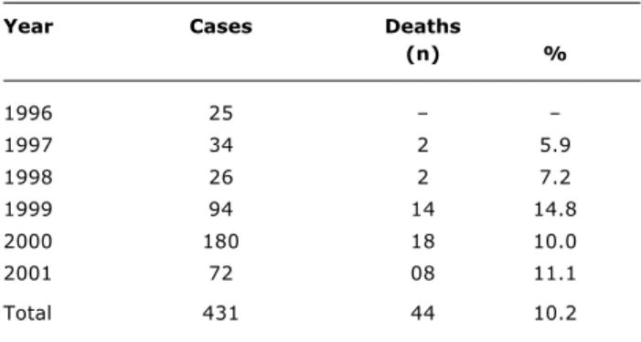
The distribution of cases over time is shown in Table 1. The greatest number of cases in any one year (180) occurred in 2000, although peak lethality observed in 1999 (14.8%).
Fifty point three percent of the children were female. The average age was 4.2 years (SD = ±3) and the youngest child was 4 months old. The worst affected age group was the under-fives (68.2% of cases) and 9% of the children were less than one year old. Of the 317 cases in which information was available on the mothers educational level, it was found that the average length of time spent at school was 3.1 years (SD = ±3). Three hundred and fifty-eight medical records yielded information about sewerage and sanitation and of these around 70% of patients did not have access to these services at home.
Table 1 - Distribution of children admitted to IMIP with diagnosis of leishmaniasis from May 1996 to December 2001
Source: Pesquisa HGP/IMIP.
Year Cases Deaths
(n) %
1996 25
1997 34 2 5.9
1998 26 2 7.2
1999 94 14 14.8
2000 180 18 10.0
2001 72 08 11.1
The majority were from Pernambuco State itself (99%), three were from Alagoas and one was from Paraíba. Eighty-two point five percent of the sample was from the interior of Pernambuco state while 14.8% were from the metropolitan region of the state capital Recife.
Nutritional evaluation revealed that 44.5% of the population under study was under-nourished, while 26.9% of the children were severely malnourished graves.
The patients had suffered from the disease before hospitalization for periods varying from 2 to 365 days, giving an average of 42.7 days(SD = ±45). Eighty-eight point seven percent of the patients had endured symptoms for less than 60 days before seeking treatment. The average hospital stay was 11.2 days (SD = ±5,7), varying from zero to 47 days. Among the symptoms referred to by family members, the following stand out as the most common: fever, increased abdominal volume, pallor, anorexia and coughing (Table 2). Approximately 50% of the patients mothers had tried some type of medication, chiefly antibiotics. Fifteen parents reported having used leishmaniasis-specific treatments.
Table 2 also contains the findings of physical examinations at the point of admission, recording that splenomegaly, pallor and hepatomegaly were the most common abnormalities. Some type of infection was detected on admission in 47 (10.9%) of the children, of which the most common were: pneumonia in 66% of cases, otitis in 18.4% and sepsis in 8.2%.
Hematological data from the point of admission on red and white blood cell and platelet tests are listed in Table 3. The average hemoglobin level was 6 g/dl, and erythrocyte transfusions were necessary for 170 patients while interned. Average white blood cell count was 3,516/mm³ and was below 5,000/mm³ in 367 cases (85,3%). The average neutrophil level was 1,215/mm³ and values below 500/mm³ were found in 15.9% of the patients. Average platelet count was 118,641/mm³. Forty-three platelet transfusions were required and 68.4% of the patients had < 150.000/mm³ platelets on admission.
Table 2 - Main symptoms and clinical findings of children admitted to IMIP with diagnosis of leishmaniasis from May 1996 to December 2001
Source: Pesquisa HGP/IMIP.
Symptoms n %
Fever 412 95.6
Abdominal volume 277 64.3
Pallor 250 58.0
Anorexia 215 49.9
Coughing 181 42.0
Weight loss 163 37.8
Asthenia 163 37.8
Abdominal pain 73 16.9
Diarrhea 55 12.8
Edema 71 16.5
Infection 50 11.6
Clinical findings n %
Splenomegaly 418 97.0
Pallor 393 91.2
Hepatomegaly 376 87.2 Malnutritrion 191 44.5
Edema 69 16.0
Dyspnea 53 12.2
Infection 47 10.9
Hemorrhage 49 11.4
Jaundice 19 4.4
Value
Parameters Mean±SD Minimum Maximum
Red blood cells (g/dl) 6.1±1.7 1.8 11.2 White blood cells (mm³) 3,516±1,923 600 16,300 Neutrophils (mm³) 1,215±1,052 32 9,682 Platelets (mm³) 118,641±88,359 2,540 808,000
Source: Pesquisa HGP/IMIP.
Table 3 - Hematological data of children admitted to IMIP with diagnosis of leishmaniasis from May 1996 to December 2001
It was possible to confirm diagnosis in 79.4% of cases. Four hundred and twelve patients were investigated for the parasite using myelograms, of whom 311 (75.5%) had positive results. DAT was positive in 28 cases and IIF in three. In the remaining cases, in which none of the above methods gave definitive proof, analysis of epidemiological, clinical and laboratory data was enough to justify treatment.
The treatment of choice for visceral leishmaniasis was glucantime (meglumine antimoniate) for 98% of the patients. Seven of these received glucantime in association with allopurinol. Seven patients continued to suffer symptoms after the first course of treatment and were given amphotericin B. The average time taken for fever to recede was 3 days (SD = ±2.7) and 50 patients did not exhibit fever while hospitalized. Seven patients died before receiving treatment specifically directed at the disease.
different infectious diseases including VL, as can be seen in this sample where, despite the majority of patient residences being brick-built, the availability of running water and sewerage is still at an unacceptably low level. It is rare for the classic form of the disease to affect middle-class people, even in endemic areas.3 According to Bezzerra,17 the level to which parents have been educated has a protective relationship with this infection, as can be corroborated by the results of our investigation, in which around 80% of the mothers had only received elementary education.
When confirmed cases were compared with probable cases it was found that there was no statistically significant difference between the two groups in terms of sex, age, origin or nutritional status. However, when the time during which the disease had been evolving before admission was evaluated we noted that the probable cases had been progressing for a significantly shorter period of time (30.4 days) than had the confirmed cases (459 days).
The great variation in disease duration before admission (2 days to one year), is in line with published data. Pastorino11 reports an average period of 6 months, while Marzochi18 describes a range of 1 to 5 months. Leishmaniasis is generally an insidious disease, with non-specific initial symptoms. This, in conjunction with potential bias due to faulty memory and the low educational level of the populations of endemic areas makes this variable particularly difficult to interpret. Furthermore, the high proportion of cases (at least 30%) in this series, in which medical attention had been sought previously and in which other medications, including antibiotics, had been used, suggests that opportunities for early diagnosis may have been lost. This must be a cause for concern in areas in which leismaniasis is endemic since late diagnosis is a risk factor for death.19
Nutritional evaluation revealed the diseases wide range of clinical variation, demonstrated by the presence of patients within the normal weight percentiles (55% of the children), while approximately 27% were severely malnourished. It should be noted that a majority of the children (83%) had suffered from the disease for less than 60 days; a period which may well not be sufficient for chronic nutritional problems to develop and which may explain the presence of well-nourished patients, as Pastorino11 also observed. It is possible that under-n o u r i s h m e under-n t m a y s u p p r e s s t h e c e l l - m e d i a t e d immunoresponse and thus be responsible for the development of progressive visceral leishmaniasis.
The clinical manifestations of leishmaniasis exhibited by the children in this sample were similar to those in published descriptions1-3,5,7,11 both in terms of symptoms described by family members and physical findings. Fever, hepatosplenomegaly and wasting are the classic signs of the disease and were presented by almost all of the patients at the point of admission. This is because it is
Causes Death
n %
Associated infections 32 72.7
Hemorrhage 26 59.0
Hepatic insufficiency 14 31.8 Hemorrhage + infection 10 22.7 Hemorrhage + infection + hepatic insufficiency 10 22.7 Severe anemia 8 18.2 Infection + hepatic insufficiency 4 9.0
Table 4 - Main death causes in children admitted to IMIP with diagnosis of leishmaniasis from May 1996 to December 2001
Source: Pesquisa HGP/IMIP.
Discussion
One important characteristic of visceral leishmaniasis is that the greater the incidence of the disease, the greater the risk to the youngest children. This fact has already been documented in Brazil, where the diseases preference for the infant population has remained constant over the years.3,5,7,11 The characteristics of the current study are similar, with VL predominating among under-fives, in which age group 68.2% of the sufferers are to be found. Since lasting immunity develops with age 1 it is probable that the higher incidence of and death rate among the lower age group is due to increased susceptibility to infection and the reduced levels of immunity observed within this age group.
Extant literature suggests that the male sex is more prone to this disease.3,5,12 In this series, children of both sexes were equally affected. We would point out that the issue of higher rates of prevalence among males has not yet been completely understood. It has been suggested that there may be a hormonal factor linked to gender or exposure.13
The distribution of the disease across Brazil reveals a cyclical tendency with a peak recorded in 2,000 also reflected in our sample. The increase in the number of cases during the study period is probably the result of endemic areas expanding which has led to the appearance of the disease in the outskirts of large cities, an occurrence hitherto unknown, which has made diagnosis more difficult and increased lethality rates. In fact, since the last few years of the eighties, leishmaniasis has been expanding into previously unaffected rural areas and into the peripheral regions of large urban centers.3,14 In Pernambuco, VL was traditionally found on the coast and the high sierra; nowadays it affects almost the entire State,15 including the metropolitan area of the State capital Recife,16 which fact was highlighted in this study where almost 15% of the children came from the metropolitan area of Recife.
precisely at this point in the clinical course that the majority of patients arrive at the clinic or hospital and it becomes possible to confirm diagnosis. The fact that many mothers describe previous attempts at medication is of no surprise as the initial symptoms are common to many childhood diseases leading to diagnostic confusion. Previous use of drugs specifically for the treatment of leishmaniasis by 3.5% of the sample should be taken as a warning of the possibility of resistance to therapy or of its failure. Cases refractory to treatment have been described before in Brazil by Badaró20 and, in the majority of cases, are the result of inappropriate treatment.
Infection is one of the principal complications associated with VL and it has even been described in relation to subclinical forms of the disease. It affects people of all ages and in its classic form is associated with a fatal outcome in around 50% of cases.11,21,22 In this investigation, infection was found to have been present in 10.9% of the patients at the time of admission, developed in 24.4% of cases during hospitalization and was associated with leishmaniasis in 72.7% of the patients who died. The most common infections were pneumonia, otitis, skin infections and sepsis. Reduced length hospital stays and the resulting reductions in exposure to nosocomial germs may have had an influence on the incidence of nosocomial infections; an aspect which should be better explored ion future studies. As has been explained by other authors,20-22 in this sample, a number of different factors, in association or isolation, may have been associated with infection. Of these factors, average hemoglobin, leukocyte and neutrophil levels deserve special attention.
Descriptions of liver involvement contain percentages varying from 2 to 28% of patient samples,23,24 with higher frequencies occurring when VL diagnosis is late and indicating a higher degree of severity. Liver problems are often resolved during the course of treatment. Moderate forms of hepatitis are the most frequent liver complaints and, in the majority of cases, are only diagnosed by means of laboratory test results.23 In our study, 19 (4.4%) children were admitted with jaundice. There is a possibility that liver compromise has been underestimated since hepatic enzymes were not routinely measured and only the clinical criterion (jaundice) was used for diagnosis of liver involvement. The pentavalent antimony (glucantime), that was used to treat the disease, is known for its principle side effect of liver toxicity7,21,25 and can contribute to liver failure. However, because jaundice was calculated only at the time of admission and as none of these patients had previously been given glucantime, liver involvement was probably the result of leishmania hepatitis, as Jerônimo observed in Natal.25
In the current study, hemorrhagic phenomena were observed in 12.3% of the patients at the time of admission and in around 60% of the patients who died. It is, therefore, an important warning sign as to the severity of the disease.
According to the WHO, anemia is recorded in 98% of cases diagnosed in Brazil and, when severe (< 5 g/dl), is an indication for hospitalization. An early study performed at the IMIP7 found that 88% of the patients were anemic, while in the present sample this occurred in 99.5% and in 25% hemoglobin levels were < 5 g/dl. It is probable that the anemia has multiple origins and it could be the result of cessation of production by bone marrow, splenic sequestration, immune hemolysis, hemorrhage, intestinal parasites or iron deficiencies. We draw attention to the fact that severe anemia should be considered one of the most import factors in the management of and vigilance over these patients, and erythrocyte transfusions should be given whenever necessary. Leukopenia and neutropenia (< 1,500/mm³) are found with great frequency among LV patients,2,11,26 in common with what we have found in our work, where 85% of the children progressed to leukopenia and 74% to neutropenia, probably because of hypersplenism with or without hypoplasia or marrow depression and hemophagocytosis. Thrombocytopenia is a common finding with VL patients and is exhibited by between 50 and 70% of patients.11,26,27 Alves,7 in an earlier study at the IMIP, found thrombocytopenia in 64.7% of the patients. This is similar to the results of the current study, in which 68.4% of the children had platelet counts below 150,000/mm³. Platelet counts can be a predictive factor for severe hemorrhage which was one of the causes of death, and so should be monitored carefully.
Corresponding author:
Márcia Jaqueline Alves de Queiroz Rua Januário Barbosa, 160/201 CEP 50610-060 - Recife, PE, Brazil
Tel.: +55 (81) 3227.5625 Fax: +55 (81) 3228.1215 E-mail: [email protected]
References
1. Badaró R, Jones TC, Lourenço B. A Prospective study of visceral leishmaniasis in an endemic area of Brazil. J Infect Dis. 1986;154:639-49.
2. Berman JD. Human leishmaniasis: clinical, diagnostic and chemotherapeutic developments in the last 10 years. Clin Infect Dis. 1996;24:684-703.
3. Ministério Nacional de Saúde. Fundação Nacional de Saúde (FUNASA). Controle, diagnóstico e tratamento da leishmaniose visceral (calazar): Normas Técnicas. Brasília; Ministério Nacional da Saúde; 1999. 85p.
4. Simplício ACR, Furtado JBV, Monteiro OS, Garret D. Leishmaniose visceral no brasil: análise epidemiológica nos últimos 16 anos.
Rev Soc Bras Med Trop. 2002;35:298.
5. Correia JB. Epidemiology of visceral leishmaniasis in Pernambuco, north-east of Braziland the use of a latex agglutination test in urine for its diagnosis [dissertation]. Liverpool: Liverpool School of Tropical Medicine; 1998.
6. Vieira JBF, Simplício ACR, Monteiro PS. A letalidade por leishmaniose visceral no Brasil. Rev Soc Bras Med Trop. 2002;35:322.
7. Alves JGB. Calazar. In: Figueira F, Ferreira OS, Alves JGB. Pediatria - Instituto Materno infantil de Pernambuco. 2ª ed. Rio de Janeiro: Medsi; 1996. p. 320-27.
8. Arias JR, Monteiro OS, Zicker F. The reemergence of visceral leishmaniasis in Brazil. Emerg Infect Dis. 1996;2(2):145-6. 9. Organização Pan Americana de Saúde (OPAS)/Organização
Mundial de Saúde (OMS). Manual de Controle da Leishmaniose Visceral. Brasília; 1997. 89p.
10. Berman J. Miltefosine, an oral agent for the treatment of India visceral leishmaniasis. N Engl J Med. 1999;341(24):1795-800. 11. Campos Jr D. Características do calazar na criança. Estudo de
75 casos. Pediatr (Rio J). 1995;71:261-5.
12. Pastorino AC, Jacob CMA, Oselka GW, Sampaio MMC. Leishmaniose visceral: aspectos clínicos e laboratoriais. J Pediatr (Rio J). 2002;78:120-7.
13. Costa HNC, Pereira HF, Araújo MV. Epidemia de leishmaniose visceral no estado do Piauí, Brasil 1980-1986. Rev Saúde Pública. 1990;24(5):361-72.
necessary to empower health workers, nursing professionals and doctors to recognize and treat the disease as early as possible.
Further complicating the problem is the fact that a large proportion of the municipalities affected encounter operational difficulties due to deficiencies in the basic health system, still in the process of creation, in particular in terms of VL diagnosis, treatment and notification. All this is proof of the necessity of greater integration between the activities of disease control and health care.
14. Ministério da Saúde. Fundação Nacional de Saúde. Boletim epidemiológico. Evolução Temporal das Doenças de Notificação Compulsória no Brasil de 1980 a 1998. Brasília: CENEPI/ FUNASA; 1999.
15. Silva AO, Silva PB, Silva OV, Melo AA, Leite JA, Pinheiro AJ, et al. Leishmaniose visceral no agreste Pernambucano: casos humanos. Rev Bras Med Trop. 2001;34 Supl:221.
16. Andrade PP. A situação das leishmanioses em Pernambuco. Consulta para o Governo do Estado de Pernambuco; 1996. 46p. 17. Bezerra GFB, Souza EC, Silva MH, Bandeira Neto AP, Barbosa R, Campelo FCC, et al. Estudo dos fatores de risco por anticorpos anti-leishmania em área endêmica de leishmaniose visceral na Ilha de São Luís - MA. Rev Soc Bras Med Trop. 2002; 35 Supl:310.
18. Marzochi MCA, Sabroza PG, Toledo LM, Marzochi KB, Tramontano NC, Rangel Filho FB. Leishmaniose visceral no Rio de Janeiro -Brasil. Cad Saúde Pública. 1985;l(1):5-17.
19. Seaman J, Mercer AJ, Sondorp HE, Herwaldt BL. Epidemic visceral leishmaniasis in Southern Sudan: treatment of severely patients under wartimate conditions and with limited resources. Ann Intern Med. 1996;124:664-72.
20. Badaró R, Duarte MI, Luz KG. Leishmaniose visceral. In: Farhat CK, Carvalho LM, Succi RC. Infectologia Pediátrica. 2ª ed. São Paulo: Atheneu; 1998. p. 563-78.
21. Andrade TM, Carvalho EM, Rocha H. Bacterial infectious in patients with visceral leishmaniasis. J Infect Dis. 1990;162: 1354-9.
22. Guerreiro J, Ribeiro S, Carvalho DM, Badaró R, Rocha H. Infecção bacteriana em pacientes portadores de leishmaniose visceral. Memórias do Instituto Oswaldo Cruz, Rio de Janeiro. 1985;80:447-52.
23. Khaldi F, Bennaceur H, Othoman BH, Achouri E, Ayachi R, Regaig R. Les formes sévères datteinte hépatique au cours de la leishmaniose viscérale. Arch Fr Pediatr. 1990;47:257-60. 24. Harrat Z, Berrouane Y, Abdesslam SB, Belkaid M, Tabet-Derraz
O. La leishmaniose viscerale em Algerie evolution de la leishmaniose viscerale dans le Foyer de Grande Kabyle. Arch Inst Pasteur Algérie. 1992;58:255-72.
25. Jerônimo SMB, Oliveira RM, Mackay S, Costa RM, Sweet J, Nascimento ET, et al. An urban outbreak of visceral leishmaniasis in Natal, Brazil. Trans R Soc Trop Med Hyg. 1994;88:386-8. 26. Nasir AM, Nasser MN, Jurayyan NAM, Fawaz IM,
Al-Ayed IH, Al-Herbish AS, et al. The haematological manifestations of visceral leishmaniasis in infancy and childhood. J Trop Pediatr. 1995;41:143-8.