154 1. Resident physician, Department of Child Care and Pediatrics, Hospital
das Clínicas, Faculdade de Medicina de Ribeirão Preto, Universidade de São Paulo (USP), Ribeirão Preto, SP, Brazil.
2. Assistant physician, Department of Child Care and Pediatrics, Service of Pediatric Oncology, Hospital das Clínicas, Faculdade de Medicina de Ribeirão Preto, Universidade de São Paulo(USP), Ribeirão Preto, SP, Brazil.
3. Professor, Department of Surgery and Traumatology, Service of Head and Neck Surgery, Hospital das Clínicas, Faculdade de Medicina de Ribeirão Preto, Universidade de São Paulo (USP), Ribeirão Preto, SP, Brazil.
4. Professor, Department of Child Care and Pediatrics, Service of Pediatric Oncology, Hospital das Clínicas, Faculdade de Medicina de Ribeirão Preto, Universidade de São Paulo (USP), Ribeirão Preto, SP, Brazil. Manuscript received Jun 25 2003, accepted for publication Jan 07 2004.
Abstract
Objective: To report the experience with OK-432 therapy for lymphangioma in children. Methods: Retrospective study of 19 children with lymphangioma treated with OK-432 in Ribeirão Preto, state of São Paulo, Brazil, between 1999 and 2003.
Results: All patients presented response to OK-432, 12 had total shrinkage and seven had partial shrinkage varying from 50 to 80%. Patients had fever after injections of OK-432 for 2 to 10 days, no damage to the overlying skin was observed.
Conclusion: OK-432 is safe, effective and can be used as primary choice of treatment of patients with lymphangiomas because of the excellent response. In these cases surgery should not be necessary. In patients with partial regression new injections of OK-432 must be used to shrink the lesion. Thereby safely surgery could be made.
J Pediatr (Rio J). 2004;80(2):154-8: Lymphangiomas, OK-432, children, sclerosing agents.
OK-432 therapy for lymphangioma in children
Everaldo Ruiz Jr.1, Elvis T. Valera2, Francisco Veríssimo3, Luiz G. Tone4
Copyright © 2004 by Sociedade Brasileira de Pediatria
O
RIGINALA
RTICLEIntroduction
Lymphangiomas are tumors that are generally diagnosed in children less than two years old, the majority of which are located in the cervical and facial areas.1 They occur as a result of abnormal lymph vessel development which impedes lymph flow resulting in the formation of cysts, the membranes of which are lined with vascular endothelium. Depending on the size of these cysts, they are classified as macrocystic (cystic
hygroma), microcystic (cavernous and capillary) and intermediary, in which cases both forms are presented. 1-6 The natural history of a lymphangioma is characterized
by progressive growth, compression and infiltration of adjacent structures, resulting in an overall clinical picture which depends on location. Spontaneous remission can occur although it is rare. It is precipitated by infection with vascular endothelium damage.1 The pathophysiology of these lesions is not fully established, having been classified as hamartomas, lymphatic malformations or benign tumors.1,2,4-6
hypotonic salt solutions that provoke an inflammation of the vascular endothelium leading to total or partial remission of the lymphangioma.1-6,8 These substances can be diffused, via the walls of the cyst, to adjacent tissues, which can provoke an inflammatory reaction and scar retraction, which may extend beyond the limits of the original lymphangioma, with unsatisfactory aesthetic results and increased difficulties for future surgery.4
Despite the limitations that make sclerosing agents unsatisfactory for the treatment of lymphangioma, a new agent, OK-432, produced by lyophilization of a culture of low virulence Su strains of group A
Streptococcus pyogenes treated with penicillin G potassium, has been used with good results.1-3,5-10 It has been approved for use by the Japanese Health Ministry for use as a biological response modifier and was initially used in clinical trials for neoplasms of the head and neck.11 Since 1987, OK-432 has been used for lymphangioma treatment in counties such as Japan.12
Objectives
To describe our experience of the use of OK-432 for the treatment of lymphangioma in 19 children at the Hospital das Clínicas in Ribeirão Preto.
Patients and methods
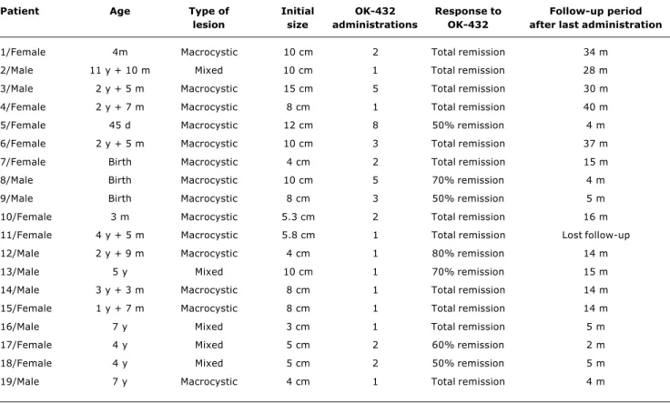
A retrospective study of 19 children diagnosed with lymphangioma and treated at the Pediatric Oncology and Head and Neck Surgery Unit of the Hospital das Clínicas at the Ribeirão Preto Medical Faculty USP, during the years from 1999 to 2003. We analyze 10 female patients and nine male patients, with an average age on diagnosis of 37 months, varying from neonates to 11 years and 10 months. All of the patients presented lesions in the head or neck area. Fourteen were macrocystic lesions, three were mixed and two were microcystic. Forty-three applications of OK-432 were performed, giving an average of 2.4 administrations per patient, varying from one to eight (Table 1).
Follow-up time after the last OK-432 administration varied from 2 to 40 months, with an average of 15.8 months. Patient number 11 dropped out of follow-up after 5 months. The average period of observation of patients in total remission was 23.7 months after the last application, and for patients in partial remission it was 7 months. Patients 8, 9, 12, 13 and 17 are scheduled for further OK-432 applications and patients 5 and 18 have been started on alpha interferon after the OK-432 applications.
Table 1 - Clinical aspects and evolution of patients treated with OK-432
Patient Age Type of Initial OK-432 Response to Follow-up period lesion size administrations OK-432 after last administration
1/Female 4m Macrocystic 10 cm 2 Total remission 34 m
2/Male 11 y + 10 m Mixed 10 cm 1 Total remission 28 m
3/Male 2 y + 5 m Macrocystic 15 cm 5 Total remission 30 m
4/Female 2 y + 7 m Macrocystic 8 cm 1 Total remission 40 m
5/Female 45 d Macrocystic 12 cm 8 50% remission 4 m
6/Female 2 y + 5 m Macrocystic 10 cm 3 Total remission 37 m
7/Female Birth Macrocystic 4 cm 2 Total remission 15 m
8/Male Birth Macrocystic 10 cm 5 70% remission 4 m
9/Male Birth Macrocystic 8 cm 3 50% remission 5 m
10/Female 3 m Macrocystic 5.3 cm 2 Total remission 16 m
11/Female 4 y + 5 m Macrocystic 5.8 cm 1 Total remission Lost follow-up
12/Male 2 y + 9 m Macrocystic 4 cm 1 80% remission 14 m
13/Male 5 y Mixed 10 cm 1 70% remission 15 m
14/Male 3 y + 3 m Macrocystic 8 cm 1 Total remission 14 m
15/Female 1 y + 7 m Macrocystic 8 cm 1 Total remission 14 m
16/Male 7 y Mixed 3 cm 1 Total remission 5 m
17/Female 4 y Mixed 5 cm 2 60% remission 2 m
18/Female 4 y Mixed 5 cm 2 50% remission 5 m
The time passed between injections varied from 1 month to 2 years with an average of 3.5 months between administrations. The interval between administrations was determined by the clinical condition of the patient and drug availability.
Patient number 2 received alpha interferon, prednisone, epsilon-aminocaproic acid and had undergone surgery twice before OK-432, Patient 3 received prednisone before treatment with OK-432, patient 6 received interferon, patient 13 received prednisone, alpha interferon and epsilon-aminocaproic acid before OK-432, patient 16 received prednisone and patients 17 and 18 were subjected to surgery before being treated with the sclerosing agent. These patients had not responded to systemic treatment and/or had suffered lymphangioma relapse after surgery.
Applications were performed with the child under sedation or general anesthetic, and some applications were guided by ultrasound. The drug was prepared at a dilution of 0.1 mg OK-432 to 10 ml saline at 0,9%. The lesion was aspirated as far as possible. If the aspirated volume was less than 20 ml, this same volume was replaced with an equal volume of dilute OK-432. The maximum infused volume did not pass 20 ml. If aspiration from within the cyst proved difficult, an amount of the solution was injected into varying points until the lymphangioma internal tension increased.
Results
All patients presented some sort of a response to OK-432, with the least significant reduction in volume being 50% of the volume of the lesion. Twelve patients exhibited complete remission from the lesion (63% of cases). Of these patients, one received five injections, one received three, six received two injections and four received a single injection each giving an average of two injections per patient. The size the lesions affecting the patients that presented complete remission varied from 3 to 15 cm with an average size of 7.6 cm. Ten lymphangioma were macrocystic and two were mixed.
Seven patients presented a partial response to OK-432, varying from 50% to 80%. Among these last, one received eight applications, one received five, one three, two received two applications and two patients received a single application giving an average of 3.1 applications per patient. The size of the partially responding lesions varied from 4 to 12 cm with an average of 7.7 cm. One patient presented mixed lymphangioma, four had macrocystic and two microcystic lymphangioma.
After administration, the majority of the children were observed at home (13 children). Four patients remained interned under observation for 24 to 48 hours. Patient number 6 was hospitalized for 10 days because of having a sustained fever and being just 3 months old at the time of first administration, patient 18 was
hospitalized for 14 days after the first administration because of edema of the tongue which made the oral route impossible.
The patients presented adverse reactions in the form of fever varying from 38 ºC to 38.8 ºC lasting from 2 to 10 days. The lesion became infected in one case after the first application. There was a local inflammatory reaction and the lesion developed erythema lasting until the tenth day after administration. No skin damage adjacent to the lesion or scarring were observed (Figures 1 e 2). There were no allergic reactions after OK-432 application.
Over an average follow-up period of 23.7 months after the final dose none of the patients who had exhibited a complete response to OK-432 relapsed.
Discussion
The results achieved are in line with published data; macrocystic and mixed lesions, with few septa, exhibited an excellent response to the drug. Among the lesions that have so far responded only partially, many had so far received only a few OK-432 doses and it is hoped that further injections will attain complete remission.
In use OK-432 should diffuse and come into contact with the greatest possible surface area of lymphangioma endothelium, which is more difficult with microcystic lesions. Authors who have injected stains into lesions before the OK-432 administration observed that, in macrocystic lesions, the dye spread along three or four large cavities and with microcystic lesions few points of contrast were found after administration.1 Microcystic lymphangioma (capillary or cavernous) exhibited a poorer response OK-432.1-3,7,8,10
Even in the absence of a complete response, there was a significant volume reduction in two of the patients suffering from microcystic lymphangioma (50-60%). This could facilitate surgery in the case it becomes necessary.
The lesions that were subjected to OK-432 in these patients were in the head and neck, but no airway obstructions were described. In more recent work, authors have indicated OK-432 even for lesions where there is a risk of airway obstruction, the support of an intensive care team is necessary in this situation.8,10
All of this drugs side effects are reversible. In a sample taking in 30,000 cases treated with OK-432 there were no reported deaths and remission from fever and local inflammation was achieved in a maximum of 14 dias.1 No renal or hepatic toxicity was observed.1,9
None of our cases presented relapse during the follow-up period. Research ahs demonstrated that patients cured with OK-432 do not present lesion relapses over more than 7 years of follow-up observation.3
Despite reports of previously treated patients presenting less favorable response,8,10 in our sample 57% of such patients presented complete remission.
Figure 1 - Patient suffering from macrocystic lymphangioma before treatment with OK-432
Figure 2 - Patient with macrocystic lymphangioma after treatment with OK-432
Corresponding author: Luiz Gonzaga Tone
Departamento de Puericultura e Pediatria, Hospital das Clínicas, Faculdade de Medicina de Ribeirão Preto USP
CEP 14049-900 - Ribeirão Preto, SP, Brazil Fax: +55 (16) 633.6695
E-mail: lgtone@fmrp.usp.br and for whom surgery is ruled out by the chance of
mutilation or by lesions that cannot be resectioned.
We therefore conclude that OK-432 is a safe and effective drug which can be indicated as the first-choice treatment for patients with lymphangioma since it offers advantages for later surgery, lower morbidity, a better cost/benefit ratio, a lower rate of relapse and the chance of less destructive surgery in those patients who respond only partially resulting in less mutilation.1-3,6,7,10
References
1. Smith RJH, Burke DK, Sato Y, Poust RI, Kimura K, Bauman NM. OK 432 therapy for lymphangiomas. Arch Otolaryngol Head Neck Surg. 1996;122:1195-9.
2. Ogita S, Tsuto T, Nakamura K, Deguchi E, Iwai N. OK 432 therapy in 64 patients with lymphangioma. J Pediatr Surg. 1994;29(6):784-5.
3. Schmidt B, Schimpt G, Höllwarth ME. OK 432 therapy of lymphangiomas in children. Eur J Pediatr. 1996;155:649-52. 4. Molich HI, Unger EC, Witte CL, van Sonnenberg E. Percutaneous
sclerotherapy of lymphangiomas. Radiology. 1995;194:343-7. 5. Giguere CM, Bauman NM, Smith RJ. New treatment options for lymphangioma in infants and children. Ann Otol Rhinol Laryngol. 2002;111(12 Pt 1):1066-75.
6. Souza RJSP, Tone LG. Tratamento clínico do linfangioma com alfa-2a-interferon. J Pediatr (Rio J). 2001;77(2):139-42.
7. Giguere CM, Bauman NM, Sato Y, Burke DK, Greinwald JH, Pranskelley P, et al. Treatment of lymphangiomas with OK-432 (Picibanil) sclerotherapy: a prospective multi-institutional trial. Arch Otolaryngol Head Neck Surg. 2002;128(10):1137-44. 8. Sanlialp I, Karnak I, Tanyel FC, Senocak ME, Buyukpamukcu N.
Sclerotherapy for lymphangioma in children. Int J Pediatr Otorhinolaryngol. 2003;67(7):795-800.
9. Ishida N, Hoshino T. A streptococcal preparation as a potent biological response modifier OK 432. 2nd ed. Amsterdam: Excerpta Medica; 1985.
10. Rautio R, Keski-Nisula L, Laranne J, Laasonen E. Treatment of lymphangiomas with OK-432 (Pincibanil). Cardiovasc Intervent Radiol. 2003;26(1):31-6.
11. Tyota B. Clinical effects of a streptococcal preparation, OK 432, against head and neck cancers. Jpn J Otol. 1967;72:1332-8. 12. Ogita S, Tsuto T, Tokiwa K, Takashi T: Intracystic injection of OK
432: a new sclerosing therapy for cystic hygroma in children. Br J Surg. 1987;74:690-1.