ISSN 1806-3713 © 2016 Sociedade Brasileira de Pneumologia e Tisiologia
http://dx.doi.org/10.1590/S1806-37562016000000226
ABSTRACT
The role of tuberculosis as a public health care priority and the availability of diagnostic tools to evaluate functional status (spirometry, plethysmography, and DLCO determination), arterial blood gases, capacity to perform exercise, lesions (chest X-ray and CT), and quality of life justify the effort to consider what needs to be done when patients have completed their treatment. To our knowledge, no review has ever evaluated this topic in a comprehensive manner. Our objective was to review the available evidence on this topic and draw conclusions regarding the future role of the “post-tuberculosis treatment” phase, which will potentially affect several million cases every year. We carried out a non-systematic literature review based on a PubMed search using speciic keywords (various combinations of the terms “tuberculosis”, “rehabilitation”, “multidrug-resistant tuberculosis”, “pulmonary disease”, “obstructive lung disease”, and “lung volume measurements”). The reference lists of the most important studies were retrieved in order to improve the sensitivity of the search. Manuscripts written in English, Spanish, and Russian were selected. The main areas of interest were tuberculosis sequelae following tuberculosis diagnosis and treatment; “destroyed lung”; functional evaluation of sequelae; pulmonary rehabilitation interventions (physiotherapy, long-term oxygen therapy, and ventilation); and multidrug-resistant tuberculosis.The evidence found suggests that tuberculosis is deinitively responsible for functional sequelae, primarily causing an obstructive pattern on spirometry (but also restrictive and mixed patterns), and that there is a rationale for pulmonary rehabilitation. We also provide a list of variables that should be discussed in future studies on pulmonary rehabilitation in patients with post-tuberculosis sequelae.
Keywords: Tuberculosis/complications; Tuberculosis/rehabilitation, Tuberculosis/therapy; Quality of life; Diagnostic imaging; Respiratory function tests.
Is there a rationale for pulmonary
rehabilitation following successful
chemotherapy for tuberculosis?
Marcela Muñoz-Torrico1, Adrian Rendon2, Rosella Centis3, Lia D’Ambrosio3,4, Zhenia Fuentes5, Carlos Torres-Duque6, Fernanda Mello7, Margareth Dalcolmo8, Rogelio Pérez-Padilla9, Antonio Spanevello10,11, Giovanni Battista Migliori3
Correspondence to:
Giovanni Battista Migliori. World Health Organization Collaborating Centre for Tuberculosis and Lung Diseases, Fondazione Salvatore Maugeri, Istituto di Ricovero e Cura a Carattere Scientiico, Via Roncaccio, 16, 21049, Tradate, Italia.
Tel.: 39 0331 829404; Fax: 39 0331 829402. E-mail: [email protected] Financial support: None.
INTRODUCTION
The World Health Organization (WHO) estimated that 3.3% of the new cases of tuberculosis and 20% of the previously treated cases of the disease are due to multidrug-resistant tuberculosis (MDR) strains of Mycobacterium tuberculosis worldwide in 2014. The highest prevalences of MDR tuberculosis (MDR-TB) have been reported in Eastern European and Central Asian countries, although relatively high prevalence rates have been described in Latin America. As of today, the “world record” MDR-TB prevalence has been described in Belarus (34% among new cases and 69% among retreatment cases), where 29% of the cases are reported to be extensively drug-resistant tuberculosis (XDR-TB).(1)
It is unfortunately well known that outcomes of MDR-TB and XDR-TB cases (particu-larly those with a resistance pattern beyond XDR-TB) are poor, since the treatment success rate is below 20% and the failure and death rates combined are 49%.(2,3)
The WHO has recently published two core documents addressing the critical importance of preventing the emergence of drug resistance, both underlining the relevance of managing MDR-TB adequately.(1,4-8) The WHO action framework “Towards tuberculosis elimination for low-incidence countries” presents eight priority action areas, two of which are focused on, namely, (action #5) optimizing the management of MDR-TB and (action #7) investing in research on new diagnostic tools and drugs. (1,4,6,9) 1. Clínica de Tuberculosis, Instituto
Nacional de Enfermedades Respiratorias – INER – Ciudad de México, México.
2. Centro de Investigación, Prevención y Tratamiento de Infecciones Respiratorias, Hospital Universitario, Universidad de Monterrey, Monterrey, México.
3. WHO Collaborating Centre for TB and Lung Diseases, Fondazione Salvatore Maugeri, Istituto di Ricovero e Cura a
Carattere Scientiico – IRCCS – Tradate,
Italia.
4. Public Health Consulting Group SAGL, Lugano, Switzerland.
5. Servicio de Neumología, Hospital General Dr. José Ignacio Baldó, El
Algodonal, Caracas, Venezuela. 6. Fundación Neumológica Colombiana,
Universidad de La Sabana, Bogotá, Colombia.
7. Instituto de Doenças do Tórax, Universidade Federal do Rio de Janeiro, Rio de Janeiro (RJ) Brasil.
8. Centro de Referência Hélio Fraga, Escola Nacional de Saúde Pública Sergio Arouca, Fundação Oswaldo Cruz, Rio de Janeiro (RJ) Brasil.
9. Clínica del Sueño, Instituto Nacional de Enfermedades Respiratorias – INER – Ciudad de México, México.
10. Unità di Pneumologia, Fondazione Salvatore Maugeri, Istituto di Ricovero
e Cura a Carattere Scientiico – IRCCS –
Tradate, Italia.
11. Dipartimento di Medicina Clinica e Sperimentale, Università dell’Insubria,
Varese, Italia.
Submitted: 28 July 2016.
Accepted: 1 September 2016.
Study carried out under the auspices of the World Health Organization Collaborating Centre for Tuberculosis and Lung Diseases, Fondazione Salvatore Maugeri, Istituto di Ricovero e Cura a Carattere Scientiico – IRCCS – Tradate, Italia.
J Bras Pneumol. 2016;42(5):374-385
374
Muñoz-Torrico M, Rendon A, Centis R, D’Ambrosio L, Fuentes Z, Torres-Duque C, et al.
However, the scientiic and programmatic focus is presently on diagnosis and treatment of the disease, whereas post-cure follow-up is seen as an approach to evaluate the proportion of relapse, particularly in MDR-TB/XDR-TB cases.
The role that tuberculosis plays as a public health care priority, as well as the importance of diagnostic tools being available in order to evaluate the patients thoroughly, by means of their functional status—via spirometry, plethysmography, and determination of DLCO —arterial blood gas analyses, their capacity to perform exercise—via the six-minute walk test (6MWT)— the description of their lesions—via chest X-rays (CXRs) and CT—and their quality of life (QoL)—via the Saint George’s Respiratory Questionnaire (SGRQ)—justiies the effort to consider what needs to be done when patients have completed their treatment successfully. This vision has ethical, clinical, organizational, programmatic, and economic implications.
To our knowledge, the follow-up of tuberculosis patients who completed their treatment has never been reviewed in a comprehensive manner in the literature. Therefore, the objective of the present study was to review the available evidence on this topic and to draw some conclusions regarding the future role of the “post-tuberculosis treatment” phase, which will potentially have an impact on several million cases every year around the globe.
METHODS
We carried out a non-systematic review of the literature based on a PubMed search using speciic keywords, including various combinations of the terms “tuberculosis”, “rehabilitation”, “MDR-TB”, “pulmonary disease”, “obstructive lung disease”, and “lung volume measurements”. The reference lists of the most important studies were also retrieved in order to improve the sensitivity of the research. Manuscripts written in English, Spanish, and Russian were selected. The main areas of interest that we identiied in order to describe the topic were as follows:
1. Tuberculosis sequelae following diagnosis and treatment of tuberculosis
2. Destroyed lung
3. Functional evaluation of sequelae
4. Pulmonary rehabilitation (PR) interventions, such as physiotherapy, long-term oxygen therapy (LTOT), and ventilation
5. MDR-TB
After describing each of these areas of interest, we will provide a summary of the evidence compiled from the literature search (Table 1) and concluding remarks.
TUBERCULOSIS SEQUELAE FOLLOWING DIAGNOSIS AND TREATMENT OF
TUBERCULOSIS
Although the potential role of PR has been clearly underlined in a study discussing the role of the new WHO recommendations on shorter treatment
regimens,(10) the concept that rehabilitation is a component of tuberculosis treatment is as old as that of sanatoria. (11,12) In 1964, Chapman and Hollander wrote that, based on their experience with 454 patients with active tuberculosis “placed on a program of intensive physical exercise, combined with chemotherapy,” “the concept of minimum exercise and prolonged bed rest in the hospital and a prolonged convalescent period after discharge is no longer justiied.”(12)
In 2006, a group of authors in India(13) prospectively studied the clinical presentation and predictors of outcome in 116 patients with acute exacerbations of COPD who had to be admitted to the ICU and found that 28.4% of those had had pulmonary tuberculosis previously. Among those patients, some required invasive mechanical ventilation and a few died. The authors concluded that “an intriguing relationship” existed among smoking, pulmonary tuberculosis, and COPD “which merits further study.”(13)
In 2010, Jordan et al.(14) wrote that “the global prevalence of bronchiectasis, a recognized sequela of tuberculosis, is unknown, but is by no means insigniicant. The pathophysiology of chronic airlow obstruction in both of these diseases is poorly under-stood, but it is associated with an accelerated rate of loss in pulmonary function.”
Hassan and Al-Jahdali(15) reported that “in addition to its acute clinical consequences, patients with pulmonary tuberculosis may be left with signiicant long-term sequelae,” “associated with considerable morbidity, mortality, and health expenditure,” and commented that both obstructive and restrictive functional abnormalities were present.
Shah and Reed(16) described, among the commonest complications of tuberculosis, “mycetomas developing within residual tuberculosis cavities, impaired pulmonary function, or focal neurologic deicits from tuberculomas,” and, therefore, “public health tuberculosis programs and health systems require additional resources to provide comprehensive tuberculosis and post-tuberculosis care.”
Bansal and Prasad(17) commented that “COPD, intersti-tial lung disease, tuberculosis, and lung cancer together are the leading causes of morbidity and mortality,” which are “increasing all over the world”; they also stated that “early fatigue and breathlessness” make patients “socially isolated and depressed”. Functional disability and repeated hospitalizations reduce the eficiency of the patients at home and at work place, being associated with increased expenditures and utilization of health care systems, which results in a socioeconomic burden. PR, an evidence-based, multidisciplinary, and comprehensive non-pharmacological intervention, has emerged as a recommended standard of health care for patients suffering from respiratory diseases. PR is advised for patients with chronic lung conditions who have dyspnea or other respiratory symptoms, reduced exercise tolerance, restriction in activities, or impaired health status despite optimal pharmacological treatment. Early leaders observed two centuries ago
Is there a rationale for pulmonary rehabilitation following successful chemotherapy for tuberculosis?
Table 1. Summary of the major studies that performed functional evaluation of patients with pulmonary tuberculosis.
Authors, year Study location (name)a
Participantsb (n)
Cases investigated Investigations performed
Major functional findings Conclusions
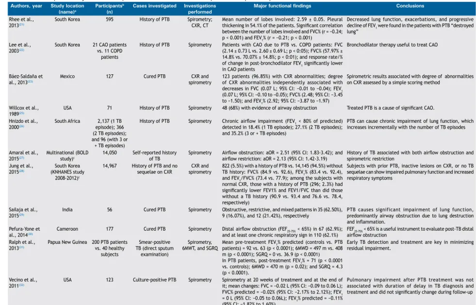
Rhee et al., 2013(21)
South Korea 595 History of PTB Spirometry;
CXR, CT
Mean number of lobes involved: 2.59 ± 0.05. Pleural thickening in 54.1% of the patients. Signiicant correlation between the number of lobes involved and FVC% (r= −0.24; p< 0.001) and FEV1% (r = −0.21; p < 0.001)
Decreased lung function, exacerbations, and progressive decline of FEV1 were found in the patients with PTB “destroyed lung”
Lee et al., 2003(22)
South Korea 21 CAO patients vs. 11 COPD
patients
History of PTB Spirometry Patients with CAO due to PTB vs. COPD patients: FVC (2.14 ± 0.73 L vs. 2.60 ± 0.69 L; p < 0:05); FVC% (57.97% ± 14.8% vs. 70.07% ± 14.8%; p < 0:01); and response rate/% of change in post-bronchodilator FEV1 signiicantly lower in CAO patients
Bronchodilator therapy useful to treat CAO
Báez-Saldaña et al., 2013(23)
Mexico 127 Cured PTB CXR and
spirometry
123 patients (96.85%) with CXR abnormalities; degree of CXR abnormalities independently associated with decreases in FVC (0.07 L; 95% CI: −0.01 to −0.04); FEV1 (0.07 L; 95% CI: −0.10 to −0.05); FVC% (2.48; 95% CI: −3.45 to −1.50); and FEV1% (2.92; 95% CI: −3.87 to −1.97)
Spirometric results associated with degree of abnormalities on CXR assessed by a simple scoring method
Willcox et al., 1989(25)
USA 71 History of PTB Spirometry 48 (68%) with evidence of airway obstruction Treated PTB is a cause of signiicant CAO.
Hnizdo et al., 2000(26)
South Africa 2,137 (1 TB episode); 366 (2 TB episodes); and 96 (with 3 or + TB episodes)
History of PTB Spirometry Chronic airlow impairment (FEV1 < 80% of predicted) detected in 18.4% (1 TB episode); 27.1% (2 TB episodes); and 35.2% (3 or + TB episodes)
PTB can cause chronic impairment of lung function, which increases incrementally with the number of TB episodes
Amaral et al., 2015(27)
Multinational (BOLD study)c
14,050 Self-reported history of TB
Spirometry Airlow obstruction: aOR = 2.51 (95% CI: 1.83-3.42); and
airlow restriction: aOR = 2.13 (95% CI: 1.42-3.19) History of TB associated with both airlow obstruction and spirometric restriction Jung et al.,
2015(28)
South Korea (KNHANES study
2008-2012)c
14,967 History of PTB and no sequelae on CXR
CXR and spirometry
822 (5.5%) with a history of PTB vs. 14,145 (94.5%) without TB history: FVC% (84.9 vs. 92.6), FEV1% (83.4 vs. 92.4), and FEV1/FVC% (73.4 vs. 77.9); among the subjects with normal CXR, those with a history of PTB (296; 2.3%) had signiicantly lower FEV1% and FEV1/FVC than did those without a TB history (90.9 vs. 93.4 and 76.6 vs. 78.4, respectively)
Subjects with prior PTB, inactive lesions on CXR, or no TB sequelae can show impaired pulmonary function and increased respiratory symptoms
Sailaja et al., 2015(29)
India 56 Cured PTB Spirometry Obstructive, restrictive, and mixed patterns in 35 (62.50%), 9 (16.07%), and 12 (21.42%), respectively
PTB causes significant impairment of lung function, predominantly airway obstruction due to lung destruction and inlammation.
Pefura-Yone et al., 2014(30)
Cameroon 177 Cured PTB Spirometry Distal airlow obstruction (FEF25-75% < 65%) in 67 (62.9%); and at least one chronic respiratory sign in 110 (62.1%)
FEF25-75% < 65% is a useful instrument to evaluate post-TB distal airlow obstruction
Ralph et al., 2013(31)
Papua New Guinea 200 PTB patients vs. 40 healthy
subjects
Smear-positive TB (direct sputum
examination)
Spirometry, 6MWT, and SGRQ
Mean pre-treatment FEV1% predicted (controls vs. PTB patients) = 92 vs. 63 (p < 0.0001); 6MWD = 497 m vs. 408 m (p < 0.0001); SGRQ = 0 vs. 36.9 (p < 0.0001)
In PTB patients, post-treatment FEV1% = 71 (p < 0.0001 vs. controls); 6MWD = 470 m (p = 0.02); and SGRQ = 4.3 (p < 0.0001).
Early TB detection and treatment are key in minimizing residual impairment.
Vecino et al., 2011(32)
USA 123 Culture-positive PTB Spirometry Spirometry at 20 weeks of treatment and at the end of it; mean changes: FVC = −0.02 L (95% CI: −0.09 to 0.06 L); FVC% predicted = −0.02% (95% CI: −2.17% to 2.12%); FEV1 = 0 L (95% CI: −0.05 to 0.06L); FEV1% predicted = −0.11% (95% CI: −1.82% to 1.60%)
Pulmonary impairment after PTB treatment was not associated with duration of delay in TB diagnosis and treatment and did not signiicantly change during follow-up
376
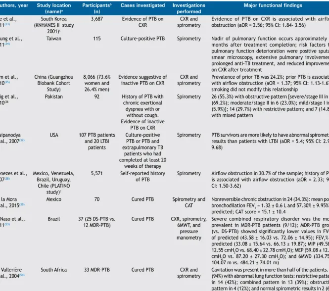
Muñoz-Torrico M, Rendon A, Centis R, D’Ambrosio L, Fuentes Z, Torres-Duque C, et al. Authors, year Study location
(name)a
Participantsb (n)
Cases investigated Investigations performed
Major functional findings Conclusions
Lee et al., 2011(33)
South Korea (KNHANES II study
2001)c
3,687 Evidence of PTB on CXR
CXR and spirometry
Evidence of PTB on CXR is associated with airflow obstruction (aOR = 2.56; 95% CI: 1.84- 3.56)
PTB is an independent risk factor for obstructive lung disease in never-smokers.
Chung et al., 2011(34)
Taiwan 115 Culture-positive PTB Spirometry Nadir of pulmonary function occurs approximately 18 months after treatment completion; risk factors for pulmonary function deterioration were positive sputum smear microscopy, extensive pulmonary involvement, prolonged anti-TB treatment, and reduced improvement on CXR after treatment
In patients with signiicant respiratory symptoms and multiple risk factors, pulmonary function tests should be used to monitor progression of functional impairment
Lam et al., 2010(35)
China (Guangzhou Biobank Cohort
Study)
8,066 (73.6% women and 26.4% men)
Evidence suggestive of inactive PTB on CXR
CXR and spirometry
Prevalence of prior TB was 24.2%; prior PTB is associated with airlow obstruction (aOR = 1.37; 95% CI: 1.13-1.67); smoking did not modify this relationship
Prior TB is an independent risk factor for airlow obstruction, regardless of smoking status
Baig et al., 2010(36
Pakistan 92 History of PTB with
chronic exertional dyspnea with or
without cough. Evidence of inactive
PTB on CXR
Spirometry 26 (55.3%) with obstructive pattern [severe/stage III in 18 (69.2%); moderate/stage II in 6 (23.0%); mild/stage I in 2 (5.9%)]; 14 (29.7%) with restrictive pattern; and 7 (14.8%) with mixed pattern
COPD can occur as one of the chronic complications of PTB
Pasipanodya et al., 2007(37)
USA 107 PTB patients and 20 LTBI
patients
Culture-positive PTB or PTB and extrapulmonary TB
patients who had completed at least 20
weeks of therapy
Spirometry PTB survivors are more likely to have abnormal spirometric results than patients with LTBI (aOR = 5.4; 95% CI: 2.98-9.68)
Pulmonary impairment after TB is associated with disability worldwide and demands more aggressive prevention strategies and post-treatment evaluation
Menezes et al., 2007(38)
Mexico, Venezuela, Brazil, Uruguay,
Chile (PLATINO study)c
5,571 Self-reported history of PTB
Spirometry Airlow obstruction in 30.7% of the sample; history of PTB is associated with airlow obstruction (aOR = 2.33; 95% CI: 1.50-3.62)
History of PTB is associated with airway obstruction
de la Mora et al., 2015(39)
Mexico 70 Cured PTB Spirometry and
CAT
Nonreversible chronic obstruction in 24 (34.3%): mean post-bronchodilation FEV1 = 1.32 ± 0.6 L and 57.30% ± 9.95% of predicted; CAT score = 15.1 ± 10.4
Functional abnormalities are frequent in PTB patients
Di Naso et al., 2011(53)
Brazil 37 (25 DS-PTB vs. 12 MDR-PTB)
Cured PTB CXR, spirometry, 6MWT, and
pressure manometry
Severe combined respiratory disorder was the more prevalent in MDR-PTB patients (9/12); MDR-PTB group (vs. DS-PTB) showed signiicantly lower values in FVC% of predicted (43.58 ± 16.03 vs. 72.06 ± 14.95); FEV1% of predicted (33.08 ± 15.64 vs. 66.13 ± 19.87); MIP (49.58 ± 12.55 cmH2O vs. 68.40 ± 22.78 cmH2O); MEP (59.08 ± 12.23 cmH2O vs. 87.20 ± 27.30 cmH2O); and 6MWD (334.75 ± 104.07 m vs. 484.21 ± 74.01 m)
Patients with MDR-PTB who have undergone multiple treatments have more severe respiratory and functional impairment than do patients who have had just a single treatment
de Vallerière et al., 2004(54)
South Africa 33 MDR-PTB Cured PTB CXR and
spirometry
Cavitation was present in more than half of the patients. 31 (94%) with abnormal lung function tests: restrictive pattern in 14 (42%); combined pattern in 13 (39%); obstructive pattern in 4 (12%); and normal spirometric results in 2 (6%)
Residual lung damage in MDR-PTB patients who completed treatment is common and extensive
PTB: pulmonary tuberculosis; CXR: chest X-ray; CAO: chronic airway obstruction; TB: tuberculosis; aOR: adjusted odds ratio; 6MWT: six-minute walk test; SGRQ: Saint George’s Respiratory Questionnaire; 6MWD: six-minute walk distance; LTBI: latent TB infection; CAT: COPD assessment test; DS-PTB: drug-susceptible PTB; and MDR-PTB: multidrug-resistant PTB. aBOLD: Burden of Obstructive lung disease; KNHANES:
Korean National Health and Nutrition Examination Surveys; and PLATINO: Proyecto Latinoamericano de Investigación en Obstrucción Pulmonar. bAll studies included both men and women. cPopulation-based studies.
Table 1. Continued...
377
Is there a rationale for pulmonary rehabilitation following successful chemotherapy for tuberculosis?
that exercise is an important element in the care of patients with lung and heart diseases, especially in tuberculosis.(17)
In a recent American Thoracic Society (ATS)/European Respiratory Society (ERS) statement, as well as in a guideline from South Africa for the management of COPD, tuberculosis is clearly among the diseases that require the use of PR.(18,19)
“DESTROYED LUNG”
Late diagnosis is often responsible for extensive bilateral lesions, usually due to bronchiectasis, scarring, parenchymal deformation, lung volume loss, and pleural thickening, which might develop to the so-called “destroyed lung” (Figure 1).(20)
Two studies described the effect of “destroyed lung” on the pulmonary function of patients treated for pulmonary tuberculosis, both carried out in South Korea.(21,22)Rhee et al.(21) studied 595 tuberculosis patients from 21 hospitals between 2005 and 2011. The mean extension of the lesions was 2.59 ± 0.05 lobes, and pleural thickening was observed in 54.1% of the patients. Various lung function parameters were reduced (mean values): FVC = 2.06 ± 0.03 L (61.26% ± 0.79% of predicted); FEV1 = 1.16 ± 0.02 L (49.05% ± 0.84% of predicted); FEV1/FVC ratio = 58.0% ± 0.70%; bronchodilator response = 5.70% ± 0.34%; and number of exacerbations/year = 0.40 ± 0.04. The number of lobes involved signiicantly correlated with FVC, FEV1, and the number of exacerbations/ year. The use of long-acting muscarinic antagonists or long-acting β2 agonists plus inhaled corticosteroids achieved bronchodilator effects. Initial FEV1% and the number of exacerbations during follow-up were independent factors affecting FEV1 deterioration in the multivariate analysis.
Lee et al.(22) investigated lung function and post-bronchodilator response in 21 patients with
“destroyed lung”-related chronic airlow obstruction against a cohort of COPD patients matched by sex, age, and pulmonary function parameters. The mean FVC values (both in L and in % of predicted) of the patients with “destroyed lung” were signiicantly lower than those of the COPD patients (2.14 ± 0.73 L vs. 2.60 ± 0.69 L and 57.9% ± 14.8% vs. 70.0 ± 14.8%, respectively). The tuberculosis patients presented with signiicantly lower FVC and post-bronchodilator FEV1 than did the COPD patients. In addition, among the tuberculosis patients, those with wheezing symptoms showed significantly lower FEF25-75% and higher airway resistance than did those without wheezing. Tuberculosis patients with wheezing responded better to the bronchodilator than did those without it. The authors concluded that bronchodilator therapy could be useful in those patients.
In Mexico, 127 cured tuberculosis patients underwent spirometry and CXR; 123 (96.85%) exhibited some degree of radiographic abnormalities.(23) The extent of lung damage was measured by dividing the lung parenchyma into four quadrants and scoring it from 0 to 5; the mean number of radiographic abnormalities was 6.45 ± 4.14. In that sample, 30 patients (24%) showed an obstructive spirometric pattern, and 22 (17%), a restrictive pattern; only 15 (12%) had a positive bronchodilator test, and 21 (17%) had an SpO2 < 90%. The adjusted multilinear regression model showed that the degree of radiographic abnormalities was independently associated with a decrease in the absolute values of FVC (0.07 L; 95% CI: −0.01 to −0.04) and FEV1 (0.07 L; 95% CI: −0.10 to −0.05; p < 0.001); as well as in their % of predicted values (FVC = 2.48%; 95% CI: −3.45 to −1.50; and FEV1 = 2.92%; 95% CI: −3.87 to −1.97). That study showed that spirometric values were associated with the degree of radiographic abnormalities assessed by a simple scoring method.
FUNCTIONAL EVALUATION OF SEQUELAE Several studies investigated the mechanical lung function in tuberculosis patients (Figure 2).
Already in 1961, Hallet and Martin(24) described the diffuse obstructive pulmonary syndrome (measured via the maximal expiratory low rate) in 34% of 710 tuberculosis patients admitted to a sanatorium during a one-year period. The factors signiicantly associated with the incidence of that syndrome were age, severity of tuberculosis, and some comorbidities (bronchial asthma, pulmonary malignancy, frequent and protracted chest colds, and silicosis). The authors concluded that the measurement of maximal expiratory low rate is a useful tool in determining diffuse obstructive pulmonary disease.
Willcox and Ferguson(25) investigated 71 patients previously treated for tuberculosis up to 16 years prior. Evidence of airway obstruction was found in 48 (68%) of the patients. An inverse relationship between the extent of the disease on the original CXRs and FEV1 Figure 1. Chest X-ray of a 39-year-old male patient with
a history of pan-susceptible tuberculosis treated for six months in 2007. The patient was considered cured. Later in time, he reported a six-month history of cough, mild dyspnea, but no fever. Tuberculosis relapse was ruled out; sputum smear microscopy and culture were negative. The image shows a giant cavity in the right upper lobe and some ibrotic changes.
Muñoz-Torrico M, Rendon A, Centis R, D’Ambrosio L, Fuentes Z, Torres-Duque C, et al.
was identiied. The authors identiied a similar inverse relationship of the amount of sputum produced with FEV1 and with the extent of the disease on the CXRs. The authors concluded that treated tuberculosis is a cause of COPD.
In a large study in South Africa,(26) a cohort was followed in order to study the chronic effect of initial and recurrent pulmonary tuberculosis: 27,660 black South African gold miners who had reliable pulmonary function test results between January of 1995 and August of 1996 were retrospectively followed for the incidence of tuberculosis to 1970. In that cohort of miners, 2,137; 366; and 96 had had, respectively, one, two, and three or more episodes of tuberculosis. The mean time between the diagnosis of the last episode of tuberculosis and lung function testing was 4.6 years (range: 1-372 months). The loss of lung function was the highest within the irst 6 months after tuberculosis being diagnosed and stabilized after 12 months, when the loss was considered chronic. The estimated mean deicits in FEV1 after one, two, and three or more episodes of tuberculosis were 153 mL, 326 mL, and 410 mL, respectively, whereas the corresponding deicits in FVC were 96 mL, 286 mL, and 345 mL. The loss of lung function was similar in HIV-positive and HIV-negative individuals. The proportion of individuals with chronic airlow impairment (FEV1 < 80% of predicted) was 18.4%, 27.1%, and 35.2%, respectively, in those with one, two, and three or more episodes of tuberculosis. The authors concluded that tuberculosis can cause chronic impairment of lung function, which increases with the number of episodes of the disease, and that early diagnosis and treatment of tuberculosis coupled with the prevention of HIV, silica dust exposure, silicosis, and poverty are important interventions.
In a recent multicenter, cross-sectional, general population-based study,(27) the association of having a history of tuberculosis with airlow obstruction and spirometric abnormalities was studied in adults. A self-reported history of tuberculosis was associated
with airlow obstruction (adjusted OR = 2.51; 95% CI: 1.83-3.42) and spirometric restriction (adjusted OR = 2.13; 95% CI: 1.42-3.19). The authors concluded that a history of tuberculosis was associated with both airlow obstruction and spirometric restriction, and should be considered as an important cause of obstructive disease and impaired lung function, particularly where tuberculosis is common.
In a study performed in South Korea between 2008 and 2012,(28) lung function impairment and persistency of respiratory symptoms were studied in 14,967 adults with and without a history of pulmonary tuberculosis. The adults were also divided into two groups: those showing residual sequelae on CXR and those without showing it in order to determine the risk factors for airlow obstruction. Among the population studied, 822 participants (5.5%) had been treated for pulmonary tuberculosis (mean) 29.0 years prior to study initiation. The individuals with a history of tuberculosis, when compared with those without that, presented with signiicantly lower FVC% (84.9 vs. 92.6), FEV1% (83.4 vs. 92.4), and FEV1/FVC% (73.4 vs. 77.9). Among the 12,885 subjects with no sequelae on CXR, those with a history of pulmonary tuberculosis (n = 296; 2.3%) had signiicantly lower FEV1% (90.9 vs. 93.4) and FEV1/FVC% (76.6 vs. 78.4). Subjects with a history of pulmonary tuberculosis but no sequelae on CXR reported a signiicantly higher frequency of cough and physical activity limitations due to pulmonary symptoms than those without that history (p < 0.001 for both). A history of pulmonary tuberculosis (OR = 2.314), along with older age, male gender, asthma, and smoking were independent risk factors for airlow obstruction. Finally, the study suggested that inactive tuberculosis lesions on CXR (OR = 2.3) were risk factors for airlow obstruction in subjects with a history of pulmonary tuberculosis. The authors concluded that the patients treated for tuberculosis should undergo regular lung function testing and stop smoking in order to prevent chronic airway disease.
--ESPIROMETRÍA--FVC (L) FEV1 (L) FEV1/FVC (%)
Pre-Bronch Post-Bronch
Real
1.99 1.05 53
Real
2.04 1.06 52 Teórico
2.85 1.63 71
%Teórico
69 64 74
%Teórico
71 65 73
%Cambio
+2 +1 -1
Pre Post
Teórico 6
4 2
0 -2
-4 -6
2
0 0
1 2 3 4 5 6 7
1 2 3
Figure 2. Spirometry of the same patient shown in Figure 1. FEV1/FVC ratio was below 70%. FEV1 was decreased and unresponsive to bronchodilator. FVC was also diminished. Fixed airway obstruction was detected, and mild restriction was considered. The inal diagnosis was pulmonary sequelae of tuberculosis. Espirometría: spirometry; teórico: predicted; pre/post bronch: pre-/post-bronchodilator; real: observed; and cambio: change.
Is there a rationale for pulmonary rehabilitation following successful chemotherapy for tuberculosis?
In a recent study,(29) 56 treated tuberculosis patients who were considered cured underwent simple spirome-try, and pre- and post-bronchodilator FEV1, FVC, FEV1/ FVC ratio were recorded. Obstructive, restrictive, and mixed patterns were identiied in 62.50%, 16.07%, and 21.42% of the patients, respectively.
In a cross-sectional study(30) involving 177 individuals who had been previously treated for tuberculosis in Cameroon between 2012 and 2013, spirometry was performed in order to evaluate the clinical impact of low FEF25-75%. Distal airlow obstruction (DAO) was deined by an FEF25-75% < 65% and a FEV1/FVC ratio ≥ 0.70. At least one chronic respiratory sign was present in 110 (62.1%) of the participants, and DAO was identiied in 67 (62.9%). Duration of symptoms prior to the diagnosis of tuberculosis > 3 months (adjusted OR = 2.91) and presence of DAO (OR = 2.22) were independent determinants that were signiicantly associated with persisting respiratory signs. The authors concluded that FEF25-75% < 65% is a useful instrument to evaluate post-tuberculosis DAO.
In Papua New Guinea, presently a hot spot for MDR-TB, a study(31) evaluated morbidity during treatment and residual pulmonary disability in pulmonary tuberculosis cases undergoing spirometry, 6MWT, and evaluation of QoL (SGRQ). The authors evaluated 200 pulmonary tuberculosis patients (at baseline and after 6 months of treatment) and 40 healthy volunteers. The distance walked in the 6MWT (6MWD) was 497 m in controls vs. 408 m in tuberculosis patients at baseline (p < 0.0001) and 470 m after 6 months (p = 0.02), whereas the SGRQ score was zero in controls vs. 36.9 in tuberculosis patients at baseline (p < 0.0001) and 4.3 after 6 months (p < 0.0001). The mean predicted FEV1 was 92% in controls vs. 63% among tuberculosis patients at baseline (p < 0.0001) and 71% after 6 months (p < 0.0001). After six months of treatment, 27% of the tuberculosis patients still showed at least moderate-to-severe pulmonary function impairment, and 57% had respiratory symptoms, although most of them achieved “successful” treatment outcomes and self-reported good QoL. More advanced disease at baseline (longer illness duration and worse results on CXR at baseline) and HIV-positive status predicted residual disability. The authors concluded that early detection and treatment of tuberculosis are key in minimizing residual impairment.
Post-pulmonary tuberculosis impairment was studied after 20 weeks of tuberculosis treatment and again on, or after, treatment completion.(32) The median duration between the irst and the second spirometry was 15 weeks. The mean change in FVC was −0.02 L (95% CI: −0.09 to 0.06 L), and that in FVC% of predicted was −0.02% (95% CI: −2.17% to 2.12%), whereas that in FEV1 was 0 L (95% CI: −0.05 to 0.06), and that in FEV1% of predicted was −0.11% (95% CI: −1.82 to 1.60). Pulmonary impairment was not related to the delay in tuberculosis diagnosis or treatment, older age, or smoking habits.
The relationship between previous tuberculosis and the risk of COPD was studied in South Korea in a population-based investigation(33) involving 3,687 individuals performing spirometry and CXR. Among those, 294 subjects had radiological evidence of previous tuberculosis with no evidence of active disease. Radiological evidence of previous tuberculosis was independently associated with airlow obstruction (adjusted OR = 2.56) after adjustments for sex, age, and smoking history. Previous tuberculosis was still a risk factor (adjusted OR = 3.13) with the exclusion of ever-smokers or subjects with advanced radiological lesions. Among the never-smokers, the proportion of subjects with previous tuberculosis on CXR increased as obstructive lung disease became more severe. The authors concluded that previous tuberculosis is an independent risk factor for COPD, even in never-smokers.
The trends toward deterioration of pulmonary function and its risk factors were studied in 115 patients with pulmonary tuberculosis after treatment completion.(34) A model with a locally weighted scatterplot smoothing technique was used in order to evaluate the trends toward changes in pulmonary function. The median interval between the end of antituberculosis treatment and pulmonary function testing was 16 months. The nadir of pulmonary function occurred approximately 18 months after completion of the treatment. The risk factors associated with pulmonary function deterioration included positive sputum smear microscopy, extensive pulmonary involvement prior to antituberculosis treatment, prolonged antituberculosis treatment, and poor radiographic improvement after treatment. The authors concluded that pulmonary function testing should be used as a follow-up tool in order to monitor the progression of functional impairment, especially within the irst 18 months after the completion of antituberculosis treatment.
A study in China(35) investigated the relationship between history of tuberculosis, smoking, and airlow obstruction in a population sample of 8,066 participants in the Guangzhou Biobank Cohort Study. The participants underwent spirometry, CXR, and a structured interview on lifestyle and exposures. Prior tuberculosis was deined as the presence of radiological evidence suggestive of inactive tuberculosis. In that sample, 24.2% of the individuals had a history of tuberculosis. After controlling for sex, age, and smoking exposure, prior tuberculosis remained independently associated with an increased risk of airlow obstruction (OR = 1.37; 95% CI: 1.13-1.67). Further adjustments for exposure to passive smoking, biomass fuel, or dust did not alter that association. Smoking did not modify the association between prior tuberculosis and airlow obstruction. The authors concluded that prior tuberculosis is an independent risk factor for airlow obstruction, which might partly explain the high prevalence of COPD in China.
In a study in Pakistan,(36) the prevalence of COPD was studied in 47 patients previously treated for pulmonary
Muñoz-Torrico M, Rendon A, Centis R, D’Ambrosio L, Fuentes Z, Torres-Duque C, et al.
tuberculosis and reporting chronic exertional dyspnea with no other apparent cause. Of the 47 patients, 26 (55.3%) showed an obstructive pattern on spirometry (severe in 18, moderate in 6, and mild in 2), whereas 14 (29.7%) were found to have a restrictive pattern, and 7 (14.8%) revealed a mixed obstructive and restrictive pattern.
In a case-control study,(37) the lung function of 107 prospectively identiied patients with pulmonary tuberculosis who had completed at least 20 weeks of therapy was compared with that of 210 patients with latent tuberculosis infection (LTBI). Impairment was present in 59% of the tuberculosis patients and in 20% of the LTBI control subjects. In comparison with the controls, the pulmonary tuberculosis patients showed signiicantly lower FVC, FEV1, FEV1/FVC ratio, and mid-expiratory phase of FEF. A VC < 50% of predicted was found in 10 (9.40%) and 1 (0.53%) of the patients, respectively, in the pulmonary tuberculosis and LTBI groups. In addition, a VC between 20% and 50% of the predicted was found in 42 (39%) and in 36 (17%) of the patients in the same groups, respectively. After adjusting for risk, tuberculosis survivors were 5.4 times more likely to have abnormal lung function test results than were LTBI patients (p > 0.001; 95% CI: 2.98-9.68). Lung damage was more common in cigarette smokers; however, after adjusting for demographic and other risk factors, that difference was not signiicant. The authors concluded that “microbiological cure is the beginning, not the end of their illness”.
In a population-based multicenter study conducted in ive Latin American cities including 5,571 subjects, a self-reported history of pulmonary tuberculosis was clearly associated with varying degrees of airlow obstruction, deined by a post-bronchodilator FEV1/FVC ratio < 0.7. In that study, 30.7% of subjects with a history of tuberculosis presented with airlow obstruction vs. 13.6% of those without that history. The association between a self-reported history of tuberculosis and the presence of airlow obstruction remained unchanged even after adjustments for confounding variables (adjusted OR = 2.33; 95% CI: 1.50-3.62).(38)
In Tijuana, Mexico,(39) 70 cured pulmonary tuber-culosis patients were evaluated in order to determine the prevalence and the severity of COPD and its impact on QoL. Among those patients, 24 (34.3%) had nonreversible chronic airway obstruction (mean post-bronchodilation FEV1 = 1.3 ± 0.6 L). In addition, patients with chronic airway obstruction had a COPD assessment test score of 15.1 ± 10.4—a score ≥ 10 points indicates a signiicant impact on QoL. The authors concluded that functional abnormalities are frequent in tuberculosis patients, and those with chronic airway obstruction are often symptomatic and experience a signiicant impact on their QoL.
PR INTERVENTIONS
The mechanisms behind lung damage following tuberculosis and its treatment have been described by
Zhuk,(40) who underlined the advantages of PR and who identiied that about 50% of those patients undergo PR programs during hospital admissions in Russia.
In an experience in Japan,(41) the effectiveness of PR was evaluated for a mean period of 3.9 weeks in 37 inpatients with pulmonary tuberculosis sequelae. The PR program included relaxation, breathing retraining, exercise training, respiratory muscle training, and educational support. Mean VC improved signiicantly (n = 37), from 1.48 L to 1.59 L, whereas FEV1 (n = 37) improved from 0.93 L to 1.02 L, as well as did PaO2 (n = 35), from 67.1 Torr to 72.4 Torr. The gain in the 6MWD (n = 29) increased from 303 m to 339 m, and MIP (n = 17) increased from 38.5 cmH2O to 47.5 cmH2O. There were also improvements in activities of daily living, dyspnea symptoms, and QoL. The effects of PR were independent from previous thoracic surgery for tuberculosis, pattern of ventilatory impairment, indings on CXR, or degree of respiratory insuficiency. The study results suggested that PR is effective in improving pulmonary function, exercise tolerance, symptoms, and QoL in patients with pulmonary tuberculosis sequelae.
A group of authors in Colombia(42,43) investigated the effects of PR on aerobic capacity and health-related QoL in patients with sequelae of pulmonary tuberculosis who participated in an eight-week PR program within a public hospital. The studies included a pre- and post-test design without a control group, and it involved 8 participants intentionally selected from a public program. The program included physical training (upper and lower limb strengthening and aerobic component), education on tuberculosis, and training on activities of daily living. A treadmill-based training protocol for the lower limbs was established, starting with an initial intensity load of 60%, and then increasing up to a load of 85%; the peak oxygen consumption (VO2peak) was set at 90%. The training sessions were carried out three times a week for eight weeks, and each lasted one hour, which included initial examination, warm-up, exercise protocol, and stretching exercises. Outcome measures (VO2peak, 6MWD, and two QoL questionnaires—the Medical Outcomes Study 36-item Short-Form Health Survey [SF-36] and SGRQ)—were performed prior to the irst training session and at eight weeks. Comparing baseline and inal results, the VO2peak increased by 1.7 mL/kg/min (p = 0.039), and the 6MWD increased by 63.6 m (p = 0.014). The QoL questionnaire scores increased as well: SF-36 physical domain score increased by 6.98 points (p = 0.039), whereas the SGRQ score increased by 13 points (p = 0.039). The authors concluded that the PR program in that sample of patients with pulmonary tuberculosis sequelae resulted in signiicant improvements in both aerobic capacity and QoL.
A single-blinded randomized controlled study was performed at a clinic in Khayelitsha, Western Cape, South Africa, in order to assess the effects of a six-week home-based PR program in patients receiving treatment for pulmonary tuberculosis.(44) The program included
Is there a rationale for pulmonary rehabilitation following successful chemotherapy for tuberculosis?
baseline and post-rehabilitation measurements of lung function (spirometry), of exercise tolerance (6MWT and Borg exercise exertion scale), and of health-related QoL (EuroQoL—EQ-5 D—questionnaire) in 34 patients receiving outpatient treatment for tuberculosis and in 33 controls. When compared with the controls, there were improvements in the lung function (FEV1 and FVC), exercise tolerance, and QoL in the tuberculosis patients, although statistical signiicance was not reached at the end of the six-week PR program. The authors concluded that the rationale for using a PR program for patients with pulmonary tuberculosis is valid and that further evidence is needed.
In a prospective nonrandomized open trial over a nine-week period conducted in Japan,(45) the effect of PR on patients with post-tuberculosis lung disorders was compared with that on patients with COPD. The post-tuberculosis group comprised 32 patients (25 of whom had undergone thoracoplasty; mean age = 71 ± 5 years; and mean FEV1 = 0.84 ± 0.29 L) who were compared with 32 age-matched and FEV1-matched COPD patients (controls). First, the two groups were compared regarding their exercise tolerance (6MWT). Then, the patients were trained to undergo a nine-week outpatient PR program. Improvements were assessed by using clinical dyspnea ratings, a daily activity score, and the results of the 6MWT. When age and FEV1 were matched, the 6MWD did not differ between the study and control groups. After the PR program, signiicant improvements were observed in the two groups regarding the Medical Research Council dyspnea scale, transition dyspnea index, and daily activity scores, as well as in the 6MWD—study group = 42 m (p < 0.01) vs. control group = 47 m (p < 0.01). The gain in the various parameters was comparable between the groups. The authors concluded that the PR program is as beneicial in post-tuberculosis patients with lung disorders as in COPD patients if the severity of the disability is similar.
LTOT AND VENTILATION
The importance of LTOT was investigated in Japan, together with the relevance of tuberculosis as a disease that demands post-treatment rehabilitation.(46)The importance of ventilation to improve the performance of tuberculosis patients with sequelae was also studied.
In a study involving 7 patients with pulmonary tuberculosis sequelae and severe restrictive ventilatory defect,(47)nasal intermittent positive pressure ventila-tion (NIPPV) was applied during exercise in order to determine whether arterial blood gas measurements, breathlessness, and exercise endurance could be improved. The authors reported that NIPPV signiicantly prolonged exercise endurance time and decreased breathlessness in all of the patients, as well as it signiicantly improved arterial blood gas measurements.
Yang et al.(48) described the positive effects of respiratory support with a poncho (wraparound) ventilator and mouthpiece intermittent positive pressure
ventilation on a 44-year-old patient affected by severe restrictive lung disease secondary to right phrenic nerve crush/pneumoperitoneum and left pneumonectomy/ decortication for bilateral lower lobe tuberculosis. The patient developed dyspnea, coryza, and somnolence. With the assistance of the two respiratory devices, the patient was able to complete her education, get married, and lead a fulilling life in the community.
PHYSIOTHERAPY
In 2004, Strelis et al.(49) proposed a vibration massage-based method toprevent early postresection complications after surgical interventions due to tuberculosis. The method included the use of a light vibromassage apparatus that allowed systemic physio-therapy involving electric vibroacupressure of the whole circumference of the chest. In that case-control study,(49) early postresection complications were signiicantly less frequently observed in the study group than in the control group (60 vs. 50 patients). The procedure reduced the likelihood of development of a number of pleuropulmonary events (atelectasis, nonspeciic pneumonia, residual postresection pleural cavity, and bronchial istulas) and enhanced the functional status of the patients.
MDR-TB
The majority of the studies included in the present review article reported drug-susceptible cases. Only three studies discussed PR interventions in MDR-TB patients.
In a cross-sectional cohort study in Brazil,(50) respiratory function, functional capacity, and QoL were investigated in 18 patients who had been treated for pulmonary MDR-TB for 18 months or more. The subjects underwent the following assessments: forced spirometry, CXR, 6MWT, bioelectrical impedance analysis, MIP, and MEP. They also completed a health-related QoL questionnaire. Spirometric evaluation showed that 78% of the subjects had abnormal ventilatory patterns. All of the subjects presented with signiicantly decreased MIP and MEP, despite the fact that their nutritional status was within the normal range. In 72% of the subjects, the 6MWD was lower than expected, and residual lesions were present in 100%, whereas 78% reported a worsening in their QoL. The authors concluded that patients achieving MDR-TB cure present impaired respiratory function, as well as mildly reduced functional capacity and QoL, suggesting that a portion of these patients might require PR.
In a cross-sectional study in India,(51) 130 MDR-TB patients who had initiated treatment were evaluated between 2002 and 2006. During the study period, 24 patients died, and 63 (59%) could be traced, of whom 51 were alive. Those patients had completed a mean post-treatment period of 24.0 ± 14.7 months (range, 6-63 months), 40 (78%) had persistent respiratory symptoms, and 50 (98%) had residual sequelae on CXR (40% of which being severe). Abnormal pulmonary
Muñoz-Torrico M, Rendon A, Centis R, D’Ambrosio L, Fuentes Z, Torres-Duque C, et al.
function test results were observed in 45 (96%) of the patients, predominantly with a mixed type ventilatory impairment in 31 (66%), a pure restrictive pattern in 9 (19%), and a pure obstructive pattern in 5 (11%). The authors concluded that functional impairment and radiological lesions are common in patients after completing MDR-TB treatment.
In a case report in Colombia,(52) a patient with MDR-TB underwent a PR program. After the completion of the program, there was improvement in the 6MWD (from 240 m to 350 m), in the Medical Research Council dyspnea scale score (from 4 to 1), and in the Borg scale (from 7 to 0). Furthermore, his upper and lower limb muscle strength increased from 3 to 4. The authors concluded that a period of PR lasting 8-10 weeks was suficient to improve his functionality.
A cross-sectional study carried out in Brazil(53) compared functional and respiratory changes between patients with a single episode of tuberculosis and MDR-TB patients who had had multiple episodes before receiving an effective treatment. The MDR-TB group showed signiicantly lower values in FVC (72.06% ± 14.95% vs. 43.58% ± 16.03% of predicted), FEV1 (66.13% ± 19.87% vs. 33.08% ± 15.64% of predicted), and 6MWD (484.21 m ± 74.01 m vs. 334.75 m ± 104.07 m). The study(53) demonstrated the existence of signiicant functional limitations in MDR-TB patients who had undergone multiple tuberculosis treatments and strengthened the importance to prevent treatment noncompliance and subsequent rescue regimens.
In a study in Limpopo Province, South Africa,(54) 33 MDR-TB patients performed spirometry: 14 (42%) had a restrictive pattern, 4 (12%) had obstructive disease, and 13 (39%) showed a combined pattern, although no further studies were performed to corroborate the presence of restriction. In the linear regression analysis, FEV1 and FVC (both in % of predicted) were negatively associated with the time between the irst diagnosis of tuberculosis and the completion of treatment (mean time, 51.8 months). The authors concluded that residual lung damage in MDR-TB patients is common and that extensive efforts should be made in order to ensure rapid diagnosis and treatment.(54)
FINAL CONSIDERATIONS
The present review of the evidence available in the literature suggests that tuberculosis is deinitively responsible for lung function sequelae, most of which causing an obstructive pattern, although restrictive and mixed patterns are also present.
Unfortunately, few studies are available in the literature investigating the physiopathology of obstruction, the potential need for PR, and the effects of a PR program. The majority of the studies investigated the functional status by spirometry, a few of them by plethysmography, whereas evidence based on determination of DLCO, arterial blood gas analyses, walk tests, and QoL is anecdotal.
The vast majority of the available studies included patients with drug-susceptible tuberculosis. Details on the characteristics of tuberculosis are rarely complete; in particular, information on the microbiological conirmation of the cases (culture or, at least, sputum smear microscopy) is rarely reported, since most studies tend to focus on the physiopathological aspects of the patients studied. We have found no studies in which a diagnosis of tuberculosis was based on rapid molecular assays, such as Xpert™ MTB/RIF as of today.(55)
Very few studies reported MDR-TB cases, and we have found no studies that investigated whether there is any difference in sequelae between drug-susceptible tuberculosis and MDR-TB cases. The latter patients need a much longer period of treatment (18-24 months in comparison with the tentative 6 months for drug-susceptible cases) and have usually completed more than one previous course of treatment with irst or second-line antituberculosis drugs. The impact of the shorter tuberculosis regimen (known as the Bangladesh regimen), which has a duration comparable to that for drug-susceptible tuberculosis (9 months), could not be evaluated since it was recommended by WHO only in May of 2016.(10,56,57)
Interestingly, although all studies identiied that tuberculosis plays a signiicant role in deteriorating pulmonary function, the additional role of smoking as a factor that creates additional lung function impairment needs to be studied further.
It is recommended that any future evaluation of tuberculosis and MDR-TB sequelae include complete information on
a) The characteristics of the patients (age, sex, ethnicity, etc.)
b) A complete description of the disease, including history of previous treatments, bacteriological status, pattern of drug resistance, and history of current treatment (drugs and regimen) with an emphasis on adverse events and their management
c) A complete description of the physiopathological status of the patients, including spirometry (and bronchodilator response), assessment of lung volumes (plethysmography or others), determina-tion of DLCO, arterial blood gas analysis, 6MWT, radiological evaluation (ideally including CXR), and QoL evaluated with a general instrument and a speciic respiratory instrument (SGRQ or others)
d) Rationale and consistence of the proposed PR plan, with clear pre- and post-test comparisons and evaluation of costs
e) Ideally, further studies should include the number of patients who need PR, since this will help to estimate the need for PR planning
ACKNOWLEDGEMENTS
The present review article was developed within the Asociación Latinoamericana del Tórax (ALAT)/ERS Collaborative LATSINTB project.
Is there a rationale for pulmonary rehabilitation following successful chemotherapy for tuberculosis?
REFERENCES
1. World Health Organization. Global tuberculosis report 2015. WHO/ HTM/TB/2015.22. Geneva: World Health Organization; 2015. 2. Migliori GB, Sotgiu G, Gandhi NR, Falzon D, DeRiemer K, Centis R, et
al. Drug resistance beyond extensively drug-resistant tuberculosis: individual patient data meta-analysis. Eur Respir J. 2013;42(1):169-79. http://dx.doi.org/10.1183/09031936.00136312
3. Sotgiu G, Mauch V, Migliori GB, Benedetti A. Evidence-based, agreed-upon health priorities to remedy the tuberculosis patient’s economic disaster. Eur Respir J. 2014;43(6):1563-6. http://dx.doi. org/10.1183/09031936.00064314
4. Lönnroth K, Migliori GB, Abubakar I, D’Ambrosio L, de Vries G, Diel R, et al. Towards tuberculosis elimination: an action framework for low-incidence countries. Eur Respir J. 2015;45(4):928-52. http:// dx.doi.org/10.1183/09031936.00214014
5. D’Ambrosio L, Dara M, Tadolini M, Centis R, Sotgiu G, van der Werf MJ, et al. Tuberculosis elimination: theory and practice in Europe. Eur Respir J. 2014;43(5):1410-20. http://dx.doi. org/10.1183/09031936.00198813
6. Migliori GB, Zellweger JP, Abubakar I, Ibraim E, Caminero JA, De Vries G, et al. European union standards for tuberculosis care. Eur Respir J. 2012;39(4):807-19. http://dx.doi.org/10.1183/09031936.00203811
7. Schito M, Migliori GB, Fletcher HA, McNerney R, Centis R, D’Ambrosio L, et al. Perspectives on Advances in Tuberculosis Diagnostics, Drugs, and Vaccines. Clin Infect Dis. 2015;61 Suppl 3:S102-18. http://dx.doi.org/10.1093/cid/civ609
8. Falzon D, Jaramillo E, Schünemann HJ, Arentz M, Bauer M, Bayona J, et al. WHO guidelines for the programmatic management of drug-resistant tuberculosis: 2011 update. Eur Respir J. 2011;38(3):516-28. http://dx.doi.org/10.1183/09031936.00073611
9. Pontali E, Sotgiu G, D’Ambrosio L, Centis R, Migliori GB. Bedaquiline and multidrug-resistant tuberculosis: a systematic and critical analysis of the evidence. Eur Respir J. 2016;47(2):394-402. http:// dx.doi.org/10.1183/13993003.01891-2015
10. Sotgiu G, Tiberi S, D’Ambrosio L, Centis R, Zumla A, Migliori GB. WHO recommendations on shorter treatment of multidrug-resistant tuberculosis. Lancet. 2016;387(10037):2486-7 http://dx.doi. org/10.1016/S0140-6736(16)30729-2
11. DANIELS M. Tuberculosis in Europe during and after the second world war. Br Med J. 1949;2(4637):1135-40. http://dx.doi. org/10.1136/bmj.2.4637.1135
12. CHAPMAN CE, HOLLANDER AG. TUBERCULOSIS AND REHABILITATION: DYNAMIC PHYSICAL RESTORATION OF PATIENTS WITH ACTIVE DISEASE. Calif Med. 1964;100:88-91.
13. Mohan A, Premanand R, Reddy LN, Rao MH, Sharma SK, Kamity R, et al. Clinical presentation and predictors of outcome in patients with severe acute exacerbation of chronic obstructive pulmonary disease requiring admission to intensive care unit. BMC Pulm Med. 2006;6:27. http://dx.doi.org/10.1186/1471-2466-6-27
14. Jordan TS, Spencer EM, Davies P. Tuberculosis, bronchiectasis and chronic airlow obstruction. Respirology. 2010;15(4):623-8. http:// dx.doi.org/10.1111/j.1440-1843.2010.01749.x
15. Hassan IS, Al-Jahdali HH. Obstructive airways disease in patients with signiicant post- tuberculous lung scarring. Saudi Med J. 2005;26(7):1155-7.
16. Shah M, Reed C. Complications of tuberculosis. Curr Opin Infect Dis. 2014;27(5):403-10. http://dx.doi.org/10.1097/ QCO.0000000000000090
17. Bansal V, Prasad R. Pulmonary rehabilitation in chronic respiratory diseases. Indian J Chest Dis Allied Sci. 2014;56(3):147-8.
18. Spruit MA, Singh SJ, Garvey C, ZuWallack R, Nici L, Rochester C, et al. An oficial American Thoracic Society/European Respiratory Society statement: key concepts and advances in pulmonary rehabilitation. Am J Respir Crit Care Med. 2013;188(8):e13-64. Erratum in: Am J Respir Crit Care Med. 2014;189(12):1570. http:// dx.doi.org/10.1164/rccm.201309-1634ST
19. Abdool-Gaffar MS, Ambaram A, Ainslie GM, Bolliger CT, Feldman C, Geffen L, et al. Guideline for the management of chronic obstructive pulmonary disease--2011 update. S Afr Med J. 2011;101(1 Pt 2):63-73.
20. Subotic D, Yablonskiy P, Sulis G, Cordos I, Petrov D, Centis R, et al. Surgery and pleuro-pulmonary tuberculosis: a scientiic literature review. J Thorac Dis. 2016;8(7):E474-85. http://dx.doi.org/10.21037/ jtd.2016.05.59
21. Rhee CK, Yoo KH, Lee JH, Park MJ, Kim WJ, Park YB, et al. Clinical
characteristics of patients with tuberculosis-destroyed lung. Int J Tuberc Lung Dis. 2013;17(1):67-75. http://dx.doi.org/10.5588/ ijtld.12.0351
22. Lee JH, Chang JH. Lung function in patients with chronic airlow obstruction due to tuberculous destroyed lung. Respir Med. 2003;97(11):1237-42. http://dx.doi.org/10.1016/S0954-6111(03)00255-5
23. Báez-Saldaña R, López-Arteaga Y, Bizarrón-Muro A, Ferreira-Guerrero E, Ferreyra-Reyes L, Delgado-Sánchez G, et al. A novel scoring system to measure radiographic abnormalities and related spirometric values in cured pulmonary tuberculosis. PLoS One. 2013;8(11):e78926. http://dx.doi.org/10.1371/journal.pone.0078926 24. HALLETT WY, MARTIN CJ. The diffuse obstructive pulmonary
syndrome in a tuberculosis sanatorium. I. Etiologic factors. Ann Intern Med. 1961;54:1146-55. http://dx.doi.org/10.7326/0003-4819-54-6-1146
25. Willcox PA, Ferguson AD. Chronic obstructive airways disease following treated pulmonary tuberculosis. Respir Med. 1989;83(3):195-8. http://dx.doi.org/10.1016/S0954-6111(89)80031-9
26. Hnizdo E, Singh T, Churchyard G. Chronic pulmonary function impairment caused by initial and recurrent pulmonary tuberculosis following treatment. Thorax. 2000;55(1):32-8. http://dx.doi. org/10.1136/thorax.55.1.32
27. Amaral AF, Coton S, Kato B, Tan WC, Studnicka M, Janson C, et al. Tuberculosis associates with both airlow obstruction and low lung function: BOLD results. Eur Respir J. 2015;46(4):1104-12. http:// dx.doi.org/10.1183/13993003.02325-2014
28. Jung JW, Choi JC, Shin JW, Kim JY, Choi BW, Park IW. Pulmonary Impairment in Tuberculosis Survivors: The Korean National Health and Nutrition Examination Survey 2008-2012. PLoS One. 2015;10(10):e0141230. http://dx.doi.org/10.1371/journal. pone.0141230
29. Sailaja K, Nagasreedhar Rao H. Study of pulmonary function impairment by spirometry in post pulmonary tuberculosis. J Evolution Med Dent Sci. 2015;4(42):7365-70. http://dx.doi.org/10.14260/ jemds/2015/1068
30. Pefura-Yone EW, Kengne AP, Tagne-Kamdem PE, Afane-Ze E. Clinical signiicance of low forced expiratory low between 25% and 75% of vital capacity following treated pulmonary tuberculosis: a cross-sectional study. BMJ Open. 2014;4(7):e005361. http://dx.doi. org/10.1136/bmjopen-2014-005361
31. Ralph AP, Kenangalem E, Waramori G, Pontororing GJ, Sandjaja, Tjitra E, et al. High morbidity during treatment and residual pulmonary disability in pulmonary tuberculosis: under-recognised phenomena. PloS One. 2013;8(11):e80302. http://dx.doi.org/10.1371/journal. pone.0080302
32. Vecino M, Pasipanodya JG, Slocum P, Bae S, Munguia G, Miller T, et al. Evidence for chronic lung impairment in patients treated for pulmonary tuberculosis. J Infect Public Health. 2011;4(5-6):244-52. http://dx.doi.org/10.1016/j.jiph.2011.08.005
33. Lee SW, Kim YS, Kim DS, Oh YM, Lee SD. The risk of obstructive lung disease by previous pulmonary tuberculosis in a country with intermediate burden of tuberculosis. J Korean Med Sci. 2011;26(2):268-73. http://dx.doi.org/10.3346/jkms.2011.26.2.268 34. Chung KP, Chen JY, Lee CH, Wu HD, Wang JY, Lee LN, et al. Trends
and predictors of changes in pulmonary function after treatment for pulmonary tuberculosis. Clinics (Sao Paulo). 2011;66(4):549-56. http://dx.doi.org/10.1590/S1807-59322011000400005
35. Lam KB, Jiang CQ, Jordan RE, Miller MR, Zhang WS, Cheng KK, et al. Prior TB, smoking, and airlow obstruction: a cross-sectional analysis of the Guangzhou Biobank Cohort Study. Chest. 2010;137(3):593-600. http://dx.doi.org/10.1378/chest.09-1435
36. Baig IM, Saeed W, Khalil KF. Post-tuberculous chronic obstructive pulmonary disease. J Coll Physicians Surg Pak. 2010;20(8):542-4.
37. Pasipanodya JG, Miller TL, Vecino M, Munguia G, Garmon R, Bae S, et al. Pulmonary impairment after tuberculosis. Chest. 2007;131(6):1817-24. http://dx.doi.org/10.1378/chest.06-2949
38. Menezes AM, Hallal PC, Perez-Padilla R, Jardim JR, Muiño A, Lopez MV, et al. Tuberculosis and airlow obstruction: evidence from the PLATINO study in Latin America. Eur Respir J. 2007;30(6):1180-5. http://dx.doi.org/10.1183/09031936.00083507
39. de la Mora IL, Martínez-Oceguera D, Laniado-Laborín R. Chronic airway obstruction after successful treatment of tuberculosis and its impact on quality of life. Int J Tuberc Lung Dis. 2015;19(7):808-10. http://dx.doi.org/10.5588/ijtld.14.0983
40. Zhuk NA. Respiratory function rehabilitation: a component of
Muñoz-Torrico M, Rendon A, Centis R, D’Ambrosio L, Fuentes Z, Torres-Duque C, et al.
treatment for tuberculosis [Article in Russian]. Probl Tuberk Bolezn Legk. 2007;(6):25-8.
41. Tada A, Matsumoto H, Soda R, Endo S, Kawai H, Kimura G, et al. Effects of pulmonary rehabilitation in patients with pulmonary tuberculosis sequelae [Article in Japanese]. Nihon Kokyuki Gakkai Zasshi. 2002;40(4):275-81.
42. Rivera Motta JA, Wilches EC, Mosquera RP. Pulmonary rehabilitation on aerobic capacity and health-related quality of life in patients with sequelae of pulmonary TB [abstract]. Am J Respir Crit Care Med. 2016;193:A2321.
43. Rivera JA, Wilches-Luna EC, Mosquera R, Hernandez NL, Hernandez Orobio OM. Pulmonary rehabilitation on aerobic capacity and health-related quality of life in patients with sequelae of pulmonary TB [abstract]. Physiotherapy. 2015;101:(Suppl 1):e1288. http://dx.doi. org/10.1016/j.physio.2015.03.1203
44. de Grass D, Manie S, Amosun SL. Effectiveness of a home-based pulmonary rehabilitation programme in pulmonary function and health related quality of life for patients with pulmonary tuberculosis: a pilot study. Afr Health Sci. 2014;14(4):866-72. http://dx.doi. org/10.4314/ahs.v14i4.14
45. Ando M, Mori A, Esaki H, Shiraki T, Uemura H, Okazawa M, et al. The effect of pulmonary rehabilitation in patients with post-tuberculosis lung disorder. Chest. 2003;123(6):1988-95. http://dx.doi.org/10.1378/ chest.123.6.1988
46. Kida K, Motegi T, Ishii T, Hattori K. Long-term oxygen therapy in Japan: history, present status, and current problems. Pneumonol Alergol Pol. 2013;81(5):468-78.
47. Tsuboi T, Ohi M, Chin K, Hirata H, Otsuka N, Kita H, et al. Ventilatory support during exercise in patients with pulmonary tuberculosis sequelae. Chest. 1997;112(4):1000-7. http://dx.doi.org/10.1378/ chest.112.4.1000
48. Yang GF, Alba A, Lee M. Respiratory rehabilitation in severe restrictive lung disease secondary to tuberculosis. Arch Phys Med Rehabil. 1984;65(9):556-8.
49. Strelis AA, Strelis AK, Roskoshnykh VK. Vibration massage in the prevention of postresection complications and in the clinical rehabilitation of patients with pulmonary tuberculosis after surgical interventions [Article in Russian]. Probl Tuberk Bolezn Legk. 2004;(11):29-34.
50. Godoy MD, Mello FC, Lopes AJ, Costa W, Guimarães FS, Pacheco AG, et al. The functional assessment of patients with pulmonary multidrug-resistant tuberculosis. Respir Care. 2012;57(11):1949-54. http://dx.doi.org/10.4187/respcare.01532
51. Singla N, Singla R, Fernandes S, Behera D. Post treatment sequelae of multi-drug resistant tuberculosis patients. Indian J Tuberc. 2009;56(4):206-12.
52. Wilches EC, Rivera JA, Mosquera R, Loaiza L, Obando L. Pulmonary rehabilitation in multi-drug resistant tuberculosis (TB MDR): a case report. Colomb Med. 2009;40(4):436-41.
53. Di Naso FC, Pereira JS, Schuh SJ, Unis G. Functional evaluation in patients with pulmonary tuberculosis sequelae [Article in Portuguese]. Rev Port Pneumol. 2011;17(5):216-21. http://dx.doi. org/10.1016/j.rppneu.2011.06.010
54. de Vallière S, Barker RD. Residual lung damage after completion of treatment for multidrug-resistant tuberculosis. Int J Tuberc Lung Dis. 2004;8(6):767-71.
55. Weyer K, Mirzayev F, Migliori GB, Van Gemert W, D’Ambrosio L, Zignol M, et al. Rapid molecular TB diagnosis: evidence, policy making and global implementation of Xpert MTB/RIF. Eur Respir J. 2013;42(1):252-71. http://dx.doi.org/10.1183/09031936.00157212
56. World Health Organization. WHO treatment guidelines for drug-resistant tuberculosis 2016 update. WHO/HTM/TB/2016.04. Geneva: World Health Organization; 2016
57. Sotgiu G, Tiberi S, D’Ambrosio L, Centis R, Alffenaar JW, Caminero JA, et al. Faster for less: the new ‘shorter’ regimen for multidrug-resistant tuberculosis. Eur Respir J. 2016. Epub 2016 Sep 1. pii: ERJ-01249-2016. http://dx.doi.org/ 10.1183/13993003.01249-2016