CASE REPORT
634 Bras J Rheumatol 2009;49(5):630-7
Received on 06/16/2009. Approved on 08/27/2009. We declare no conflict os interest. 1. Medical Student at PUC-PR
2. Medical Student at UFPR 3. Pathology Resident at UFPR 4. Rheumatologist from UFPR 5. PhD from UFPR
Correspondence to: Valderilio Feijó Azevedo. Rua Lamenha Lins 1110 ap. 11 a. Rebouças – Curitiba – PR, Brasil. CEP: 80250-020. E-mail: [email protected]
Pulmonary fungal infection with hyalohyphomycosis
associated with zygomycosis and
Actinomyces
spp. in a patient with ankylosing spondylitis
Valderílio Feijó Azevedo1, Lúcio Ricardo Hiurko Felippe2, Larissa Luvison Gomes da Silva3,Carlos Frederico Rodrigues Parchen4, Flávio Queiros Telles5
ABSTRACT
Ankylosing spondylitis (AS) can be associated with extra-articular manifestations, among which we ind pulmonary
disorders. Fibrosis of the pulmonary apices is seen in up to 30% of the cases, and cyst formation is less common, being seen in advanced cases. Colonization of those cavities is a rare complication. A patient with a diagnosis of AS since 1998 with axial involvement and history of pulmonary tuberculosis treated in 2002 and 2007, developed bilateral aspergillosis of the pulmonary apices associated with zygomycosis and Actinomyces spp. The patient had been hospitalized to investigate complaints of weight loss, nocturnal diaphoresis, productive cough, and lesion in both lung apices. He was
submitted to right upper pulmonary lobectomy after identiication of a fungus ball on chest X-ray and CT scan, which was conirmed by a ibrobronchoscopy and biopsy for anatomopathological exam. The patient evolved without expansion of the right lung and underwent another ibrobronchoscopy that suggested occlusion of the middle lobar bronchus. Repeat thoracotomy did not conirm the indings of the last ibrobronchoscopy, but the right lung failed to expand. The patient
developed septic shock refractory to treatment and died.
Keywords: ankylosing spondylitis, hyalohyphomycosis, zygomycosis, actinomycosis.
INTRODUCTION
Ankylosing spondylitis (AS) is a chronic autoimmune inlammatory disease affecting mainly the axial skeleton, but it can also affect peripheral joints. It can be associated with different extra-articular manifestations, such as uveitis, enthesitis, and gastrointestinal and pulmonary manifestations, among others. Fibrotic infiltrative changes simulating tuberculosis might be seen in the apices of the lungs. The development of cysts may be followed by fungal colonization, such as Aspergillus, especially in advanced disease. We report the case of a patient with AS with pulmonary manifestation who died due to severe pulmonary fungal infection with hyalohyphomycosis associated with zygomycosis and Actinomyces spp.
CASE REPORT
A 49-year old male with a history of AS since 1998 was referred to the Rheumatology outpatient clinic of the Hospital das Clínicas of UFPR in May 2006.
Pulmonary fungal infection with hyalohyphomycosis associated with zygomycosis and Actinomyces spp. in a patient with Ankylosing Spondylitis
635
Bras J Rheumatol 2009;49(5):630-7
In April 2007, the patient still had the same complaints, but he denied paroxysmal nocturnal dyspnea, orthopnea, fever, and weight loss. The chest X-ray showed bilateral reticular opacities in the apices. A CT scan showed bilateral apical ibro retractile lesion with multiple cystic, aerated cavities, and an iniltrate in the left posterior apical segment. Sputum was once more negative for AAFB, and the Mantoux test was positive (10 mm). Serology for HIV was negative. Treatment for tuberculosis, with rifampin, isoniazid, and pyrazinamide, was instituted.
In July 2007, patient also complained of dry cough. All drugs were maintained. In October 2007, he still complained of dyspnea, the cough became productive and worse at night, and he presented a 3-kg weight loss in one month.
In July 2008, persistent dyspnea and cough productive of yellowish secretion, associated with nocturnal diaphoresis, but no weight loss. He was admitted to the Pneumology Service to be investigated. A CT scan of the chest showed cavitations in both lung apices with a material inside suggestive of a fungus ball. Fibrobronchoscopy conirmed the radiologic indings. Once more, AAFB were not present in four different sputum samples. Direct exam of the lung biopsy showed the presence of septate hyaline hyphae with 45° ramiication. Culture of the lung biopsy was positive for A. fumigates. Anatomopathological exam of the lung biopsy was suggestive of a fungus ball by hyalohyphomycosis. It was decided to refer the patient to be evaluated by the Thoracic and Cardiovascular Surgery Department to plan the therapeutic strategy.
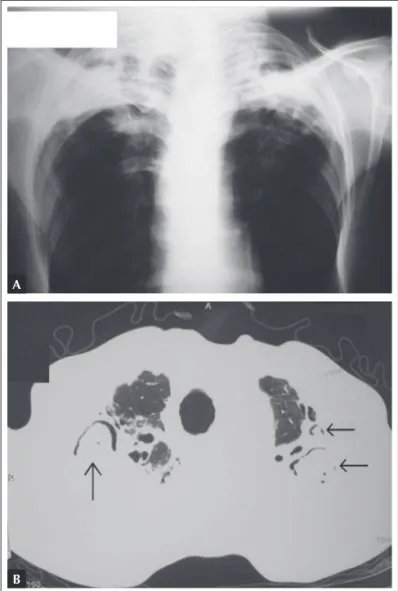
In August 2008 his symptoms worsened. He was waiting to be admitted by the Thoracic and Cardiovascular Surgery Service. In October 2008 the condition of the patient deteriorated and he was admitted to the Cardiac Surgery ICU. Preoperative posteroanterior chest X-ray (Figure 1A) showed chronic lesions in both lung apices. Preoperative axial chest CT scan (Figure 1B) showed the presence of a cavitary lesion in the right lung apex with an ovoid mass, and a crescent of air between the mass and the wall of the cavity. Two other masses were identiied in the left apex (aspergilloma).
It was decided to perform bilateral lobectomy at different surgical times. Firstly, the patient underwent a right upper lobectomy without intercurrences. Macroscopic anatomopathological study identiied reddish purple lung tissue with foci of anthracosis and a 9.0 x 3.0 x 2.5 cm cavity with a pasty, dark, fetid material. Microscopic analysis identiied an extensive ibrous pneumopathy with chronic and acute inlammation and fungal infection with hyalohyphomycosis associated with zygomycosis and Actinomyces spp. (Figure 2).
On the third postoperative day, the patient developed right lung atelectasis. An emergency fibrobronchoscopy was suggestive of occlusion of the middle lobar bronchus; however, a new thoracotomy did not conirm this inding. On subsequent days, the right lung failed to expand and the patient developed septic shock refractory to treatment, and the patient eventually died.
DISCUSSION
We presented a rare case of AS with a severe mixed fungal lung disease. The prevalence of lung disease in patients with AS ranges from 0 to 30%1. Apical lung ibrosis, such
Figure 1.A) Preoperative posteroanterior chest X-ray showing chronic lesions in both apices. B) Preoperative axial chest CT scan showing a mass in the right lung apex and two in the left apex.
A
Azevedo etal.
636 Bras J Rheumatol 2009;49(5):630-7
as the one presented by this patient, is the main pulmonary manifestation. Cavitary lesions are not as common2. Lung
disease has been attributed to mechanical factors (the rigidity of the thoracic spine is responsible for a restrictive disorder) and interstitial lung inflammation3. For those reasons,
patients with AS on immunosuppressors have a higher risk of developing lung infections. Tuberculosis is the lung infection more common in patients with AS with pulmonary ibrosis4.
Aspergillus spp. infection (aspergilloma) develops inside a
preexisting pulmonary cavity, such as that resulting from prior tuberculosis. Our patient was treated for tuberculosis in another service and had apical ibrosis and cavitations. Although, to our understanding, the presence of tuberculosis was never conirmed, a new treatment with three drugs was reinstituted because the possibility of pulmonary mycosis had not been considered. The association of AS and aspergillosis is rare. Rosenow et al.5 described aspergilloma in just ive (0.2%) out
of 2,080 patients with AS.
In hyalohyphomycosis, the causative agents present as hyaline, septate hyphae with closed angle ramiications, while in zygomycosis ramiications show a 90° angle5.
On radiological evaluation, aspergillomas appear as an oval to roundish mass with soft tissue opacity inside a pulmonary cavity. Typically, they are separated from the cavity wall by an aerated space known as the crescent air sign. Aspergillomas move according to the position of the body of the patient. Although patients with aspergilloma can be asymptomatic, most of them have symptoms such as cough, dyspnea, and hemoptysis5.
Besides aspergilloma, aspergillosis can also present as chronic necrotizing pulmonary aspergillosis (CNPA) and invasive aspergillosis (IA). Treatment with amphotericin B, alone or associated with lucytosine, can show excellent results in cases of IA, but aspergillomas do not show a good response to this treatment. Newer broad spectrum agents, such as itraconazole and luconazole, have shown encouraging results with less toxicity7. When pharmacotherapy is ineffective,
surgical resection is indicated5.
Franquet et al.6 described the case of a patient with AS
for 15 years with hemoptysis, progressive dyspnea, weight loss, and fever. The patient denied history of tuberculosis. Imaging exams showed extensive bilateral apical ibrosis, and a cavitary lesion with an ovoid mass inside with crescent air sign. Microbiology showed septate fungal hyphae at a 45° angle, compatible with Aspergillus spp. Clinical evolution, imaging exams, and microbiological indings of their patient was similar to ours.
Elliott et al.7 reported the case of a patient with AS for
35 years who developed CNPA and aspergilloma, which was treated successfully with systemic antifungal therapy. The patient had been admitted with consumptive syndrome, productive cough, dyspnea, and fever. He was treated with itraconazole for six months. In our patient, unlike the patient of Elliott et al.7, surgery was indicated.
At the beginning of the decade of 1970, Kennedy et al.8
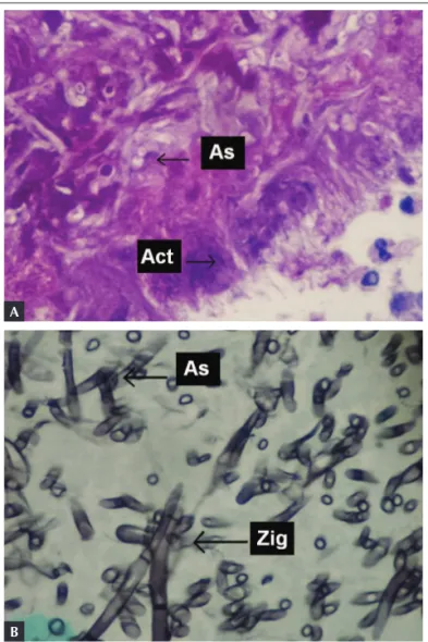
reported the case of two male patients with AS, apical lung ibrosis, and cavity, who developed fungal infection with a Figure 2.A) Histological slide of the fungus ball from the
lung biopsy of October 2008 showing hyphae and asexual spores, suggestive of Aspergillus spp. (As) associated with Actinomyces spp. (Act). (PAS staining; 400x). B) Histological slide of the fungus ball from the lung biopsy of October 2008 showing Aspergillus sp. hyphae. (As – note the 45° septa) and Zygomycetes (Zig – note thicker hyphae with 90° septa). (Grocott staining; 400x).
A
Pulmonary fungal infection with hyalohyphomycosis associated with zygomycosis and Actinomyces spp. in a patient with Ankylosing Spondylitis
637
Bras J Rheumatol 2009;49(5):630-7
combination of different agents. In this report, a combination of fungal strains suggestive of Aspergillus spp., Zygomycetes, and Actinomyces spp. was identiied.
We conclude that vigilance and follow-up of lung disease in patients with long-standing AS, especially in those patients with pulmonary symptoms, are required. The possibility of pulmonary mycosis should be raised in case of poor response to drug therapy or when the evolution is not compatible with the pulmonary involvement of AS and/or tuberculosis. We suggested individualized evaluation of each case with imaging exams (chest X-ray and CT scan), respiratory endoscopy, microbiological tests, and multidisciplinary follow-up to minimize the morbidity and mortality of those patients.
REFERÊNCIAS REFERENCES
1. Rosenow E, Strimlan CV, Muhm JR, Ferguson RH. Pleuropulmonary manifestations of ankylosing spondylitis. Mayo Clin Proc 1977 Oct;52(10):641-9.
2. Crompton GK, Cameron SJ, Langlands AO. Pulmonary ibrosis, pulmonary tuberculosis and ankylosing spondylitis. Br J Dis Chest 1974; 68(1):51-6.
3. Pamuk ON, Harmandar O, Tosun B, Yörük Y, Cakir N. A patient with ankylosing spondylitis who presented with chronic necrotising aspergillosis: report on one case and review of the literature. Clin Rheumatol 2005; 24(4):415-9.
4. Ho HH, Lin MC, Yu KH, Wang CM, Wu YJ, Chen JY. Pulmonary tuberculosis and disease-related pulmonary apical fibrosis in ankylosing spondylitis. J Rheumatol 2009; 36(2):355-60.
5. Telles FQ. Infecções por fungos ilamentosos invasores. Prática Hospitalar. 2004 Nov-Dez; vol 36: Disponível em http://www. praticahospitalar.com.br/pratica%2036/paginas/materia%2009-36. html [Acesso em 21 de agosto de 2009].
6. Franquet T, Müller NL, Flint JD. A patient with ankylosing spondylitis and recurrent haemoptysis. Eur Respir J 2004; 23(3):488-91. 7. Elliott JA, Milne LJR, Cumming D. Chronic necrotizing pulmonary
aspergillosis treated with itraconazole. Thorax 1989; 44(10):820-1. 8. Kennedy WP, Milne LJ, Blyth W, Crompton GK. Two unusual