J Bras Pneumol. 2007;33(2):234-237
Case Report
Pulmonary metastases in men:
primary tumor in an unusual location*
Daniel Brito de Araújo1, Nilton Haertel Gomes2, Décio Valente Renck3, Ricardo Bertolino Silva4,
Danise Senna Oliveira5, Fábio Eduardo Nunes Vieira6
Abstract
We report a case of breast cancer identified in a 75-year-old male as an accidental finding during the course of the investigation of a primary tumor and the search for pulmonary metastases. We address aspects related to the epidemiology, diagnosis, treatment, and prognosis of this condition in males.
Keywords: Neoplasm metastasis/lung; Breast neoplasms; Male.
* Study carried out at the Santa Casa de Misericórdia Hospital of Pelotas – Pelotas (RS) Brazil.
1. Specialist in Clinical Medicine. Resident in Rheumatology at the Hospital do Servidor Público Estadual – HSPE, Hospital for State Civil Servants – and the Instituto de Assistência Médica ao Servidor Público – IAMSPE, Institute for the Medical Treatment of Civil Servants – São Paulo (SP) Brazil.
2. Member of the Brazilian College of Surgeons. Universidade Federal de Pelotas – UFPel, Federal University of Pelotas – Pelotas (RS) Brazil. 3. Member of the Brazilian College of Radiology. Santa Casa de Misericórdia Hospital of Pelotas, Pelotas (RS) Brazil.
4. Resident in Gastroenterology at the Joinvile Regional Hospital, Joinvile (SC) Brazil.
5. Family and Community Doctor. Resident in Infectology at Heliópolis Hospital, São Paulo (SP) Brazil. 6. Resident in General Surgery, University Hospital of Western Paraná, Cascavel (PR) Brazil.
Correspondence to: Daniel Brito de Araújo. Rua Estado de Israel, 847/63, CEP 04022-002, São Paulo, SP, Brasil. E-mail: [email protected]
Submitted: 5 November 2005. Accepted, after review: 26 April 2006.
Introduction
Clinicians frequently need to evaluate patients with multiple pulmonary nodules found through imaging studies. The etiology of such lesions is typically identified based on appropriate clinical examination and on the taking of a comprehensive patient history. In the present case, however, the identification of the primary site was an accidental finding on computed tomography scans. It is likely that the necessary attention was not given to the physical examina-tion findings, which showed breast alteraexamina-tions, since breast carcinoma is unexpected in males.
Breast carcinoma in men is a rare entity, although its incidence has been increasing in the last 25 years.(1) It
accounts for less than 1% of all breast carcinoma cases and 0.2-1.5% of all malignant tumors in men.(1-3) Therefore,
there is much less information regarding factors related to the prognosis and treatment of this neoplasia in men than there is regarding such factors in women. In contrast to the increasing incidence of cases in women, the incidence of male breast cancer has been stable in the last four decades, presenting an exponential increase with aging. The
accu-mulated information suggests that this neoplasia presents the same behavior and prognosis in both genders when the stages are the same. Unfortunately, the diagnosis in male patients is delayed due to the lack of knowledge of the problem on the part of the patient and, frequently, of the doctor.(2) In addition, the proximity of the tumor to the skin
and muscle plane explains the low frequency of cases in the initial stages, which, consequently, leads to a higher inci-dence of tumors invading adjacent structures, as well as of lymph node metastases and distant metastases.
Case report
Pulmonary metastases in men: primary tumor in an unusual location
J Bras Pneumol. 2007;33(2):234-237
235
Physical examination revealed that the patient was pale, presented severe dyspnea, and had suffered significant weight loss. His left breast was discretely increased in size, painless, and hardened, without effusion or nipple retraction. Pulmonary ausculta-tion showed decreased breath sounds, principally in the left lung, with wheezing, diffuse rales and crackles in the lung bases.
Chest X-rays showed multiple nodules in both pulmonary fields consistent with metastases, which were confirmed through tomographic study. In addition to the pulmonary metastases, there was an accidental X-ray finding of a mass consistent with neoplasia in the left breast (Figure 1).
A puncture biopsy of the lesion was carried out, and a diagnosis of invasive ductal breast carcinoma was made. The patient claimed to have no family history of breast cancer and refused to be submitted to any type of treatment.
Discussion
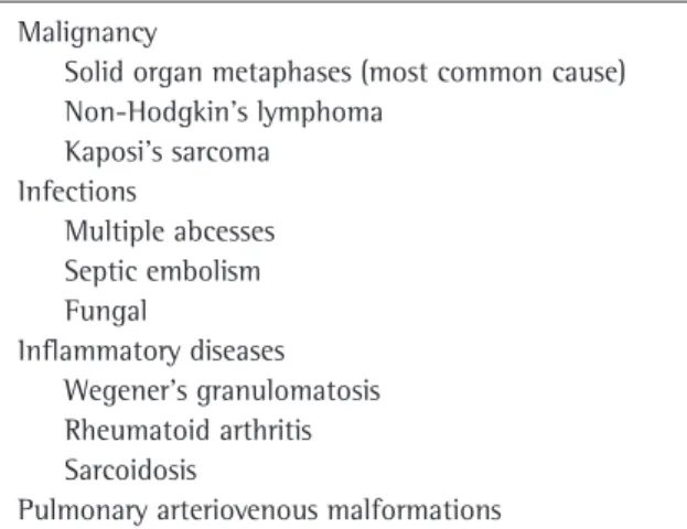
The incidence of pulmonary parenchymal metastases originating from primary extrathoracic neoplasias ranges from 20 to 54%. Metastases orig-inating from solid tumors are the most common causes of multiple pulmonary nodules, accounting for approximately 80% of such cases (Chart 1).(4)
Computed tomography scans of the chest are the test of choice for the evaluation of multiple pulmo-nary nodules. Such lesions are typically smaller than 5 mm in diameter and are found in the subp-leural region, although they can occasionally be
obscured by the mediastinal structures or by the hemidiaphragm.
Breast cancer most often metastasizes to the regional lymph nodes and subsequently to the bones (principally the pelvis and spine). Other affected sites are the liver, lungs, pleura, and brain. Pulmonary metastases from breast neoplasia occur principally through hematogenous and lymphatic dissemination.(5)
The symptomatology is related to the loca-tion and size of the tumor, and some patients are asymptomatic. Pulmonary involvement manifests as dyspnea, with or without cough, hemoptysis, or chest pain. Dyspnea results from endobron-chial involvement, carcinomatosis, pleural effusion, or pneumothorax. Progressive dyspnea, with or
b a
Figure 1 - a) Computed tomography of the chest showing the presence of multiple irregular nodules (arrows), which were heterogeneous after endovenous injection of contrast media (consistent with metastases), in both lungs; and b) The imaging shows the rate of soft-tissue attenuation. In the left breast, the soft tissue was permeable and nodular, with irregular borders and fibroglandular content (arrow).
Chart 1 - Pulmonary nodules - Differential diagnosis. Malignancy
Solid organ metaphases (most common cause) Non-Hodgkin’s lymphoma
Kaposi’s sarcoma Infections
Multiple abcesses Septic embolism Fungal
Inflammatory diseases Wegener’s granulomatosis Rheumatoid arthritis Sarcoidosis
236 Araújo DB, Gomes NH, Renck DV, Silva RB, Oliveira DS, Vieira FEN
J Bras Pneumol. 2007;33(2):234-237
without nonproductive cough, is characteristic of lymphatic dissemination. Endobronchial involve-ment or primary endobronchial metastasis can lead to cough with hemoptysis, and chest pain suggests involvement of the parietal pleura.(6,7)
Male breast carcinoma is a rare entity and, consequently, is recognized late, which delays the treatment and worsens the prognosis. Currently, due to a better understanding of this pathology, the duration of the symptomatology before diag-nosis has been decreasing, now ranging from one to eight months, whereas in the past it was as much as twenty-one months.
In contrast to in the situation for breast carci-noma involving women, the etiology of male breast carcinoma remains little understood. However, hormonal alterations are certainly implicated in the genesis of this pathology.(1,2,8) The incidence of
the disease remains higher among patients being treated with anti-androgens.(9)
From 15 to 20% of patients have a family history of breast neoplasia, which is especially important if among first-degree relatives. Gynecomastia occurs in 6 to 38% of patients, although there is no evidence that it is a predisposing factor for the development of cancer. There are other risk factors related to the appearance of this neoplasia (Chart 2).(1,2,10)
Genetic mutations, as in the case of the BRCA2 gene, are associated with having at least one male family member who has had breast cancer, and the probability of having more cases in the family can be as high as 76%.(1,11,12)
The age at the appearance of male breast cancer is from 60 to 67 years, which is at least 10 years later than that observed for female breast cancer. A painless palpable mass, typically located centrally (retroareolar), is the factor that motivates the individual to seek treatment in 85% of cases. Consequently, the principal differential diagnosis is gynecomastia. Other signs presented are nipple retraction, papillary effusion (occasionally hemor-rhagic), and mastalgia; the bilateral form of the disease is rare.(1,13) Axillary adenopathy is seen in
40-55% of patients. As in women, the involvement of the axillary lymph nodes, the size of the tumor, the histological grade, and the existence of hormonal receptors are important prognostic factors.(14-16)
The initial diagnostic procedure should be fine-needle aspiration biopsy, through which 27 to 49% of cases are diagnosed. A negative result does not rule out cancer, and the investigation should proceed with an excisional biopsy.(2) Mammography has
limited applications due to the technical difficulties involved, although it is very useful in obese patients with large breasts. It permits gynecomastia to be differentiated from carcinoma as well as the evalua-tion of the opposite breast. Mammography findings show the presence well-defined masses with spicu-lated margins and, less frequently, calcifications.
Although it is possible to find any histological type in the male breast, up to 90% of cases are of invasive ductal carcinoma. However, due to the absence of lobules in the rudimentary male breast, few cases of lobular carcinoma have been reported.(17) Estrogen and progesterone receptors
are positive in up to 81% and up to 74% of tumors, respectively, which makes it possible to obtain good results from hormonal therapy both as an adjuvant therapy and in the treatment of the metastatic form of the disease.(2)
Currently, for men with the nonmetastatic form of the disease, modified radical mastectomy with total axillary lymphadenectomy, followed by adju-vant radiotherapy, is recommended. Due to the small dimensions of the male breast and to the fact that men, unlike women, rarely present psychological
Chart 2 - Risk factors for male breast cancer. Testicular abnormalities
Cryptorchidia
Congenital inguinal hernia Orchiectomy
Orchitis
Testicular trauma Hormonal alterations
Infertility
Klinefelter’s syndrome Obesity
Cirrhosis
Family history of breast cancer Benign breast lesions
Nipple discharge Breast cysts Breast trauma
Esposure to radiation and to high temperatures Old age
Pulmonary metastases in men: primary tumor in an unusual location
J Bras Pneumol. 2007;33(2):234-237
237
problems with respect to the procedure, conserva-tive surgery is not usually performed.(15,18)
Since the rates of hormonal receptor positivity are high in male breast cancer, adjuvant hormonal therapy with tamoxifen for five years appears quite promising. The role of adjuvant chemotherapy is less established at the moment, but it is indicated in cases involving positive lymph nodes or primary tumors larger than 1 cm in diameter.(1,2)
In the metastatic form of the disease, hormonal treatment with tamoxifen is offered initially. When hormonal therapy is unsuccessful or if there are no hormonal receptors in the tumor, chemotherapy is recommended.(19,20) The metastatic dissemination is
similar to that seen in women, involving the bones, lungs, liver, lymph nodes, and skin.
More recent studies have shown that men and women with breast cancer have equivalent prognoses when compared by age and by disease stage, although the survival rate in males is lower due to the more advanced stage of the disease at diagnosis, higher age, and greater occurrence of comorbidities.(15)
The average five-year survival rate is 86%, ranging from 65% among patients presenting posi-tive lymph nodes to 90% among those presenting negative lymph nodes. The presence of positive lymph nodes is the most significant adverse prog-nostic factor.(16)
Areas for future investigation are abundant, principally concerning tumor markers, the role of hormonal therapy and chemotherapy, new agents for treatment as well as the study of genetic muta-tions in the pathogenesis of male breast cancer.
References
1. Giordano SH. A review of the diagnosis and management of male breast cancer. Oncologist. 2005;10(7):471-9.
2. Giordano SH, Buzdar AU, Hortobagyi GN. Breast cancer in men. Ann Intern Med. 2002;137(8):678-87.
3. El Omari-Alaoui H, Lahdiri I, Nejjar I, Hadadi K, Ahyoud F, Hachi H, et al. Male breast cancer. A report of 71 cases. Cancer Radiother. 2002;6(6):349-51.
4. Hirakata K, Nakata H, Nakagawa T. CT of pulmonary metastases with pathological correlation. Semin Ultrasound CT MR. 1995;16(5):379-94.
5. Connolly JE Jr, Erasmus JJ, Patz EF Jr. Thoracic manifestations of breast carcinoma: metastatic disease and complications of treatment. Clin Radiol. 1999; 54(8):487-94.
6. Kreisman H, Wolkove N, Finkelstein H, Cohen C, Margolese R, Frank H. Breast cancer and thoracic metastases: review of 119 patients. Thorax. 1983;38(3):175-9.
7. Burt M. Pulmonary Metastases. In: Fishman AP, Elias JA editors. Pulmonary Diseases and Disorders. 3rd ed. New York: McGraw-Hill Book Co; 1998.p.1851-60.
8. Thomas DB, Jimenez LM, McTiernan A, Rosenblatt K, Stalsberg H, Stemhagen A, et al. Breast cancer in men: risk factors with hormonal implications. Am J Epidemiol. 1992;135(7):734-48.
9. Karamanakos P, Mitsiades CS, Lembessis P, Kontos M, Trafalis D, Koutsilieris M. Male breast adenocarcinoma in a prostate cancer patient following prolonged anti-androgen monotherapy. Anticancer Res. 2004;24(2C):1077-81. 10. Milham S. A cluster of male breast cancer in office workers.
Am J Ind Med. 2004;46(1):86-7.
11. Palli D, Masala G, Mariani-Costantini R, Zanna I, Saieva C, Sera F, et al. A gene-environment interaction between occupation and BRCA1/BRCA2 mutations in male breast cancer? Eur J Cancer. 2004;40(16):2474-9.
12. Kwiatkowska E, Teresiak M, Filas V, Karczewska A, Breborowicz D, Mackiewicz A. BRCA2 mutations and androgen receptor expression as independent predictors of outcome of male breast cancer patients. Clin Cancer Res. 2003; 9(12):4452-9.
13. Clark JL, Nguyen PL, Jaszcz WB, Jatoi A, Niehans GA. Prognostic variables in male breast cancer. Am Surg. 2000;66(5):502-11.
14. Ribeiro GG, Swindell R, Harris M, Banerjee S, Cramer A. A review of the management of the male breast carcinoma based on an analysis of 420 treated cases. The Breast. 1996;5(3):141-6.
15. Cutuli B, Lacroze M, Dilhuydy JM, Velten M, De Lafontan B, Marchal C, et al. Male breast cancer: results of the treatments and prognostic factors in 397 cases. Eur J Cancer. 1995;31A(12):1960-4.
16. Guinee VF, Olsson H, Moller T, Shallenberger RC, van den Blink JW, Peter Z et al. The prognosis of breast cancer in males. A report of 335 cases. Cancer. 1993;71(1):154-61. 17. Goss PE, Reid C, Pintilie M, Lim R, Miller N. Male breast
carcinoma: a review of 229 patients who presented to the Princess Margaret Hospital during 40 years: 1955-1996. Cancer. 1999;85(3):629-39.
18. Yildirim E, Berberoglu U. Male breast cancer: a 22-year experience. Eur J Surg Oncol. 1998;24(6):548-52.
19. Jaiyesimi IA, Buzdar AU, Sahin AA, Ross MA. Carcinoma of the male breast. Ann Intern Med. 1992;117(9):771-7. 20. Lopez M, Di Lauro L, Lazzaro B, Papaldo P. Hormonal