CASE REPORT
Journal of Evolution of Medical and Dental Sciences/ Volume 2/ Issue 47/ November 25, 2013 Page 9150
SCHWANNOMA OF THE TONGUE - A CASE REPORT
S. Satyananda Rao1, J. Mansoor Ahmed2, Arun Ingale3
HOW TO CITE THIS ARTICLE:
S Satyananda Rao, J Mansoor Ahmed, Arun Ingale. Schwannoma of the tongue - a case report . Journal of Evolution of Medical and Dental Sciences 2013; Vol. 2, Issue 47, November 25; Page: 9150-9153.
ABSTRACT: A case report of an 18-year-old young girl presented with a slow-growing, painless
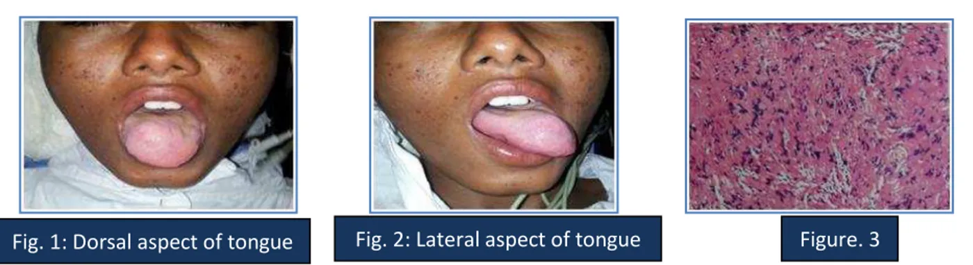
swelling on the right side of the tongue since 4 years. This was associated with disturbances in mastication. Examination revealed a 3 cm x 4 cm, globular smooth, mobile mass on right side of the tongue. There was no neurological deficit and no neck nodes palpable. She underwent excision of the mass under general anaesthesia. Complete enucleation with primary closure was carried out. The patient had an uneventful postoperative recovery and histopathological evaluation was consistent with schwannoma. The patient is recurrence free till date.
KEYWORDS: Schwannoma, Neurilemmoma, Tongue, Oral.
INTRODUCTION: Schwannoma or neurilemmoma are benign, slow growing, usually solitary and
encapsulated tumor, originating from Schwann cells of the peripheral, autonomic and cranial nerves.1-4 First, it was identified by Virchow in 1908.5 Approximately 25–45% of all schwannomas
occur in the head and neck .6 with the parapharyngeal space being the most common site .7 Intraoral
schwannoma accounts for 1% of all head and neck region tumors.8-10 and are commonly seen at the
base region of tongue.8,11,12 We report a patient with a schwannoma of the anterior part of the
tongue, that was excised intraorally.
CASE REPORT: A 18-year-old young girl presented with a slow-growing, painless swelling on the
CASE REPORT
Journal of Evolution of Medical and Dental Sciences/ Volume 2/ Issue 47/ November 25, 2013 Page 9151
DISCUSSION: Schwannoma or neurilemmoma are benign, slow growing, usually solitary and
encapsulated tumor, originating from Schwann cells of the peripheral, autonomic and cranial nerves.1-4 First, it was identified by Virchow in 1908.5 Approximately 25–45% of all schwannomas
occur in the head and neck.6 with the parapharyngeal space being the most common site.7 Intraoral
schwannoma accounts for 1% of all head and neck region tumors8-10 and are commonly seen at the
base region of tongue.8,11,12
Identification of the originating nerve may be difficult. In more than 50% of intraoral lesions, it is not possible to differentiate between tumors of the lingual, hypoglossal and glossopharyngeal nerves.13
Etiology is still unknown and the disease is generally asymptomatic.14 Schwannomas usually
present as a solitary lesion. When multiple, however, they can be associated with neurofibromatosis. The differentiation between schwannoma and neurofibroma is essential because an apparently solitary neurofibroma may be a manifestation of neurofibromatosis.15 Approximately 15% of
patients with neurofibromatosis will have malignant transformation in one or more lesion, which is in marked contrast to the typical behavior of a schwannoma.16
Histopathologically, the tumor tissue consists of so called Antoni A and B type cells. Type A tissue shows densely packed, elongated spindle cells, while type B tissue has more myxoid consistency.In addition, haemorrhage form adjacent tissue, necrosis, hyalinization and cystic degeneration, may also occur.17
Magnetic resonance imaging (MRI) is superior to other imaging modalities, for examination of the base of tongue. On MRI, a schwannoma is smooth and well demarcated .This tumor is isointense to the muscle on T1 – weighted images and homogenously hypointense on T2- weighted
images.18
The differential diagnosis of lingual schwannoma, benign lesions like granular cell tumors, salivary gland tumors, leiomyomas, rhabdomyomas, lymphangiomas, haemangiomas, dermoid cysts,lipomas, inflammatory lesions and lingual thyroid and malignant lesions like squamous cell carcinoma, sarcomas.19
Schwannomas of the tongue have been treated by surgical excision. The most common approach was the transoral route. This is an obvious choice for approaching these tumors since most are easily accessible via this route. Several other approaches have also been reported to have success including submandibular 11, suprahyoid pharyngotomy 20, and transhyoid21 approaches. All
of these approaches were used for base of tongue schwannomas that were deemed difficult to approach by the transoral route. More recently, the use of CO2 laser for excision of a base of tongue schwannoma has also been reported.22 The goal of surgical therapy is to complete resection. If this is
accomplished, recurrence is rare.21
CASE REPORT
Journal of Evolution of Medical and Dental Sciences/ Volume 2/ Issue 47/ November 25, 2013 Page 9152 Radiation therapy is not indicated because schwannomas exhibit a high degree of radioresistance.23 Prognosis is excellent as malignant transformation of schwannoma is an
exceptionally rare event and can safely be disregarded.
CONCLUSION: Although the incidence of schwannoma in the tongue is low, it should still be kept in
mind when making a diagnosis. They are most often diagnosed in adults but can also occur in younger age group although not that often. Schwannoma of the tongue is a relatively rare tumor of the head and neck. Transoral resection allows for removal of this tumor in a manner that precludes recurrence, avoids causing morbidity of tongue function, and remains the standard approach for treatment of the vast majority of these tumors. The chance of malignant transformation of these tumors is exceedingly unlikely.
REFERENCES:
1. Carinci F, Carls FP, Grasso DI, Pelucchi S, Pastore A. Schwannoma of parapharyngeal space. J Craniofac Surg 2000;11:367-70.
2. Cunningham LL Jr, Warner MR. Schwannoma of vagus nerve first diagnosed as parotid tumor. J Oral Maxillofac Surg 2003;61:141-4.
3. Hazarika P, Nooruddin SM, Nayak RG,. Neurilemmoma of the floor of the mouth. A case report. J Indian Dental Assoc 1983;55:325-6.
4. Bochlogyros PN, Kanakis PR, Tsikou-Papafrangou N, Chase D. A large, painless mass in the submandibular space. J Oral Maxillofac Surg 1992;50:1213-6.
5. Mosharraf TM, Kuppersmith RB, Porter JP, Donovan DT. Pathological quiz case 1. Malignant peripheral nerve sheath tumor of the ethmoid sinus. Arch Otolaryngol Head and Neck Surg 1997;123:654,656-7.
6. Katz AD, Passy V, Kaplan N (1971) Neurogenous neoplasms of major nerves of head and neck. Arch Surg 103:51–56.
7. Franzen A, Koegel K (1996) Neurinome im Halsbereich. Laryngorhinootologie 75:250–253. 8. Pfeifle R,Baur DA, Paullino A, Helman J. Schwannoma of the tongue:report of 2 cases. J Oral
Maxillofac Surg 2009;59:802-4.
9. Budde R, Brehmer D, Cantemir S, Laubert A. Schwannoma of the tongue. Laryngorhinootologie 2001;80:36-8.
10.Lacosta J, Zabelta M, Sanchez Del Hoyo A. Ectracranial schwannomas. Report of seven cases. Acta Otorrinolaringol Esp 1999;50:587-9.
11.de Bree R, Westerveld GJ, Smeele LF. Submandibular approach for excision of a large schwannoma in the base of the tongue. Eur Arch Otorhinolaryngol2000;257:283-6.
12.Spandow O, Fagerlund M, Bergmark L, Boquist L. clinical and histopathological feature of a large parapharyngeal neurilemmoma located at the base of the tongue. J Otorhinolaryngol Relat Spec1999;61:25-30.
13.Gutmann R, Grevers G: Extracranial schwannoma of the ENT region. Review of the literature with a case report of benign schwannoma of the base of tongue. HNO 1997;45:468-71. 14.Chiapasco M, Ronchi P, Scola G. Nerilemmoma (schwannoma) of the oral cavity. A report of 2
CASE REPORT
Journal of Evolution of Medical and Dental Sciences/ Volume 2/ Issue 47/ November 25, 2013 Page 9153 15.Sardinha SDCS, Paza AO, Vargas PA, Moreira RW, de Moraes M (2005) Schwannoma of the
oral cavity. Histological and immunohistochemical features. Braz J Oral Sci 4:806–809. 16.Wright BA, Jackson D (1980) Neural tumors of the oral cavity. A review of the spectrum of
benign and malignant oral tumors of the cavity and jaws. Oral Surg Oral Med Oral Pathol 49:509–522.
17.Batsakis JG, Tumor of Head and Neck. Clinical and pathological consideration. 2nd ed. Willams
and Wilkins: Baltimore; 1979.p.313-33.
18.Flickinger FW, Lozano RL, Yuh WT, Sachs MA. Neurilemmoma of the tongue: MR findings. J Comput Assist Tomogr 1989;13:886-8.
19.Nelson W, Chuprevich T, Galbraith DA. Enlarging tongue mass. J Oral Maxillofac Surg 1998;56:224-7.
20.Ying Y-LM, Zimmer LA, Myers EN (2006) Base of tongue schwannoma: a case report. Laryngoscope 116:1284–1287.
21.Hsu Y-C, Hwang C-F, Hsu R-F, Kuo F-Y, Chen C-Y (2006) Schwannoma (neurilemmoma) of the tongue. Acta Otolaryngol 126:861–865.
22.Mehrzad H, Persaud R, Papadimitriou N, Kaniyur S, Mochloulis G (2006) Schwannoma of the tongue base treated with transoral carbon dioxide laser. Lasers Med Sci 21:235–237.
23.Gallo WJ, Moss M, Shapiro DN, Gaul JV: Neurilemmoma: Review of the literature and report of five cases. J Oral surg 1997;35:235-6
AUTHORS:
1. S. Satyananda Rao 2. J. Mansoor Ahmed 3. Arun Ingale
PARTICULARS OF CONTRIBUTORS:
1. Professor and HOD, Department of ENT, Vijayanagar Institute of Medical Sciences. 2. Senior Resident, Department of ENT,
Vijayanagar Institute of Medical Sciences. 3. 2nd Year Post Graduate, Department of ENT,
Vijayanagar Institute of Medical Sciences.
NAME ADDRESS EMAIL ID OF THE CORRESPONDING AUTHOR:
Dr. J. Mansoor Ahmed, Consultant, ENT Surgeon,
Stall No. 7, Shadi Mahal Complex, Balgirao Road, Vadarabanda, Bellary. Email – [email protected]