Is [F-18]-fluorodeoxyglucose FDG-PET/CT better than ct
alone for the preoperative lymph node staging of muscle
invasive bladder cancer?
_______________________________________________
Mete Uttam
1, Nayak Pravin
1, Bhattacharya Anish
2, Kakkar Nandita
3, Mandal Arup
11 Department of Urology, PGIMER, Chandigarh; 2 Department of Nuclear Medicine PGIMER, Chandigarh;
3 Department of Histopathology, PGIMER, Chandigarh
ABSTRACT
ARTICLE
INFO
______________________________________________________________ ______________________
Objective: To evaluate whether the use of [F-18]-FDG-PET/CT can accurately predict pelvic lymph node metastasis in patients with muscle invasive TCC of the bladder un-dergoing radical cystectomy.
Materials and Methods: Fifteen patients with muscle invasive bladder cancer had un-dergone FDG-PET/CT scan from the skull base to the mid-thighs after IV injection of 6.5MBq (Mega-Becquerel)/Kg of FDG. After intravenous hydration IV furosemide was given to overcome the difficulties posed by urinary excretion of 18F-FDG. PET/ CT data were analyzed as PET and CT images studied separately as well as fused PET/ CT images. The imaging findings were correlated with the histopathology of the nodes (gold standard).
Results: CT and FDG-PET had demonstrated positive lymph nodes in 9 & 8 patients
respectively. Among the 15 patients 3 had documented metastasis on histopathology. Both CT and PET could detect the nodes in all these 3 patients (100% sensitivity). Nodes were histologically negative amongst 6&5 patients who had node involvement by CT
and PET respectively. Therefore, specificity, positive predictive value (PPV) & negative
predictive value (NPV) for CT and PET/CT were 50%, 33.3%, 100% and 58.3%, 37.5%, 100% respectively.
Conclusion: The theoretical advantage of this cutting edge technology for whole body imaging has not been translated into clinical practice as we found minimal advantage of combined FDG-PET/CT over CT alone for nodal staging of muscle invasive bladder cancer. This may be due to substantial overlap between standardized uptake values (SUVs) from active inflammatory processes with those of malignant lesion.
Key words:
Urinary Bladder Neoplasms; Positron-Emission Tomography; Tomography, X-Ray Computed; Carcinoma, Transitional Cell
Int Braz J Urol. 2016; 42: 234-41
_____________________
Submitted for publication: November 11, 2014
_____________________
Accepted after revision: June 28, 2015
INTRODUCTION
Approximately, 70% of transitional cell carcinomas (TCC) of the bladder present as a su-perficial lesion and nearly 20-25% of newly diag-nosed bladder cancers are muscle invasive and 5-10% are metastatic at initial presentation (1). The most frequent site of metastasis is the
meninges. Few patients with distant metastases survive 5 years. Clearly, the identification of me-tastases affects the decision-making process for tumor thought to be locally invasive (1).
Radical cystectomy is the standard surgi-cal treatment for muscle invasive urinary bladder cancer. In patients undergoing cystectomy, nodal tumor burden with pathologic evidence of lymph node metastasis is a major prognostic variable (2). Therefore, the accuracy of lymph nodal staging is crucial since treatment options differ significantly according to stage. Lymph node staging is a chal-lenge. With the imaging technologies growing fast 18F-FDG PET has gained popularity because it not only identifies the tumour site but also determines the metabolic activity of metastatic cell and hen-ce determines either regional or distant metasta-sis. Whereas CT scanning has the limitations that it can only describe the size of the lymph nodes, PET/CT identifies the metastasis in those lymph nodes, which otherwise can be a simple reactive hyperplasia. Therefore, it sounds reasonable to try this technology to improve detection of lymph node metastasis in muscle invasive transitional cell carcinoma of urinary bladder.
The reported accuracy of CT/MRI for nodal staging ranges from 70-90% with false negative rates of 25%-40%. In a study by Stojovska-Jova-novska et al. (3), it was found that primary tumor staging was correct in 55.6% with CT, 56.7% with conventional MRI and in 86.7% with dynamic MRI meaning that none of these modalities are 100% accurate. Therefore, there is a need for alter-native functional imaging modality like PET/CT. PET is the newer modality of investigation, which can be used to detect the primary tumor and its metastatic lesion, which have high metabolic acti-vity where FDG is picked up by the hypermetabo-lic cells (4-7). The current study was designed to determine whether PET/CT can accurately detect nodal metastasis, which were later confirmed by histopathology, the gold standard for nodal me-tastasis.
MATERIALS AND METHODS
A total of fifteen patients with muscle inva-sive TCC of the urinary bladder undergoing radical
cystectomy were included during the study period between August 2009 and October 2010. Radical cystectomy was performed by the same surgeon who was blinded to the PET-CT result. Informed consent was obtained from them to take part in the study. Patients with distant metastasis, high blood sugar level (>150mg%), patients who had urinary tract infection, patients who received chemotherapy and radiotherapy, who underwent partial cystectomy or diagnosed to have acute cystitis (CRP>1mg%) were excluded from the study. Patients with positive uri-ne culture were treated with appropriate antibiotics and were included once repeat urine culture had become sterile. History, clinical examination, labo-ratory tests and cystoscopy findings were recorded. All patients had undergone radical cystectomy with lymph node dissection.
FDG-PET Imaging
Patients were subjected to whole body PET--CT with dedicated BGO PETPET--CT scanner, at the department of Nuclear Medicine. All patients were asked to fast for 6-8 hours prior to the scan, blood sugar level was measured for all patients. After confirming acceptable blood sugar level (mean of 110.5mg/dL), 6.5MBq/kg of 18F-FDG was adminis-tered. In addition to oral contrast mixed with wa-ter, intravenous contrast was also administered via 18G venflon 500mL normal saline was started and 20mg of furosemide was given to all patients 10-15min after FDG injection. This was performed in order to avoid accumulation of FDG metabolite in the urinary bladder causing difficulty in analysis of lymph nodes. Patients were asked to void frequen-tly and were made to wait in the dark room for almost one hour after injection of FDG. Following this, patients were subjected to scanning from head to middle of the thigh. The image reconstruction was done with inbuilt matrix system using recons-truction algorithm.
Figure 1 - CT showing enhancing soft tissue lesion in the urinary bladder suggesting neoplastic lesion.
PET Data Analysis
The PET images were reviewed on high re-solution display monitor in sagital, coronal and transverse sections by physicians of the nuclear medicine department who had an experience of more than five years in this field of nuclear medi-cine. The observers were blinded to the prior diag-nostic work up data. Nodes greater than one cen-timetre on CT and SUVmax more than 2.5 were considered positive on CT and PET respectively. For assessment of accuracy of staging all imaging data were compared to the gold standard, which was histopathology.
Specimen Extraction, Labelling And Transport All patients were operated by the same sur-geon. Urinary bladder and regional lymph node specimen were packed in formalin containing pa-ckets separately labeled. Histopathological exami-nation was done by the same pathologist who was also blinded to the imaging result.
Correlation of imaging tests and pathological results
Any patient who had positive pelvic nodes on CT and or PET found to have histologically po-sitive nodes was considered as true popo-sitive. Any patient who had positive node on imaging but negative on histopathology was labelled as false positive for nodal staging. Positivity was conside-red for a given patient with any number of nodes positive on histology. Therefore for final statistical analysis any patient found to have either single node or multiple node positive on histology were considered gold standard positive.
Statistical analysis
The sensitivity, specificity, positive predic-tive value and negapredic-tive predicpredic-tive value were cal-culated using standard definition.
RESULTS
Age of our patients ranged from 34 to 67 years, with mean age of 53.4 years. Out of 15 patients, 14 were men (93.3%). Twelve patients (60%) were smokers. None of them was diabetic.
All of them had muscle invasive TCC on histologi-cal examination of TURBT specimens. All the pa-tients had undergone open radical cystectomy and standard lymphadenectomy. A total of 150 pelvic lymph nodes from 15 patients were removed out of which 68 nodes were from patients who had positive nodes on PET-CT.
Analysis of CT and Lymph Nodes Examination In all the 15 patients CT detected the pri-mary lesion as it enhanced after giving contrast because of its vascularity (Figure-1). Five patients showed perivesical stranding or loss of fat planes around the urinary bladder and on final histopa-thology only one patient showed involvement of the serosa of the bladder and surrounding tissues.
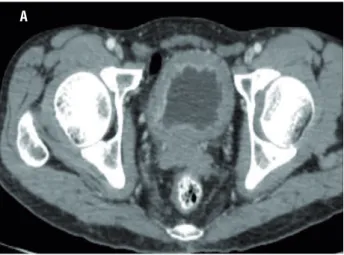
Nine patients showed enlarged regional lymph node on CT scan suggestive of metastasis (Figure-2). The result of CT was compared with
the gold standard of the lymph nodes obtained from the radical-cystectomy specimen of the same patients. However, only 3 out of the 9 patients actually had metastasis in the lymph nodes as evi-denced on histopathology (Figure-3). The other patients (n=6) showed reactive lymph node hyper-plasia. Sensitivity, specificity, positive predictive value and negative predictive value of CT alone was 100%, 50%, 33.5% and 100% (Tables 1 and 2).
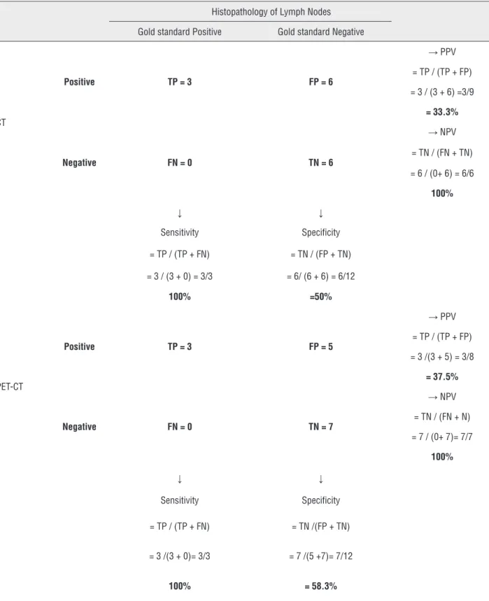
Table 1 - CT and PET-CT accuracy for lymph nodes.
Histopathology of Lymph Nodes
Gold standard Positive Gold standard Negative
CT
Positive TP = 3 FP = 6
→ PPV
= TP / (TP + FP)
= 3 / (3 + 6) =3/9
= 33.3%
Negative FN = 0 TN = 6
→ NPV
= TN / (FN + TN)
= 6 / (0+ 6) = 6/6
100%
↓ ↓
Sensitivity Specificity
= TP / (TP + FN) = TN / (FP + TN)
= 3 / (3 + 0) = 3/3 = 6/ (6 + 6) = 6/12
100% =50%
PET-CT
Positive TP = 3 FP = 5
→ PPV
= TP / (TP + FP)
= 3 /(3 + 5) = 3/8
= 37.5%
Negative FN = 0 TN = 7
→ NPV
= TN / (FN + N)
= 7 / (0+ 7)= 7/7
100%
↓ ↓
Sensitivity Specificity
= TP / (TP + FN) = TN /(FP + TN)
= 3 /(3 + 0)= 3/3 = 7 /(5 +7)= 7/12
100% = 58.3%
TP = true positive; TN = true negative; FP = false positive; FN = false negative; PPV = positive predictive value; NPV = negative predictive value; PET = positron emission
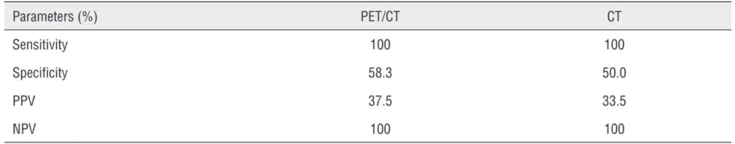
Table 2 = Comparison of PET/CT versus CT in terms of nodal detection.
Parameters (%) PET/CT CT
Sensitivity 100 100
Specificity 58.3 50.0
PPV 37.5 33.5
NPV 100 100
Figure 2 - CT showing enlarged lymph node in the pelvis.
Analysis of PET/CT and Lymph Nodes Exami-nation
In all 15 patients PET/CT detected hyper-metabolic lesion in the urinary bladder suggesting
the primary tumour which was clearly visualized after diuresis with furosemide (Figure-4).
Eight patients showed high FDG uptake by the regional lymph nodes suggestive of metasta-sis. PET along with the CT differentiation was used to find out the site of the positive lymph node. The result of PET/CT was compared with the gold standard, which was histopathology. Among the 8 patients with positive lymph nodes on PET/CT (Figure-5), only 3 had metastasis, and 5 patients showed reactive lymph node hyperplasia. Sensiti-vity, specificity, positive predictive valve and ne-gative predictive value was 100%, 58.3%, 37.5% and 100%, respectively (Tables 1 and 2).
DISCUSSION
Muscle invasive urinary bladder tumor is best treated by radical cystectomy which is a high complex procedure. Majority of these patients are elderly with comorbidities and will be subjected to increased risk of procedure related
complica-Figure 3 - Histopathology showing lymph node metastasis H&E X 20.
B
Figure 4 - High FDG uptake is seen in soft tissue mass in the bladder suggestive of primary lesion.
Figure 5 - PET / CT showing moderate FDG uptake in left internal iliac nodes (arrow).
tions. Cystectomy in the presence of nodal disease is controversial. Extent of lymph node dissection and number of nodes retrieved have important im-pact on outcome in patients with muscle invasive bladder cancer (8-10). During cystectomy more meticulous and extended node dissection impro-ves disease specific survival (11, 12). Therefore, accurate staging of lymph nodes can avoid un-necessary radical cystectomy. However, the fact remains that the most accurate method to stage regional lymph nodes remains regional lymph node dissection. Regional lymph node dissection is usually performed at the time of radical cystec-tomy at a single operative setting.
CT scan detects only enlarged lymph nodes. Most of the time these are reported on histopatho-logy as reactive lymph node hyperplasia. Therefore, it is not justified to discard the morbid yet curative procedure like radical cystectomy on the basis of tra-ditional CT findings alone. Many of these patients will be under-treated if we deny the gold standard treatment leading to reduced disease specific sur-vival. There is still a significant amount of “mis--staging” (40%) of bladder tumor with MRI. Future advances in MRI, including endorectal coil, enhan-cement and dynamic contrast imaging may make it more accurate, but they have still not been validated.
The role of PET scanning has also been in-vestigated for use in invasive TCC(Table-3). PET provides images of physiological and metabolic pro-cesses. A limitation of PET is the lack of an anato-mical reference frame. The combined PET/CT device offers optional fusion of images, which allows the localization of functional findings detected by PET in morphological structures as shown by CT during
imaging procedure. FDG, the most commonly used radiopharmaceutical for PET, is unsuitable for evalu-ation of bladder cancer due to intense accumulevalu-ation in the urine (13-17). More specific tracers like (11) carbon labeled choline, which are not excreted in the urine fared equally in terms of nodal staging.
Ahlstrom et al. (17) had compared (18) FDG PET with (11) C-methionine PET and found it sui-table for detecting primary tumour but it was not superior. Heicappell et al. (18) reported that in a case series of 8 patients, 3 had metastatic lymph nodes out of which 2 patients showed uptake by PET with a sensitivity of 66.7%.
In our study, the sensitivity was 100% but the specificity was as low as 58.3%. This low spe-cificity is the major concern for its routine use in clinical practice. In the current study CT alone was equally sensitive (100%) as PET/CT. However, it also lacks high specificity in terms of node
detec-Table 3 - Contemporary series on role of PET/CT for pelvic nodal staging.
Authors PET Tracer Number of pts Sensitivity Specificity
Drieskens et al. (19) 18F-FDG 55 60 88
Goodfellow H et al. (22) 18F-FDG 233 69 95
Liu et al. (23) 18F-FDG 46 76.9 97.1
Li et al. (24) 18F-FDG 73 75 —
Gofrit et al. (25) 11C-choline 18 100 —
Picchio M et al. (26) 11C-choline 27 62
Current study 18F-FDG 15 100 58.3
tion. The only noticeable thing was the negative predictive value which was 100%. That means if a patient has no detectable nodes on CT it will be un-likely to find histologically positive nodes. But the major concern for the physician is for the patients with positive nodes where one has to decide whether to go ahead with complex surgery like radical cys-tectomy or not. The current study reveals that PET/ CT is no better than CT alone for nodal staging. Simi-lar findings had been discussed by Greet Swinnen et al. (13). Their study involved 51 patients, 13 patients had histologically proven nodal metastasis out of which only six patients had demonstrated increased uptake on PET-CT. For the remaining seven patients PET-CT could not diagnose the nodes. The authors related that accuracy, sensitivity, and specificity of FDG-PET/CT for the diagnosis of node positive di-sease were 84%, 46%, and 97%, respectively. When analysing the results of CT alone, it was observed ac-curacy of 80%, sensitivity of 46%, and specificity of 92%. The authors found no advantage for combined FDG-PET/CT over CT alone for lymph node staging of invasive bladder cancer or recurrent high-risk su-perficial disease. In that study the sample size was large and hence more lymph node positive patients were involved, when compared to our study. We had only three patients with metastatic involvement of lymph nodes which was detected by PET/CT hence sensitivity was high.
On the contrary, Drieskens et al. (19) in their series of 55 patients found a sensitivity of 60% and specificity of 88% and they concluded that PET/CT has an advantage over CT in detec-ting distant lymph nodes. Most investigators feel that the use of FDG-PET has a limited utility for diagnosis and monitoring of urological tumors due to the erratic uptake of the isotope (20, 21). Goodfellow et al. (22) reported that sensitivity and specificity of the CT scans for pelvic LN involve-ment were 45% and 98%, respectively (N=93).
The small sample size was the main limita-tion of our study, which can explain the 100% sensi-tivity observed. Another major limitation of this stu-dy was the number of lymph nodes retrieved during surgery. We have dissected an average of 10 lymph nodes per patient. In 8 patients who had PET/CT po-sitive for lymph nodes, 68 nodes were examined on histology, with an average of 8.4 lymph nodes per
patient, which is a bit low for accurately identifying all the metastatic lymph nodes as reported by Herr HW et al. (8). These authors concluded that at least 9 lymph nodes should be studied to define nodal sta-tus accurately.
In order to overcome the limitations of FDG PET-CT, Li H et al. (24) had used dual-phase (18)
F--FDG PET/CT with oral diuretics and achieved the
detection rate of 75.0% (6/8) for lymph nodes. Gofrit
et al. (25) noticed uptake of (11) C-choline in lymph
nodes as small as 5mm (standardized uptake value 3.8±1.4). Picchio M et al. (26) compared CT with (11)
C-choline PET amongst 27 patients. Sensitivity for lymph node involvement was 50% by CT and 62% by (11) C-choline PET. The authors concluded that (11) C-choline PET is superior to CT for nodal sta-ging. However, it is obvious that majority of studies confirm that PET-CT with FDG or choline has limited advantage over conventional CT in staging lymph nodes in patients with bladder carcinoma. The cur-rent study confirms this point of view.
CONCLUSIONS
CT detects only enlarged lymph nodes but it tells nothing about the nature of the nodes in terms of presence or absence of metastasis. Theoretically, PET/CT could detect the lymph nodes harbouring metastasis correctly. This study reveals the sensitivi-ty of PET/CT for detection of primary lesion is 100% but in terms of nodal staging its accuracy is low. Therefore, at this stage we cannot say that PET/CT is a better imaging technique compared to CT, to detect malignant lymph nodal involvement. In addition, PET/CT is time consuming, costly and not accessible at every centre. Therefore at the moment we cannot recommend that PET/CT should be an essential tool for the urologist managing invasive bladder cancer.
CONFLICT OF INTEREST
None declared.
REFERENCES
2. Nishiyama H, Habuchi T, Watanabe J, Teramukai S, Tada H, Ono Y, et al. Clinical outcome of a large-scale multi-institutional retrospective study for locally advanced bladder cancer: a survey including 1131 patients treated during 1990-2000 in Japan. Eur Urol. 2004;45:176-81.
3. Stojovska-Jovanovska E, Mitreska N, Stojovski M, Lazarova A, Stavridis S, Dodevski A. Computed tomography or magnetic resonance imaging - our experiences in determining preoperative TNM staging of bladder cancer. Prilozi. 2013;34:63-70.
4. Bachor R, Kotzerke J, Reske SN, Hautmann R. [Lymph node staging of bladder neck carcinoma with positron emission tomography]. Urologe A. 1999;38:46-50.
5. Bachor R, Kocher F, Gropengiesser F, Reske SN, Hautmann RE. [Positron emission tomography. Introduction of a new procedure in diagnosis of urologic tumors and initial clinical results]. Urologe A. 1995;34:138-42.
6. Hoh CK, Seltzer MA, Franklin J, deKernion JB, Phelps ME, Belldegrun A. Positron emission tomography in urological oncology. J Urol. 1998;159:347-56.
7. Kosuda S, Kison PV, Greenough R, Grossman HB, Wahl RL. Preliminary assessment of fluorine-18 fluorodeoxyglucose positron emission tomography in patients with bladder cancer. Eur J Nucl Med. 1997;24:615-20.
8. Herr HW, Bochner BH, Dalbagni G, Donat SM, Reuter VE, Bajorin DF. Impact of the number of lymph nodes retrieved on outcome in patients with muscle invasive bladder cancer. J Urol. 2002;167:1295-8.
9. Stein JP, Lieskovsky G, Cote R, Groshen S, Feng AC, Boyd S, et al. Radical cystectomy in the treatment of invasive bladder cancer: long-term results in 1,054 patients. J Clin Oncol. 2001;19:666-75.
10. Poulsen AL, Horn T, Steven K. Radical cystectomy: extending the limits of pelvic lymph node dissection improves survival for patients with bladder câncer confined to the bladder wall. J Urol. 1998;160:2015-9.
11. Herr HW, Donat SM. Outcome of patients with grossly node positive bladder cancer after pelvic lymph node dissection and radical cystectomy. J Urol. 2001; 165:62-4.
12. Liedberg F, Månsson W. Lymph node metastasis in bladder cancer. Eur Urol. 2006;49:13-21.
13. Swinnen G, Maes A, Pottel H, Vanneste A, Billiet I, Lesage K, et al. FDG-PET/CT for the preoperative lymph node staging of invasive bladder cancer. Eur Urol. 2010;57:641-7. 14. Kamel EM, Jichlinski P, Prior JO, Meuwly JY, Delaloye JF,
Vaucher L, et al. Forced diuresis improves the diagnostic accuracy of 18F-FDG PET in abdominopelvic malignancies. J Nucl Med. 2006;47:1803-7.
15. Leisure GP, Vesselle HJ, Faulhaber PF, O’Donnell JK, Adler LP, Miraldi F. Technical improvements in fluorine-18-FDG PET imaging of the abdomen and pelvis. J Nucl Med Technol. 1997;25:115-9.
16. Anjos DA, Etchebehere EC, Ramos CD, Santos AO, Albertotti C, Camargo EE. 18F-FDG PET/CT delayed images after diuretic for restaging invasive bladder cancer. J Nucl Med. 2007;48:764-70.
17. Ahlström H, Malmström PU, Letocha H, Andersson J, Långström B, Nilsson S. Positron emission tomography in the diagnosis and staging of urinary bladder cancer. Acta Radiol. 1996;37:180-5.
18. Heicappell R, Müller-Mattheis V, Reinhardt M, Vosberg H, Gerharz CD, Müller-Gärtner H, et al. Staging of pelvic lymph nodes in neoplasms of the bladder and prostate by positron emission tomography with 2-[(18)F]-2-deoxy-D-glucose. Eur Urol. 1999;36:582-7.
19. Drieskens O, Oyen R, Van Poppel H, Vankan Y, Flamen P, Mortelmans L. FDG-PET for preoperative staging of bladder cancer. Eur J Nucl Med Mol Imaging. 2005;32:1412-7. 20. Shvarts O, Han KR, Seltzer M, Pantuck AJ, Belldegrun AS.
Positron emission tomography in urologic oncology. Cancer Control. 2002;9:335-42.
21. Hofer C, Kübler H, Hartung R, Breul J, Avril N. Diagnosis and monitoring of urological tumors using positron emission tomography. Eur Urol. 2001;40:481-7.
22. Goodfellow H, Viney Z, Hughes P, Rankin S, Rottenberg G, Hughes S, et al. Role of fluorodeoxyglucose positron emission tomography (FDG PET)-computed tomography (CT) in the staging of bladder cancer. BJU Int. 2014;114:389-95.
23. Liu IJ, Lai YH, Espiritu JI, Segall GM, Srinivas S, Nino-Murcia M, et al. Evaluation of fluorodeoxyglucose positron emission tomography imaging in metastatic transitional cell carcinoma with and without prior chemotherapy. Urol Int. 2006;77:69-75.
24. Li H, Wu H, Wang Q, Han Y, Wang Q. [Value of dual-phase (18)F-FDG PET/CT in preoperative staging of bladder cancer]. Nan Fang Yi Ke Da Xue Xue Bao. 2014;34:500-3. 25. Gofrit ON, Mishani E, Orevi M, Klein M, Freedman N, Pode
D, et al. Contribution of 11C-choline positron emission tomography/computerized tomography to preoperative staging of advanced transitional cell carcinoma. J Urol. 2006;176:940-4.
26. Picchio M, Treiber U, Beer AJ, Metz S, Bössner P, van Randenborgh H, et al. Value of 11C-choline PET and contrast-enhanced CT for staging of bladder cancer: correlation with histopathologic findings. J Nucl Med. 2006;47:938-44.
_______________________ Correspondence address: