TECHNICAL NOTE
I School of Pharmaceutical Sciences, Universidade de São Paulo - São Paulo/
SP, Brazil
II Servico de Terapia Intensiva, Hospital do Servidor Publico Estadual - São
Paulo/SP, Brazil
III Plastic Surgery and Burns, Hospital das Clinicas da Faculdade de Medicina
da Universidade de São Paulo - São Paulo/SP, Brazil Email: [email protected]
Tel.: 55 11 3069.2189
FLUCONAZOLE PLASMA CONCENTRATION
MEASUREMENT BY LIQUID CHROMATOGRAPHY
FOR DRUG MONITORING OF BURN PATIENTS
doi: 10.1590/S1807-59322010000200017
Silvia Regina Cavani Jorge Santos,I Edvaldo Vieira Campos,II Cristina Sanches,I David Souza Gomez,III Marcus Castro FerreiraIII
INTRODUCTION
Fluconazole (2-(2,4-diluorophenyl)-1,3-bis(1H-1,2,4-triazol-1-yl)-2-propanol) is a triazole antifungal agent that has been available since 1990. Fluconazole is a broad-spectrum triazole antifungal agent that has emerged as a suitable alternative to amphotericin B inthe treatment of a wide variety of superficial and systemic fungal infections.1 Triazole drugs act by inhibiting the enzyme
14-α-demethylase, which belongs to the microsomal CYP
system; triazoles also act by blocking the biosynthesis of ergosterol, resulting in the accumulation of 14-α-methyl
sterols and producing a fungistatic action. In general, luconazole is well tolerated, with side effects including nausea, headache, skin rash, vomiting, abdominal pain, and diarrhea occurring during long-term therapy.2
Excellent bioavailability has been reported after oral dosing, and a linear pharmacokinetics has been demonstrated at a dose of 200-800 mg daily. The elimination half-life of luconazole ranges from 27 to 37 hours, the apparent volume of distribution ranges from 0.5 to 0.7 L/kg, and approximately 80% of unchanged drug is excreted in the urine.3-6
Fluconazole is now used prophylactically in the management of various immune-compromisedpatient subsets1. High morbidity and mortalityin patientswith severe
thermal injury were associated with fungal sepsis; thus, a study of the eficacy of this drug to prevent fungal sepsis in burn patients is necessary. It is well-known that patients with thermal injuries show many changes in the pharmacokinetics of several drugs, but data on the clinical efficacy of luconazole related to its kinetics are very limited.7,8 Since
changes in the pharmacokinetics of luconazole are still unknown, it is imperative to investigate burn patients’ drug levels with a controlled clinical protocol.
A n a l y t i c a l m e t h o d s u s i n g m i c r o b i o l o g i c a l , spectrophotometric, and chromatographic techniques have all been shown to determine fluconazole in biological samples. High-performance liquid chromatography (HPLC) is preferred due to its selectivity and the specificity of the assay. Methods for the determination of luconazole in biological samples by HPLC-UV have been described previously.9-16 The objective of the present study was to
develop a simple analytical method to determine luconazole for plasma drug monitoring in patients with extensive thermal injury. An additional focus is its application to future pharmacokinetic studies. The method was validated as per FDA guidelines.17
MATERIALS AND METHODS
Experimental
Reagents and Chemicals
Instrumentation
The chromatographic system consisted of a Shimadzu model LC-10AVP solvent delivery module (Kyoto, Japan) equipped with a CBM 101 controller/software Class VP, an autosampler (model SIL-10ADVP) and a detector (UV -VIS model SPD-10AVP). The peak areas were integrated using a Shimadzu CR6A integrator with printer/plotter. The analytical column was a Shimpack CN (150 x 6.0 mm, 5 µm Shimadzu, Kyoto, Japan), with a Nova Pak C18 guard column (Waters Assoc., Milford, USA).
Chromatographic Conditions
The mobile phase was a mixture of puriied water and acetonitrile (60:40 v/v), freshly prepared on the day used and iltered through a 0.45- (µm ilter and helium degassed for 3 minutes. The chromatographic analysis was performed in an isocratic system using a low rate of 0.5 mL min-1 at room temperature. The injection volume was 5 µL,and the efluent was monitored by an ultraviolet absorbance detector at 210 nm. A run time of 15 minutes was required to guarantee the selectivity of chromatographic analysis.
Preparations of Standards and Internal Controls
The stock fluconazole solution was prepared by dissolving the appropriate amount of luconazole standard (10 mg) in ultrapure water to reach a inal drug concentration of 1.0 mg mL-1. Volumes of 5 mL of stock solution of
luconazole were transferred to a volumetric lask (50 mL). The higher standard (100.0 µg mL-1) was obtained by adding
the stock solution to drug-free human plasma (blank). Linearity was investigated by dilution of the higher standard to obtain the following concentrations of luconazole in plasma: 100.0, 50.0, 20.0, 10.0, 5.0, 2.0, 1.0, 0.4 and 0.2 µg mL-1; the calibration curve across the range 0.4 - 100.0
µg mL-1 was constructed daily. Internal quality controls
were prepared by dilution of the stock solution in drug-free plasma to obtain the following high (90.0 µg mL-1), medium
(50.0 µg mL-1) and low (1.2 µg mL-1) concentrations.
Aliquots of 0.5 mL from plasma standards and also from the internal controls were distributed into Eppendorf tubes and stored at -20 °C until assayed.
A stock solution of carbamazepine, the internal standard (IS), was prepared in methanol at 1 mg mL-1; an intermediate
solution was prepared by diluting the stock solution with methanol to reach the concentration 100 µg mL-1. Finally,
the solution of the IS was prepared freshly by diluting with methanol to reach 20 µg mL-1. Fifty µL was then added per
assay, and methanol was evaporated to dryness in a water bath before analyses.
Sample Extraction Procedure
Extraction tubes of IS (50 µL) were added, and the methanol was evaporated. Next, tubes containing dried IS were added to plasma (200 µL) and 50 µL 1.25 M sodium hydroxide, and the mixture was vortexed for 10 seconds. Drug extraction was performed by adding 3 mL dichloromethane and vortexing for 1 minute, following by centrifugation at 3000 rpm at 4°C for 30 minutes. The upper phase was aspirated and the tube containing the organic phase was immersed in a liquid nitrogen bath. The organic phase was then carefully transferred to a conic tube. Solvent was evaporated to dryness in a water bath under a nitrogen stream at 37°C. Residue was dissolved with 200 µL of a mixture of acetonitrile and ultrapure water (8:2 v/v), and under the conditions above detailed, 5 µL volumes were injected into the HPLC.
Linearity, Calibration Curve and Calculation Proce-dures
The nominal value of luconazole in plasma was plotted as a function of the peak area ratio of the internal standard to the drug concentration. A linear regression line was made, and the estimated linear correlation coeficient was applied to the calibration curve. At least six calibrators were included in the construction of the daily calibration curve of 0.4-100.0 µg mL-1. The daily curve was accepted
if at least 4/6 of the internal controls (high, medium and low concentrations, analyzed in duplicate) presented systematic error lower than 15%. At least one control of each concentration was within the acceptable variation. Once accepted, the calibration curve was applied to estimate drug concentration in samples from patients. The linearity of the method was determined in triplicate for each concentration ranging from 0.4 to 100.0 µg mL-1
Accuracy, Precision and Recovery
The precision of the quantitative method is the degree of agreement among individual tests when the procedure is applied repeatedly to multiple replicates of three different concentrations. Data were expressed as the coeficient of variation (CV%). The intra-day precision was evaluated by analysis of three replicates of the high (90.0 µg mL-1), medium (50.0 µg mL-1) and low (1.2 µg mL-1)
accuracy can be expressed as systematic error, representing also the recovery of each drug/assay. The parameter can be estimated by the value of the mean back-calculated concentrations divided by theoretical concentrations, expressed as percentage. The intra-day accuracy was evaluated by analysis of three replicates of the high (90.0 µg mL-1), medium (50.0 µg mL-1) and low (1.2 µg mL-1)
concentrations. The inter-day accuracy was determined by the analysis of three replicates of the high, medium and low concentrations of luconazole on three different days.
Absolute recovery of luconazole drugs from plasma was estimated by the peak area integrated for the drug in plasma
versus the peak area integrated for the drug after direct injection of the same nominal drug concentration, expressed as a percentage. The eficiency of relative recovery was estimated by the peak area ratio integrated for each drug in plasma as compared to that of its internal standard.
Speciicity
The speciicity of an analytical method is its ability to measure accurately an analyte in the presence of endogenous compounds. The speciicity was evaluated by the analysis of drug-free plasma samples (normal, hemolyzed, lipemic and icteric biological samples). The retention times of endogenous compounds were compared with those obtained for both luconazole and the internal standard in the puriied plasma extract.
Limit of Detection and Limit of Quantiication
The limits of detection (LOD) and of quantiication (LOQ) were determined based on the analysis of nine replicates. The LOQ was defined as the lowest plasma drug concentration on the calibration daily curve that could be determined with an accuracy of 80-120% and with a precision lower than 20%. The LOD was deined as 0.5 times the limit of quantiication. In addition, the LOD has a peak signal-to-noise ratio equivalent to 3:1, while the LOQ shows a ratio of 6:1.
Stability study
Study of short-term stability was performed at room temperature by several repetitions of a sequence of injections over 72 h. The study was done by testing a sequence of micro vials on the rack of the auto sampler containing plasma extracts in four different concentrations (2.2, 20.0, 50.0 and 90.0 µg mL-1) determined on the basis of the daily curve.
Spiked blank plasma was analyzed by HPLC after the clean-up of the plasma samples after three freeze/thaw cycles.
The same sequence as detailed above was performed using four different concentrations (2.2, 20.0, 50.0 and 90.0 µg mL -1) and analyzed in triplicate during three consecutive periods.
Data were expressed as a percentage of the systematic error. The acceptance criterion for all concentrations studied was adopted at less than 10% variation.
Robustness
The robustness of the method was determined by using two different columns with small changes in the proportion of acetonitrile in the mobile phase. The study was developed using two different concentrations (3 replicates each). Data were expressed by systematic error, as a percentage.
Therapeutic Drug Monitoring for dose adjustment
The study was designed for drug plasma monitoring of luconazole in burn patients with fungal infection plus sepsis. The six patients included in the study had the following characteristics: age: 44±15 yrs, body weight: 72.0±15.5 kg, total burned body surface area: 32.4±25.0% (TBSA %, patient #1: 70%, #2: 18%, #3: 10%, #4: 53%, #5: 8%, #6: 36%). Either the patient or the legally responsible person was informed in detail about the procedures to be performed during the treatment, and written consent was acquired. The protocol was approved by the ethics committee of the hospital Clinical Hospital, Medical School, University of Sao Paulo, Brazil (HC FMUSP). A sequential allocation number was assigned for the identiication of patients; data were described anonymously in accordance with ethical guidelines.
Antimicrobial therapy for the control of the sepsis caused by MRSA was an empirically dosed regimen of vancomycin (2g/day, 1 hour infusion). Once the opportunistic fungal infection was also documented, the Hospital Committee of Infection recommended the inclusion of luconazole (200 mg twice daily, 0.25 h infusion) in the therapy.
Fluconazole plasma monitoring was performed in different periods for each patient. Blood samples (1 mL) were collected at the trough from the arterial catheter. Each blood sample was transferred to tubes (BD, Sao Paulo, Brazil) containing sodium EDTA, and the plasma was separated by centrifugation at 3000 rpm for 30 min and stored at -20oC until analysis.
RESULTS
Experimental
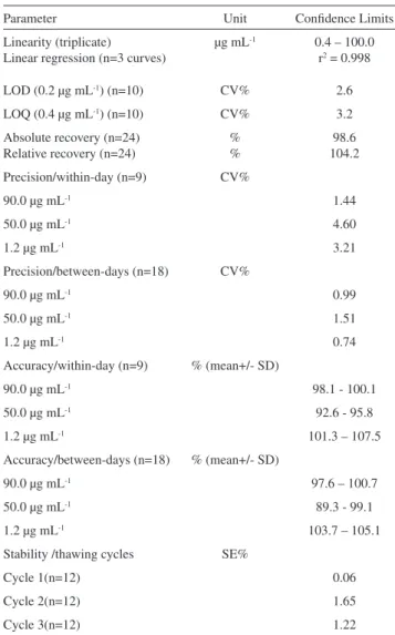
Table 1. The speciicity of the analytical method for the determination of luconazole in biological samples was evaluated. Once endogenous compounds had been eluted over the irst minute, no interference with the analysis was observed under the analytical conditions described above. Typical chromatograms of a blank plasma and spiked plasma are shown in Figure 1. The retention times were 9.3 and 13.3 minutes for luconazole and carbamazepine (IS), respectively. Peaks were monitored at 210 nm, and the total time required for each chromatographic run was 15 minutes to guarantee high selectivity and speciicity.
The analytical method exhibits excellent linearity, based on good recovery, and acceptable accuracy and precision (Table 1). Data for the linearity studies were expressed by the intercept and also the slope of the linear function. The mean, standard error of the mean (SEM) and the linear determination coeficient (r2) are: intercept 0.0146 (SEM:
0.0183), slope 0.0273 (SEM: 0.0004) and r2: 0.998 over
0.4-100.0 µg mL-1. The detection limit was 0.2 µg mL-1, and the
quantiication limit was 0.4 µg mL-1 based on the analysis of
0.2 mL plasma in 10 replicates.
The method showed good sensitivity, linearity and stability, with acceptable accuracy and precision (Table 1). In addition, small variations in the proportion of acetonitrile in the mobile phase showed acceptable precision and systematic error (Table 1). It should be emphasized that no robustness was investigated in the methods previously reported.
Analysis of the short-term stability of luconazole (time
and conditions of analysis) indicated no degradation of the drug in the plasma extract within a period up to 72 h while in vials on a tray of the chromatographic system. The short-term stability of luconazole in plasma on the bench showed good precision (4.8%) and systematic error (–1.8%). Thaw cycles showed good stability of the drug in biological samples after three consecutive freeze/thaw cycles (Table 1).
Therapeutic Drug Monitoring
The validated bioanalytical method was applied to luconazole plasma monitoring in six burn patients. Trough bloods drawn were obtained in different periods for each patient. Figure 2 shows for the six patients investigated. luconazole plasma concentrations versus time during the follow-up period.
Table 1 - Mean conidence limits of analytical method of luconazole in plasma.
Parameter Unit Conidence Limits
Linearity (triplicate) Linear regression (n=3 curves)
µg mL-1 0.4 – 100.0
r2 = 0.998
LOD (0.2 µg mL-1) (n=10) CV% 2.6
LOQ (0.4 µg mL-1) (n=10) CV% 3.2
Absolute recovery (n=24) Relative recovery (n=24)
% %
98.6 104.2 Precision/within-day (n=9) CV%
90.0 µg mL-1 1.44
50.0 µg mL-1 4.60
1.2 µg mL-1 3.21
Precision/between-days (n=18) CV%
90.0 µg mL-1 0.99
50.0 µg mL-1 1.51
1.2 µg mL-1 0.74
Accuracy/within-day (n=9) % (mean+/- SD)
90.0 µg mL-1 98.1 - 100.1
50.0 µg mL-1 92.6 - 95.8
1.2 µg mL-1 101.3 – 107.5
Accuracy/between-days (n=18) % (mean+/- SD)
90.0 µg mL-1 97.6 – 100.7
50.0 µg mL-1 89.3 - 99.1
1.2 µg mL-1 103.7 – 105.1
Stability /thawing cycles SE%
Cycle 1(n=12) 0.06
Cycle 2(n=12) 1.65
Cycle 3(n=12) 1.22
Abbreviations: coeficient of variation (CV%), standard deviation of mean (SD), systematic error (SE%), higher and lower limits (mean+/-SD) for accuracy and precision. Symbol: r2: coeficient of determination.
Figure 1 - Chromatographic proile of luconazole in plasma.
Chromato-grams: (A) Blank plasma. (B) Spiked blank plasma extracts Lower limit of detection (0.4 µg mL-1); (C) Low standard (2.2 µg mL-1) and (E) Medium
standard (25.0 µg mL-1). (D) Plasma extract of Patient #3 (15.0 µg mL-1).
An increase in the empirical daily dose was required for most patients to maintain clinical eficacy related (deined as a trough higher than the minimum effective concentration of 10 µg/mL). There was no accumulation after multiple infusions of the empirical dose regimen in patients #2 and #3, as seen by the trough lower than 1 µg/mL. Thus, their daily dose was increased 2-fold, from 400 mg/day (200 mg every 12 hrs) to 800 mg/day (200 mg every 6 hrs) to reach a trough higher than 10 µg/mL. The daily dose was increased 1.5 times for patient #5 because a trough equivalent to 5 µg/ mL was obtained after the empirical daily dose. The daily dose was increased to 600 mg daily (200 mg every 8 hrs) for patient #4, but the minimum effective concentration at the trough was not reached.3
For patient #6, a 50% reduction in the daily dose (from 400 mg/daily to 200 mg day) was required, as a trough luconazole plasma level higher than 20 µg/mL was obtained after the empirical dose regimen. After the empirical dose regimen, patient #1 showed trough plasma levels higher than 10 µg/mL in four consecutive periods. The high variability in luconazole plasma levels obtained on days 24, 25, 27 and 28 are probably due to changes in drug kinetics reported generally for antimicrobials and speciically for luconazole in burn patients.18-20
Considering the trough plasma levels for all patients monitored in the present study (Figure 2), dose adjustment was required to reach clinical eficacy in most patients due to failure of the empirical dose regimen (Figure 3).
DISCUSSION
Analytical methods reported for the determination of
Figure 2 - Antimicrobial therapy was started at an empirical dose regimen (luconazole 200 mg twice daily, fast infusion) and was altered based on
thera-peutic drug monitoring of six burn patients with fungal infection. Trough plasma levels were obtained at different days after the accident. Abbreviation: minimum effective concentration (MEC > 10 µg/mL).
luconazole in plasma9,12-16, serum,10,11,14 cerebral spinal luid14
and saliva13 have employed LC-UV after several puriication
procedures. A direct injection into the chromatographic system of small volumes of diluted plasma or extracts obtained from plasma protein precipitation showed serious problems related to speciicity and selectivity. A large number of runs were required, and the reduction in the lifetime of the analytical column and leakage as a consequence of the blockade of the injector must be considered.10-12
Therapeutic drug monitoring of antimicrobial agents contributes considerably to dose adjustments and drug eficacy in burn patients. Problems such as blood sample collection in limited-access patients, and problems related to volume of blood drawn and the frequency of sampling must be solved. In addition, it is necessary that the analytical method have a high sensitivity for drug plasma
Figure 3 - Dose adjustment of luconazole to reach clinical eficacy was
measurement. As described in the present study, only 0.2 mL of plasma was necessary for drug analysis. Analytical methods that present low sensitivity require higher plasma volumes (0.5 to 1.0 mL) and thus may be considered inadequate for luconazole plasma measurements.9,13,14
For the plasma purification done prior to the chromatographic measurement, solid-phase extraction (SPE) is a good option with high selectivity and sensitivity. Next best is the liquid-liquid extraction procedure, which is preferable due to its low cost compared to SPE9,13,14,16. Higher
recovery of luconazole was obtained by a liquid-liquid single extraction in an alkaline medium than in data reported previously.13-15 Both of the above procedures are favorable to
plasma protein precipitation using methanol or acetonitrile. In the present study, the analytical method used in the determination of fluconazole in plasma by HPLC-UV is quite simple, rapid and low in cost. Considering the selectivity, specificity, and sensitivity of the analytical method, the choice of the wavelength of the ultraviolet detector is important to guarantee the detection of low plasma levels accurately. A wavelength of 210 nm is usually applied for the detection of plasmatic luconazole analyzed by LC.11-13,16 It has been suggested that 260
nm might be a more convenient wavelength because nonspeciic substances detected at 210 nm could be co-eluted and consequently interfere with the area of peak integrated for each analyte.9,10,14-16 Nevertheless, in the
present method, no peak of any substance was co-eluted with luconazole or its internal standard; the speciicity and selectivity of the method was good, although a peak of an endogenous compound was recorded in the chromatogram at approximately 7.5 minutes of retention time.
Concerning the chromatographic conditions for fluconazole plasma measurements, the separation was performed in a reverse-phase column and with a mixture of puriied water plus acetonitrile as the mobile phase at a 0.5 mL min-1 low rate. This method was superior to the
analytical procedures reported previously.9-16
Compared to data reported previously, the linearity obtained in the present study showed a greater detectable range for luconazole plasma concentrations.9,10,12,14-16 Data
obtained in the present study were comparable to the results described by Koks et al., in spite of the higher plasma volumes required13.
Therapeutic Drug Monitoring (TDM)
The literature recommends ten times the minimum effective concentration (0.4 µg mL-1) of luconazole for
antifungal prophylaxis.4,6,21-24,26 However, it was reported that
for effective pharmacotherapy of candidiasis, the luconazole
trough must reach 4.0 µg mL-1 at low-dose intravenous
therapy of 100 mg a day.6,22,23 The length of the infusion
matters; if the drug is infused over 2 hours instead of 10 minutes, lower troughs (2.2 µg mL-1) can be expected.24
In the present study, luconazole plasma monitoring was applied to investigate six hospitalized burn patients (8-70% TBSA) from the Intensive Care Burn Unit. The patients were receiving an empirical dose regimen of 200 mg every 12 hours (400 mg daily) for systemic fungal infection. Some were dose-adjusted up to 800 mg a day on the basis of the drug plasma monitoring. Dose adjustments of luconazole to reach clinical efficacy were required in 5/6 burn patients with fungal infection. The dose regimen remained unchanged only for patient #1. The dose was increased up to 800 mg daily for patients #2, #3, #4 and #5, but the dose was reduced to 200 mg daily for patient #6 due to moderate renal insuficiency (creatinine clearance, 40 mL/min).
The fluconazole plasma troughs reported previously ranged from 20 up to 50 µg mL-125 or up to 70 µg mL-1 for
high-dose therapy (800 mg day-1).26 Additionally, because the
diffusion of the drug through the blood-brain barrier is rapid, drug levels in the cerebral spinal luid (CSF) were about 70% of luconazole plasma levels.22,23,26 Menichetti et al. reported
that at high-dose therapy, the drug level in CSF is equivalent to 36±21 µg mL-1, and a plasma/CSF ratio of approximately
1.0 was shown.26 On the other hand, the luconazole trough
ranged from 4.6 µg mL-1 to 9.0 µg mL-1 at low-dose therapy
(200 - 400 mg daily), as reported by Cousin et al.4
Fluconazole trough plasma levels during the empirical dose regimen (200 mg twice daily, or 400 mg a day) in the irst period of drug therapy were used to determine the dose adjustment required to reach clinical eficacy in burn patients investigated. The patients had a large variability in the TBSA registered: #1 (TBSA: 70%), #2 (18%), #3 (10%), #4 (53%), #5 (8%) and #6 (36%). Data obtained in the present study are in agreement with the results reported previously by Rayatt et al. based on changes in drug plasma levels as a consequence of kinetic changes in burn patients receiving the empirical dose (400 mg once daily) for the control of fungal infection.27 Controversially, Boucher et al. reported
no change in the pharmacokinetics of severe burn patients receiving luconazole (2-hour infusion, 400 mg every 24 hours) on the basis of variability of burn area.20
CONCLUSION
in burn patients, probably as a consequence of important changes in their pharmacokinetics. This may explain changes in drug accumulation over time. Therapeutic drug
monitoring will be useful in long-term antifungal therapy of burn patients to maintain clinical eficacy. High variability in plasma levels in these patients over time can be expected.
REFERENCES
1. Goa KL, Barredell LB. Fluconazole. An update of its pharmacodynamic and pharmacokinetic properties and therapeutic use in major supericial and systemic mycoses in immunocompromised patients. Drugs. 1995;50:658-90.
2. Henderson HM, Chapman SW. Infections due to Fungi, Actinomyces and Nocardia. In: Betts RF, Chapman SW, Penn RL. Editors. Reese and Betts’ a practical Approach to Infectious Diseases. 5th ed. Philadelphia:
Lippincott Williams & Wilkins; 2003;628-30.
3. Debruyne D, Ryckelynck JP. Clinical Pharmacokinetics of luconazole. Clin Pharmacokinet. 1993;24:10-27
4. Cousin L, Berre M, Launay-Vacher V, Izzedine H, Deray G. Dosing guidelines for luconazole in patients with renal failure. Nephrol Dial Transplant 2003;18:2227-31.
5. Fernandez ACM, Pimentel TT, Martinez MG, Gonzalez MM. Determination of the minimun inhibitory concentration of luconazole against Cryptococcus neoformans. Ver. Cubana Med Trop. 1999;51:55-7.
6. Gallagher JC, Lee KB. Program to restrict use of i.v. luconazole. Am J Health Syst Pharm. 2004;61:1695-8.
7. Jaehde L, Sorgel F. Clinical pharmacokinetics in patients with burns. Clin Pharmacokinet. 2000;29:15-28.
8. Fraser VJ, Jones M, Dunkel J, Strofer S, Medoff G, Dunagan WC. Candidemia in tertiary care hospital: epidemiology, risk factors, and predictors of mortality. Clin Infect Dis. 1992;15:414-21.
9. Wattananat T, Wiyada A. Validated HPLC method for the determination of luconazole in human plasma. Biomed Chromatogr. 2006;20:1-3. 10. Egle H, Trittler R, Kûmmerer K. A new, rapid, fully automated method
for determination of luconazole in serum by column-switching liquid chromatography. Ther Drug Monit. 2004;26:425-31.
11. Ng TKC, Chan RCY, Adeyemi-Doro FAB, Cheung SW, Cheng AFB. Rapid high performance liquid chromatographic assay for antifungal agents in human sera. J Antimicrob Chemother. 1996;37:465-72. 12. Cociglio M, Brandissou S, Alric R, Bressolle F. High-performance liquid
chromatographic determination of luconazole in plasma. J Chromatogr Biomed Appl. 1996;686:11-7.
13. Koks CHW, Rosing H, Meenhorst PL, Bult A, Beijnen J. H. High-performance liquid chromatographic determination of the antifungal drug luconazole in plasma and saliva of human immunodeiciency virus-infected patients. J Chromatogr Biomed Appl. 1994;663:435-51. 14. Wallace JE, Harris SC, Gallegos J, Foulds G, Chen TJ, Rinaldi MG. Assay of luconazole by high-performance liquid chromatography with a mixed-phase column. Antimicrob Agents Chemother. 1992;36:603-6.
15. Flores-Murrieta FJ, Granados-Soto V, Hong E. Simple and rapid method for determination of luconazole in human plasma samples by high performance liquid chromatography. J Liq. Chromatogr. 1994;17:3803-11.
16. Inagaki K, Takagi J, Lor E, Okamoto MP, Gill MA. Determination of luconazole in human serum by solid-phase extraction and reversed-phase high-performance liquid chromatography. Ther Drug Monit. 1992;14:306-11.
17. FDA Home page. Guidance for Industry: Bioanalytical Method Validation, http://www.fda.gov/cder/guidance/4252fnl.htm, 2008. 18. Weinbren MJ, Pharmacokinetics of antibiotics in burn patients. J
Antimicrob Chemother. 1999;44:319-27.
19. Blanchet B, Jullien V, Vinsonneau C, Tod M. Influence of burns in pharmacokinetics of drugs used in the care of burn patients. Clin pharmacokinet. 2008;47:635-54.
20. Boucher AB, King SR, Wandschneider HL, Hickerson WL, Hanes SD, Herring VL, et al. Fluconazole pharmacokinetics in Burn Patients. Antimicrob Agents Chemother. 1998;930-3.
21. Sia-Cheing L, Chang-Phone F, Jen-Seng H, Chin-Jen T, Kuo-Su C, Huang-Yang C, et al. Clinical correlates of antifungical macrodilution susceptibility test results for non-AIDS patients with severe candida infections treated with luconazole. Antimicrob Agents Chemother. 2000;44:2715-8.
22. Foulds G, Brennan DR, Wajszczuk C, Cattanzaaro A, Gaarg DC, Knopf W, et al. Fluconazole penetration into cerebrospinal luid in humans. J Clin Pharmacol. 1988; 28:363-6.
23. Tucker R, Williams PL, Araathoon EG, Levine BE, Haartstein AI, Hanson LH, et al. Pharmacokinetics of luconazole in cerebrospinal luid and serum in human coccidioidal meningitis. Antimicrob Agents Chemother. 1988;32:369-73.
24. DeMuria D, Forrest A, Rich J, Scavone JM, Cohen LG, Kazanjian PH. Pharmacokinetics and bioavailability of luconazole in patients with AIDS. Antimicrob Agents Chemother. 1993;37:2187-92.
25. Haubrich KH, Haghighat D, Bozzette AS, Tilles J, McDutchan JA. High-dose luconazole for treatment of cryptococcal disease in patients with human immunodeiciency virus infection. The California collaborative treatment group. J Infect Dis. 1994;170:238-42.
26. Menichetti F, Fiorio M, Tosti A, Gatti G, Pasticci MB, Miletich F, et al. High-dose luconazole therapy for cryptococcal meningitis in patients with AIDS. Clin Infect Dis. 1996;22:838-40.