CLINICAL SCIENCE
I Pediatric Surgery and Pediatric Liver Transplantation Divisions, Faculdade
de Medicina da Universidade de São Paulo – São Paulo/SP, Brazil.
II Laboratory of Pediatric Surgery (LIM 30), Faculdade de Medicina da
Universidade de São Paulo – São Paulo/SP, Brazil. Phone: 55 11 3061.7479
Email: [email protected]
Received for publication on June 30, 2008 Accepted for publication on July 21, 2008
IMMUNOHISTOCHEMICAL STUDIES OF STELLATE
CELLS IN EXPERIMENTAL CHOLESTASIS IN
NEWBORN AND ADULT RATS
Nelson Elias Mendes Gibelli,I,II Uenis Tannuri, I,II Evandro Sobroza de MelloI,II
doi: 10.1590/S1807-59322008000500019
Gibelli NEM, Tannuri U, Mello ES de. Immunohistochemical studies of stellate cells in experimental cholestasis in newborn and adult rats. Clinics. 2008;63(5):689-94.
BACKGROUND AND AIMS: Although there is much known about liver diseases, some aspects remain unclear, such as the nature of the differences between the diseases observed in newborn infants and those in adults. For example, how do newborns respond to duct epithelial cell injury? Do the stellate cells in newborns respond similarly to those in adults during biliary obstruction? METHODS: Ninety newborn Wistar rats aged six days, weighing 8.0 – 13.9 g each, and 90 adult rats weighing 199.7 – 357.0 g each, were submitted to bile duct ligation. After surgery, they were randomly divided and sacriiced on the 3rd, 5th, 7th, 14th, 21st
or 28th day post-bile duct ligation. Hepatic biopsies were obtained and immunohistochemical semi-quantiication of desmin and
α-SMA expression was performed in hepatic stellate cells and in myoibroblasts in the portal space, and between the portal space and the liver lobule.
RESULTS: Desmin expression in the myoibroblast cells post-bile duct ligation was higher in young rats, reaching its peak level in a shorter time when compared to the adult animals. The differences between the groups for α-SMA expression were less sig-niicant than for desmin.
CONCLUSIONS: These indings indicate that there is an increase in the number of collagen-producing myoibroblast cells in young animals, suggesting that there is more intense ibrosis in this population. This inding may explain why young animals with bile duct obstruction experience more intense portal ibrosis that is similar to the pathology observed in the livers of newborns with biliary atresia.
KEYWORDS: Biliary atresia. Liver ibrogenesis. Pediatric liver disease. Experimental cholestasis. Pediatric disorders. Biliary congenital disorders.
INTRODUCTION
Biliary atresia (BA) is a condition found in newborns that is characterized by a progressive obliteration of the bile ducts. Even in successfully-operated patients, the condition results in severe liver injury and death. Therefore, BA is the cause of more than 60% of pediatric liver transplantations in almost all medical centers. It is known that BA is not a single disease, but instead, it is a phenotype associated with
several disorders in which the infant liver responds in a well-deined manner. The complex dynamic series of responses includes inlammation, bile duct proliferation, apoptosis, and ibrogenesis. The latter process may occur in different cell types, however, the stellate cells (perisinusoidal or Ito cells) are considered to be the main producers of extracellular
matrix proteins in the liver parenchyma.1-5 After a stimulus
that causes injury and activation of the inflammatory cascade, the release of cytokines then leads to stellate cell activation and their differentiation into myofibroblasts, interactions with the extracellular matrix and the subsequent alteration of its composition, collagen production and the onset of ibrosis.6-10
There is evidence that stellate cells in rats express desmin, and when activated, they also express smooth
structural proteins of the intermediary ilaments that are present in the cytoskeleton of smooth muscle cells and other cell types. Cassiman et al.17 described the presence
of myoibroblast sub-populations in the liver parenchyma in three main areas: stellate cells in the lobule, the portal space and at the interface between the portal space and the liver lobule. In an experimental model of common bile duct ligation (BDL) with the induction of biliary-type portal fibrosis, these myofibroblast sub-populations can also be involved in the initial stages of ibrosis development, especially those populations located in the portal space and the interface zone.17,18
Although much is known about cell injury and ibrosis induction as a result of liver diseases, some aspects remain unclear, such as the nature of the differences between the diseases seen in newborn infants, and those in older children and adults. Common bile duct ligation in rats is a classic experimental model for the study of biliary obstruction. However, the response of neonatal animals to bile duct ligation has yet to be completely understood, and few reports in the literature have focused on differences in the behavior of the liver in neonatal and adult animals.
In our previous investigation using newborn and adult rat experimental models of bile duct ligation, we demonstrated that although the initial responses of ductule proliferation and inlammatory iniltration were less intense in newborn animals, portal and periportal ibrosis were
more extensive in newborns compared to adult animals.19 In
the present study, we utilized the same experimental model and immunohistochemical staining techniques to detect desmin and smooth muscle alpha-actin ( -SMA), which are classic markers of myoibroblastic cells.19,20 Our aim was
to determine if the stellate (perisinusoidal) cells and the myoibroblasts of the newborn animals respond in a different manner compared to those of adult animals during biliary obstruction. These indings will enhance the understanding of the pathophysiology of biliary-type portal ibrosis of cholestatic diseases of childhood, including biliary atresia.
METHODS
The study was approved by the Ethical Committee for Research Project Analysis of our institution, and was carried out according to the international guidelines regarding the use of laboratory animals.
Surgical operations
The experimental model of cholestasis was created through common bile duct ligation (BDL) and sectioning between ligations, as previously described.19-25 Male and
female Wistar rats were used in the experiments. The animals were divided into two groups: young six-day old rats weighing between 8 to 13.9 g (mean weight of 11.4 g ± 0.038) submitted to common BDL (n=90), and adult rats weighing between 199.7 and 357 g (mean of 257.9 g ± 1.58) submitted to common BDL (n=90). The control group consisted of 10 adult and 10 six-day old rats that were submitted to the same anesthetic procedures and liver biopsy sampling without the common bile duct ligation procedure. The young and adult animals that were submitted to surgery were randomly divided and killed on the 3rd, 5th, 7th, 14th, 21st
or 28th day post-BDL. Wedge biopsies of the left liver lobe
were collected from the young and adult animals submitted to BDL, as well as the control animals. The fragments were kept in a buffer solution with 10% formaldehyde for a period of 24 to 48 hrs, after which they were parafin-embedded. Sections 3 µm in thickness were cut from the paraffin
blocks.
Immunohistochemical studies
Desmin: Immunohistochemical staining for desmin
was performed using the mouse anti-human desmin antibody (DAKO® Corporation A/S Denmark, clone D33) at a 1/200 concentration. Antigenic recovery of the material was performed by boiling the sections at 95º C in a citrate buffer at pH 6. The secondary kit used to detect immunohistochemical staining was the Vectastain® ABC kit (Vector® laboratories). The chromogen used was 3,3-diamino benzidine (Sigma®, code D-8001), and counterstaining was performed with Harris hematoxylin. Immunohistochemistry was used to identify the populations of myofibroblast and stellate cells to investigate their location in the normal liver of young and adult rats, to assess the degree of desmin expression, and to perform a semi-quantitative analysis of desmin expression throughout the observation period after ligation in animals of different ages. The semi-quantitative evaluation was performed by the global analysis of the entire lamina by two experimental pathologists in a double-blind analysis in order to estimate the number of desmin-positive cells in the different areas under study: the liver lobule, the portal region and the interface zone. The semi-quantitative measurements were performed using scores from 0 to 4, with 0 indicating the absence of positive staining, 1 indicating the presence of mild staining (+); 2 indicating the presence of moderate staining (++); 3 indicating the presence of intense staining (+++) and 4 indicating the presence of very intense staining (++++).
alpha-actin (α-SMA) was performed using a mouse anti-human alpha-actin antibody, diluted 1/500 (DAKO® Corporation A/S Denmark, clone 1A4). Antigenic recovery of the material was performed by boiling the sections at 95º C in a citrate buffer at pH 6. The secondary kit utilized was the Vectastain® ABC kit (Vector® laboratories). The chromogen used was 3,3-diamino benzidine (Sigma®, code D-8001), and counterstaining was performed with Harris hematoxylin. Immunohistochemistry was used to identify the activated stellate cells in the liver lobule and myoibroblast populations in the portal space and interface area, to evaluate the degree of -SMA expression, and to perform a semi-quantitative analysis of -SMA expression throughout the observation period after ligation in animals of different ages, using the same criteria described for the evaluation of desmin expression.
Statistical Analysis
The histological data were recorded in a double-blind analysis by two pathologists, as previously described, and were compared at each phase of the experiment between adult and young animals. Statistical analysis was performed with the non-parametric Mann-Whitney Rank Sum Test, using Sigmastat® software. The null hypothesis that the samples were equal was rejected for a p value < 0.05.
RESULTS
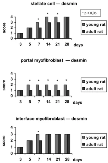
Desmin
Normal livers from animals of different ages showed desmin expression in the smooth muscle cells of blood vessel walls, in a few myoibroblasts of the portal stroma, and in certain cells located in zones 1 and 3 of the liver lobules. These cells were identiied as stellate cells (perisinusoidal or Ito cells) due to their location and characteristic star-shaped morphology with cytoplasmic extensions. The sham-operated animals showed exactly the same expression of normal livers in both age groups.
After BDL, the desmin expression increased signiicantly in young as well as in adult animals when compared to the control group. This increase was observed mainly inside the liver lobules, where the population of stellate cells increased signiicantly in both groups of animals.
On the 5th day after operation, desmin expression
increased to a similar extent in both groups. However, from the 7th day onwards, higher levels of desmin expression
were observed in the young animals, reaching a maximum value on the 14th day after the operation. In contrast, desmin
expression in the adult animals reached its maximum value
only on the 28th day. In addition, desmin expression in
myoibroblasts located in the interface zone between the portal space and the lobule reached higher levels in a shorter period of time in the young animals when compared to the adult animals. However, the portal myoibroblasts showed a very weak and constant staining intensity throughout the observation period after BDL. The expression of desmin in the portal myoibroblasts was higher in the young animals (Figure 1).
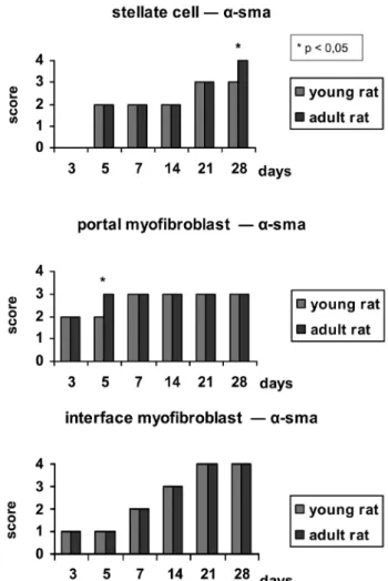
Smooth muscle alpha-actin (α-SMA)
α-SMA was expressed in the intermediary layer of portal
tract vessels and in some cells around the terminal liver
venules in the normal liver. Occasionally, α-SMA-positive
cells were found in the ibrous stroma of biliary ducts and liver sinusoids. The sham-operated animals showed exactly the same expression of normal livers in both age groups. Three days after BDL, myoibroblasts that stained intensely
for α-SMA could be easily identiied, and they formed layers around the proliferated bile ductules. In both groups
of rats, the population of α-SMA-positive cells continued to
increase throughout the post-operative period up until the
7th day, and maintained the same expression level thereafter
until the end of the experiment. The expression of α-SMA in
the portal myoibroblast population was very different from that observed for desmin expression, indicating a different pattern of marker expression in this cell population, i.e., while the myoibroblasts located in the lobule and in the interface express desmin, those located in the portal space
express α-SMA at high levels.
The expression of α-SMA by the stellate cells began
to appear on the 5th day post-CBDL, and the same pattern
of expression was maintained up until the 14th day in both
young and adult animals. From the 21st day onwards, -SMA
expression increased, reaching maximum levels on the
28th day in the adult rats compared to its expression in the
young rats. A constant inding in all the animals on the 5th
day post-BDL was the presence of stellate cells with -SMA expression distributed along zone 1 of the lobule, the most distal zone of the centrolobular vein (zone 3), thus “bridges” of myoibroblast cells joining the portal spaces were present. With time, the stellate cells subsequently occupied all zones of the liver lobule.
Myoibroblasts located in the interface between the portal space and the lobule also expressed -SMA, with similar increases in the expression levels in animals of different ages. The interface myoibroblasts showed higher expression
of α-SMA than desmin at the end of the observation period
(Figure 2).
In summary, desmin expression in the myoibroblast cells post-BDL was higher in the young rats, reaching its peak value in a shorter time compared to the adult animals. This observation indicates there is an actual increase in the number of collagen-producing myoibroblast cells in young animals, which suggests that more extensive fibrosis is
occurring in the younger rats. α-SMA expression translates
into the activation of the stellate cells. The differences in
α-SMA expression between the groups were smaller, and
in certain situations, the α-SMA expression, especially in stellate cells, was higher in adult animals.
DISCUSSION
The main population of cells involved in extracellular matrix protein synthesis, mesenchymal myofibroblast cells, was studied by immunohistochemical analysis. Of the sub-populations of these cells, stellate cells have been the focus of several studies as they are the major source of
collagen production.1-4 However, in an experimental model
of common bile duct ligation (BDL), other myoibroblast cell populations can also be involved with the initial stages of ibrogenesis, especially the portal myoibroblasts and myoibroblasts located in the interface zone, between the portal space and the lobule.18,19 In this experimental model,
as well as in several clinical forms of cholestatic diseases, the type of ibrosis that occurs is known as biliary-type portal ibrosis.
In this study, the “sectorial” expression of desmin and smooth cell alpha-actin by stellate cells, portal myoibroblasts and interface myoibroblasts were evaluated in a semi-quantitative analysis. The results obtained showed that there was discordance in the expression of markers in different myoibroblast populations. In particular, the myoibroblasts located in the portal space showed weak
desmin expression and strong α-SMA expression. These
observations indicate that there is a different pattern of marker expression in this cell population. In a previous study of liver injury caused by carbon tetrachloride and
BDL in adult rats, the authors studied the populations of myoibroblasts in the liver, and obtained similar results
regarding the expression of desmin and α-SMA in the portal
cells.17 In our investigation, the expression of desmin after
BDL was higher in the young rats, with all myoibroblast populations reaching the peak desmin expression more quickly than the adult animals. This detail indicates that there is an actual increase in the number of myoibroblast cells in young animals, which suggests that a more extensive ibrosis process is occurring.
The differences in α-SMA expression between the
age groups were not so apparent. In some instances,
the expression of α-SMA was higher in adult animals,
especially in stellate cells (28th day). A possible explanation
for this observation could be the fact that in this model of biliary-type portal ibrosis, the most important cells in the initial stages of ibrogenesis are the portal and interface myoibroblasts, and not the stellate cells located in the liver lobule. There were no statistical differences in the activation
(α-SMA expression) of the interface myofibroblasts in
animals of different ages.
In conclusion, this study demonstrates that in young rats with BDL, the population of collagen-producing cells increased to a greater extent compared to adult animals. This inding may explain why young animals with bile duct obstruction experience a more intense portal ibrosis that is similar to the pathology observed in the livers of newborns with biliary-type portal ibrosis, such as biliary atresia.
REFERENCES
1. Mann DA, Mann J. Epigenetic regulation of hepatic stellate cell activation. J Gastroenterol Hepatol. 2008;23(Suppl 1):S108-111. 2. Bataller R, Brenner DA. Hepatic stellate cells as a target for the treatment
of liver ibrosis. Semin Liver Dis. 2001;21:437-51.
3. Abdel-Aziz G, Rescan PY, Clement B, Lebeau G, Rissel M, Grimaud JA, et al. Cellular sources of matrix proteins in experimentally induced cholestatic rat liver. J Hepatol. 1991;164:167-74.
4. Ramm GA, Nair VG, Bridle KR, Shepherd RW, Crawford DH. Contribution of hepatic parenchymal and nonparenchymal cells to hepatic ibrogenesis in biliary atresia. Am J Pathol. 1998;153:527-35. 5. Maher JJ, McGuire R. Extracellular matrix gene expression increases
preferentially in rat lipocytes and sinusoidal andothelial cells during hepatic ibrosis in vivo. J Clin Invest. 1990;86:1641-48.
6. Hautekeete ML, Geerts A. The hepatic stellate (Ito) cell: its role in human liver disease. Virchows Arch. 1997;430:195-207.
7. Tao LH, Toi M, Naruse K, et al. Ito cell’s migration into ibrous areas is an essential prerequisite to their myoibroblastic transformation. J Gastroenterol Hepatol. 2000;15(Suppl ):F59.
8. Hellerbrand C, Wang SC, Tsukamoto H, Brenner DA, Rippe RA. Expression of intercellular adhesion molecule 1 by activated hepatic stellate cells. Hepatology. 1996;24:670-6.
9. Kinnman N, Goria O, Wendum D, Gendron MC, Rey C, Poupon R, et al. Hepatic stellate cell proliferation is an early platelet-derived growth factor-mediated cellular event in rat cholestatic liver injury. Lab Invest 2001;81:1709-16.
10. Marra F. Hepatic stellate cells and the regulation of liver inlammation. J Hepatol.1999;31:1120-30.
11. Maher JJ. Interations between hepatic stellate cells and the immune system. Semin Liver Dis. 2001;21:417-26.
12. Yokoi Y, Namihisa T, Hiroyuki K, Komatsu I, Miyazaki A, Watanabe S. Immunocytochemical detection of desmin in fat-storing cells (Ito cells). Hepatology. 1984;4:709-14.
13. Ballardini G, Groff P, De Giorgi LB, Schuppan D, Bianchi FB. Ito cell heterogeneity: desmin-negative Ito cells in normal rat liver. Hepatology. 1994;19:440-446.
14. Ballardini G, Fallani M, Biagini G, Bianchi FB, Pisi E. Desmin and actin in the identiication of Ito cells and monitoring their evolution to myoibroblasts in experimental liver ibrosis. Virchows Arch B Cell Pathol. 1988;56:45-9.
15. Burt AD, Robertson JL, Heir J, Macsween RNM. Desmin-containing stellate cells in rat liver; distribution in normal animals and response to experimental acute liver injury. J Pathol. 1986;150:29-35.
16. Schmitt-Gräff A, Krüger S, Bochard F, Gabbiani G, Denk H. Modulation of alpha smooth muscle actin and desmin expression in perisinusoidal cells of normal and disease human livers. Am J Pathol. 1991;138:1233-42.
17. Cassiman D, Libbrecht L, Desmet V, Denef C, Roskams T. Hepatic stellate cell/myoibroblast subpopulations in ibrotic human and rat livers. J Hepatol 2002; 36: 200-209.
18. Tuchweber B, Desmouliére A, Bochaton-Piallat ML, Rubbia-Brandt L, Gabbiani G. Proliferation and phenotypic modulation of portal ibroblasts in the early stages of cholestatic ibrosis in the rat. Lab Invest.1996;74:265-78.
19. Gibelli NE, Tannuri U, de Mello ES, Rodrigues CJ. Bile duct ligation in neonatal rats: Is it a valid experimental model for biliary atresia studies? Pediatr Transpl 2008; Apr 28 [Epub ahead of print].
21. Desmoulière A, Darby I, Costa AMA, Raccurt M, Tuchweber B, Sommer P, et al. Extracellular matrix deposition, lysyl oxidase expression, and myoibroblastic differentiation during the initial stages of cholestatic ibrosis in the rat. Lab Invest 1997;76:765-78.
22. Cameron GR, Oakley CL. Ligation of the common bile duct. J Pathol Bact. 1932;35:769-98.
23. Chou ST, Gibson JB. A histochemical study of the bile ducts in long-term biliary obstruction in the rat. J Pathol 1971; 103: 163-175.
24. Schaffner F, Bacchin PG, Hutterer F, Scharnbeck HH, Sarkozi LL, Denk H, et al. Mechanism of cholestasis: structural and biochemical changes in the liver and rats after bile duct ligation. Gastroenterology. 1971;60:888-97.
25. Johnstone JMS, Lee EG. A quantitative assessment of the structural changes in the rat’s liver following obstruction of the common bile duct. Br J Exp Pathol. 1976;57:85-94.