Vascular dementia
Current concepts and nomenclature harmonization
Lea Tenenholz Grinberg
ABSTRACT. Several types of cerebrovascular lesions are associated with cognitive decline, but the role of each type in dementia manifestation has yet to be determined. One of the greatest barriers of conducting clinicopathological studies in vascular dementia concerns the overlapping of nomenclature for these lesions. The aim of the present review was to discuss current nomenclature for cerebrovascular lesions and suggest modifications to allow better diagnostic reproducibility in this field
Key words: pathology, Alzheimer’s disease, cerebrovascular diseases, vascular dementia.
DEMÊNCIAS VASCULARES: CONCEITOS ATUAIS E HARMONIZAÇÃO DE NOMENCLATURA
RESUMO. Diversas lesões cerebrovasculares estão associadas à perda cognitiva. Entretanto ainda não se conhece qual a contribuição exata dessas lesões para quadros os demenciais. Um dos maiores empecilhos para estudos de correlação clinicopatológica em demências vasculares é a heteregeonidade de termos usados para definir essas lesões. A presente revisão discute a nomenclatura neuropatológica atual para lesões cerebrovasculares e sugere alternativas para melhorar a acurácia e reprodutividade do diagnóstico das mesmas.
Palavras-chave: patologia, doença de Alzheimer, demência vascular, doenças cerebrovasculares.
INTRODUCTION
C
erebral arteriosclerosis was considered the main cause of senile dementia in the beginning of the twentieth century.1How-ever, the demonstration of Alzheimer’s dis-ease (AD) type as very frequent in control elderly and demented subjects led to such an extreme shift in this view that for many de-cades dementia of vascular origin was virtu-ally dismissed in the diferential diagnosis of dementia. More recently, vascular dementia again became a focus of attention following clinicopathological studies showing that even a small degree of vascular brain change may cause cognitive decline if occurring in a stra-tegic area.2-3
One of the most important barriers pre-venting further advances in the understand-ing of vascular dementia is a lack of a reliable way of determining which element of the cognitive decline is due to vascular changes. Unlike other common causes of dementia,
vascular dementia is not easily identiiable by a speciic neuropathological hallmark, such as neuritic plaques in AD or Lewy bodies in Par-kinson’s disease. In addition, vascular chang-es are also found frequently in brains of cog-nitively normal elderly,4 making it diicult to
establish a causal relationship between brain lesions and cognitive decline. Moreover, vas-cular changes are likely to have a synergistic rather than an additive efect to primary neu-rodegenerative processes.5-8 herefore, a
path-ological diagnosis of vascular dementia (VaD) is often reached by excluding other causes.
A drastic change in the way cerebrovascu-lar lesions are deined is critical for the cre-ation of a new set of pathological diagnostic criteria.2 Vascular dementia is not a single
entity, but an umbrella term to describe cognitive decline due to a series of diferent vessel disorders, frequently seen in combina-tion with other non-vascular changes. hese vessel disorders can induce various types of
Department of Neurology, University of California San Francisco - 675 Nelson Rising Lane, San Francisco – CA – 94158 – USA. Departamento de Patologia da FMUSP – Av. Dr. Arnaldo,455 / sala 1353 – 01246903 São Paulo SP, Brazil.
Figure 1. Example of small vessel disease. [A] Subcortical white matter exhibiting hyaline arteriolosclerosis. Note how the arterial wall is thickened by hya-line material. HE. 200x. [B] Cor-tex exhibiting cerebral amyloid angiopathy. Amyloid-β peptide infiltrates the vessel wall. 200x. Immunohistochemistry against amyloid-β.
A
A
cerebral tissue lesions such as hemorrhage, infarction, hippocampal sclerosis, and white matter lesions.9
Currently, vascular lesions are classiied based on their morphological characteristics rather than by their pathogenesis. he aims of this review article were to review characteristics of cerebrovascular lesions associ-ated with cognitive decline, discuss overlapping clature and propose strategies to harmonize the nomen-clature adopted for vascular brain disorders.
CURRENT CLASSIFICATION AND NOMENCLATURE
OF VASCULAR BRAIN DISORDERS
Current classiications distinguish vessel disorders from parenchymal lesions. here is a great deal of overlapping among the diferent categories.10-12,14
VESSEL DISORDERS
Vessel disorders are divided into those involving large or small vessels.
Large vessel disorders. Atherosclerosis refers to age-re-lated degenerative vessel disorder of medium- to large-sized arteries, in which the circle of Willis is the most vulnerable site. Atherosclerosis progression follows a predictive sequence starting with intima proliferation and accumulation of blood-derived lipids and proteins, especially cholesterol within vessel walls, resulting in atherosclerotic plaques and further degeneration and ibrosis of vessel walls.15,16 Atherosclerotic plaques often
break up leading to thrombosis or emboli.9,17
Small vessel disorders (Figure 1). Small vessel disease (SVD) encompasses distinct changes such as small vessel arte-riosclerosis, arteriolosclerosis, arteriohyalinosis, and li-pohyalinosis.9,18,19 Arteriosclerosis resembles large vessel
atherosclerosis, with the exception of calciication.12,20
Arteriolosclerosis occurs by concentric hyaline thicken-ing of vessel walls with stenosis of arterioles.20
Lipohya-linosis is characterized by asymmetric areas of ibrosis, hyalinosis associated with foam cells, and accumulation of blood-derived lipids and proteins.9,21,22 White matter
arteries often show loss of smooth muscle cells, ibro-sis, and thickening of the basement membrane, as well as enlarged perivascular spaces with leakage of plasma proteins.23
Preferential sites of involvement for SVD are basal ganglia, followed by peripheral white matter and lep-tomeningeal arteries, thalamus and cerebellar white matter. Cortical vessels are usually spared.24 SVD is an
important cause of white matter destruction25,26 and
Wallerian degeneration.27 More details about SVD
pro-gression can be found elsewhere.24
Sporadic cerebral amyloid angiopathy (CAA) is char-acterized by amyloid protein, mainly amyloid-β 1-40, deposits in cerebral and leptomeningeal artery, vein and capillary walls. Typically, these deposits are located near the basement membrane or in the smooth muscle cell layer.28,29 CAA rarely leads to lethal hemorrhage,28 and
more often to microbleeds,30 capillary occlusion, blood
low disturbances31 and microinfarcts.32 CAA is
fre-quently found together with AD-type changes, but this inding is not universal.
VASCULAR-ASSOCIATED PARENCHYMAL LESIONS
COMMONLY SEEN IN COGNITIVE DECLINE
Parenchymal disorders are mainly divided into ischemic or hemorrhagic.
Ischemic. Brain infarcts are subclassiied by size into large (greater than 1.5 cm3 or 1 cm in diameter), lacunar
(0.5–1.5 cm3 in volume or 0.5–1.0 cm in diameter) and
microinfarcts (not seen macroscopically, usually less than 0.5 cm in diameter).9,12
arteries are called watershed or borderzone infarcts. Clinical manifestation depends on location and ranges from motor impairment to language and cognitive dif-iculties.33 A recent meta-analysis showed that 10% of
stroke patients had dementia before their irst episode, and more than a third developed dementia after recur-rent strokes.34
Lacunar infarcts are visible radiologically and upon gross examination. hey are largely conined to cerebral white matter and subcortical structures because the lack of anastomoses makes these regions more vulner-able. It is believed, but not proven, that lacunar infarcts are either caused by SVD-related vessel occlusion or by embolic events. Certainly, lacunar infarcts are associ-ated with hypertension9 and may evolve with cognitive
decline.35,36 he special terms etat lacunaire or status lacunaris (when seen in gray matter) and etat crible or
status cribrosus (when seen in white matter) indicate a
large number of lacunes in the same region, and have no pathogenic meaning.37
Microinfarcts are often associated with SVD and CAA, but can also be caused by thromboembolism.38,39
hey are invisible on gross or imaging examinations and most commonly found in the watershed areas of cortex. Besides their apparent irrelevance, microinfarcts con-tribute to cognitive decline.40
Cortical laminar necrosis or pseudolaminar necro-sis is characterized by neuronal loss and glionecro-sis in the neocortex as a consequence of global hypotension or hy-poxaemia. herefore, they are most evident at arterial borderzones.41
Hippocampal sclerosis (HS) describes abundant neu-ronal loss without pseudocystic cavitation in the CA1 sector of the hippocampus and subiculum. Although these cells are very sensitive to ischemia, making it logi-cal to associate cognitive decline to vascular problems, they predominate in epilepsy cases, while HS-associated frontotemporal lobar degeneration is the form most fre-quently seen in dementia.42
White matter lesions are found in up to 65% of sub-jects over 65 years of age, and their frequency increases in patients with cerebrovascular disease or cardiovascu-lar risk. WMLs usually comprise, to varying degrees, de-myelination, axonal loss, mild reactive astrocytosis, ede-ma, macrophage reaction, and microangiopathy of the penetrating arteries. As a rule, the subcortical U-ibers are spared.26,27,43 Binswanger was the irst to suggest
that such changes evolve with cognitive impairment.44
Hemorrhagic. Hemorrhagic infarcts occur after reperfu-sion of an ischemic infarct or when remaining or
collat-Figure 2. Hemorrhagic infarcts. [A] Coronal section across the thalamus. The boxed area encompasses an hemorrhagic infarct. Note how the tissue color is darker at this site due to hemosiderin. [B] Histological slide of the infarcted area stained for iron. The blue staining represents iron deposits, an indirect marker of bleeding. [C] Histological slide of the infarcted area stained with HE. The whole area is saturated by hemosiderin (in brown).
A
B
C
eral blood low is insuicient to keep the infarcted area viable and spills into the damaged area.9 Ischemic
dam-age of the vessel walls in the infarct area and impaired coagulation mechanisms (e.g. lysis therapy) may facili-tate blood leakage under the above-mentioned condi-tions leading to hemorrhagic infarcts.
Large hemorrhages displace brain tissue and are often fatal due to brain edema, increased intracranial pressure and herniations. Hypertension in arteries involved by SVD is the most frequent cause of cerebral hemorrhage, followed by CAA.45-48 Aneurysms and vascular
malforma-tions rarely cause hemorrhage in the elderly. Cognition related brain areas are usually afected in this process.
Microbleed is the term used to describe either blood leakage into perivascular or Virchow-Robin spaces, or small intracerebral hemorrhages measuring less than 10 mm in diameter.49 Microbleeds are age-related and
believed to be surrogates of microvascular disease, but their exact pathogenesis and cognitive impact has yet to be clariied50,51 Radiologically, microbleeds can be easily
detected by magnetic resonance imaging as areas of sig-nal loss.30 As such, radiologically-detected microbleeds
in the cortex are indicative of CAA whereas those seen in white matter point to SVD.52 Caution should be taken
in interpreting imaging results, since it has been dem-onstrated that striatal microbleeds are overestimated even on 7.0 T MRI.51
STRATEGIES FOR ALLOWING HARMONIZATION OF
TERMINOLOGY FOR VESSEL DISORDERS AND
THEIR ASSOCIATED TISSUE LESIONS
Current terminology for cerebrovascular lesions is based on descriptive characteristics, and has a great deal of overlap. For instance, atherosclerotic lesions in small ar-teries can be deined either as atherosclerosis or as small vessel disease. Lacunar infarcts are by deinition smaller than 1.0 cm, but giant lacunae are described in the liter-ature. Moreover, lacunar infarcts, lacunar hemorrhages, and enlarged perivascular spaces with lacunar appear-ance are all termed lacunes by some authors, despite the fact they may be caused by diferent processes.17
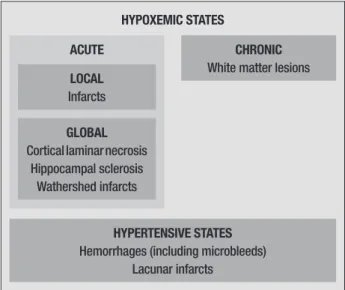
A classiication based on pathogenesis is likely to ameliorate overlapping problems and enable a more readily reproducible set of diagnosis criteria. For
in-stance, hypoxemic lesions should be classiied as global or local. his scheme would place ischemic infarcts and watershed infarcts in diferent categories (Figure 3).
An important prerequisite is required before at-tempting to reclassify cerebrovascular lesions. Current methods for neuropathological characterization of these lesions difer little to those used a century ago, in contrast to the advances seen in methods employed for detecting other brain-related conditions such neopla-sias and neurodegenerative diseases. herefore, meth-odological improvement is an essential irst step. his can be achieved through the development of antibodies against certain markers expressed during cerebrovas-cular lesions or by introducing imaging techniques into pathology labs.
A pathogenic-based classiication will be beneicial in establishing preventive and also therapeutic measures against these lesions, and should be prioritized in future investigations on vascular dementia.
REFERENCES
1. Roman GC, Sachdev P, Royall DR, et al. Vascular cognitive disorder: a new diagnostic category updating vascular cognitive impairment and vascular dementia. J Neurol Sci 2004;226:81-87.
2. Grinberg LT, Heinsen H. Toward a pathological definition of vascular dementia. J Neurol Sci 2010;299:136-138.
3. Korczyn AD, Vakhapova V. The prevention of the dementia epidemic. J Neurol Sci 2007;257:2-4.
4. Schneider JA, Arvanitakis Z, Bang W, Bennett DA. Mixed brain patholo-gies account for most dementia cases in community-dwelling older per-sons. Neurology 2007;69:2197-2204.
5. Korczyn AD. Mixed dementia--the most common cause of dementia. Ann N Y Acad Sci 2002;977:129-134.
6. Korczyn AD. The complex nosological concept of vascular dementia. J Neurol Sci 2002;203-204:3-6.
7. Langa KM, Foster NL, Larson EB. Mixed dementia: emerging concepts and therapeutic implications. JAMA 2004;292:2901-2908.
8. Iadecola C. Neurovascular regulation in the normal brain and in Al-zheimer’s disease. Nat Rev Neurosci 2004;5:347-360.
9. Grinberg LT, Thal DR. Vascular pathology in the aged human brain. Acta Neuropathol 2010;119:277-290.
10. Ferrer I. Cognitive impairment of vascular origin: neuropathology of cog-nitive impairment of vascular origin. J Neurol Sci 2010;299:139-149. 11. Gorelick PB, Scuteri A, Black SE, et al. Vascular contributions to
cogni-tive impairment and dementia: a statement for healthcare profession-als from the american heart association/american stroke association. Stroke 2011;42:2672-2713.
12. Hachinski V, Iadecola C, Petersen RC, et al. National Institute of Neurological Disorders and Stroke-Canadian Stroke Network
vascu-HYPOXEMIC STATES
ACUTE CHRONIC
White matter lesions LOCAL
Infarcts
GLOBAL Cortical laminar necrosis
Hippocampal sclerosis Wathershed infarcts
HYPERTENSIVE STATES Hemorrhages (including microbleeds)
Lacunar infarcts
lar cognitive impairment harmonization standards. Stroke 2006;37: 2220-2241.
13. Kalaria RN. Dementia comes of age in the developing world. Lancet 2003;361:888-9.
14. Roman GC, Tatemichi TK, Erkinjuntti T, et al. Vascular dementia - diag-nostic criteria for research studies - report of the NINDS-AIREN interna-tional workshop. Neurology 1993;43:250-260.
15. Kolsch H, Larionov S, Dedeck O, et al. Association of the glutathione S-transferase omega-1 Ala140Asp polymorphism with cerebrovascular atherosclerosis and plaque-associated interleukin-1 alpha expression. Stroke 2007;38:2847-2850.
16. Larionov S, Dedeck O, Birkenmeier G, Thal DR. Expression of alpha2-macroglobulin, neutrophil elastase, and interleukin-1alpha differs in early-stage and late-stage atherosclerotic lesions in the arteries of the circle of Willis. Acta Neuropathol 2007;113:33-43.
17. Liberato B, Chong JY, Sacco RL. Focal brain ischemia. Clinical features, epidemiology, risk factors and outcome. In: Kalimo H, ed. Cerebrovas-cular Diseases. Basel: ISN Neuropath Press; 2005.
18. Jellinger KA. The enigma of vascular cognitive disorder and vascular dementia. Acta Neuropathol 2007;113:349-388.
19. Pantoni L. Cerebral small vessel disease: from pathogenesis and clinical characteristics to therapeutic challenges. Lancet Neurol 2010;9:689-701. 20. Lammie AG. Small vessel disease. . In: Kalimo H, ed. Cerebrovascular
Diseases. Basel: ISN Neuropath Press; 2005: 85-91.
21. Utter S, Tamboli IY, Walter J, et al. Cerebral small vessel disease-in-duced apolipoprotein E leakage is associated with Alzheimer disease and the accumulation of amyloid beta-protein in perivascular astro-cytes. J Neuropathol Exp Neurol 2008;67:842-856.
22. Lammie GA. Hypertensive cerebral small vessel disease and stroke. Brain Pathol 2002;12:358-370.
23. Simpson JE, Wharton SB, Cooper J, et al. Alterations of the blood-brain barrier in cerebral white matter lesions in the ageing brain. Neurosci Lett 2010;486:246-51.
24. Thal DR, Ghebremedhin E, Orantes M, Wiestler OD. Vascular pathology in Alzheimer disease: correlation of cerebral amyloid angiopathy and arteriosclerosis/lipohyalinosis with cognitive decline. J Neuropathol Exp Neurol 2003;62:1287-1301.
25. Takao M, Koto A, Tanahashi N, Fukuuchi Y, Takagi M, Morinaga S. Pathologic findings of silent, small hyperintense foci in the basal ganglia and thalamus on MRI. Neurology 1999;52:666-668.
26. Fazekas F, Kleinert R, Offenbacher H, et al. Pathologic correlates of incidental MRI white matter signal hyperintensities. Neurology 1993;43:1683-1689.
27. Leys D, Pruvo JP, Parent M, et al. Could Wallerian degeneration con-tribute to “leuko-araiosis” in subjects free of any vascular disorder? J Neurol Neurosurg Psychiatry 1991;54:46-50.
28. Vonsattel JP, Myers RH, Hedley-Whyte ET, Ropper AH, Bird ED, Richardson EP, Jr. Cerebral amyloid angiopathy without and with ce-rebral hemorrhages: a comparative histological study. Ann Neurol 1991;30:637-649.
29. Wisniewski HM, Wegiel J, Wang KC, Lach B. Ultrastructural studies of the cells forming amyloid in the cortical vessel wall in Alzheimer’s disease. Acta Neuropathol 1992;84:117-127.
30. Greenberg SM, Nandigam RN, Delgado P, et al. Microbleeds versus macrobleeds: evidence for distinct entities. Stroke 2009;40:2382-2386. 31. Thal DR, Capetillo-Zarate E, Larionov S, Staufenbiel M, Zurbruegg S,
Beckmann N. Capillary cerebral amyloid angiopathy is associated with vessel occlusion and cerebral blood flow disturbances. Neurobiol Aging 2009;30:1936-1948.
32. Cadavid D, Mena H, Koeller K, Frommelt RA. Cerebral beta amyloid
angiopathy is a risk factor for cerebral ischemic infarction. A case con-trol study in human brain biopsies. J Neuropathol Exp Neurol 2000;59: 768-773.
33. Hoffmann M, Schmitt F, Bromley E. Vascular cognitive syndromes: rela-tion to stroke etiology and topography. Acta Neurol Scand 2009;120: 161-169.
34. Pendlebury ST, Rothwell PM. Prevalence, incidence, and factors as-sociated with pre-stroke and post-stroke dementia: a systematic review and meta-analysis. Lancet Neurol 2009;8:1006-1018.
35. Koga H, Takashima Y, Murakawa R, Uchino A, Yuzuriha T, Yao H. Cog-nitive consequences of multiple lacunes and leukoaraiosis as vascular cognitive impairment in community-dwelling elderly individuals. J Stroke Cerebrovasc Dis 2009;18:32-37.
36. Jellinger KA. A critical evaluation of current staging of alpha-synuclein pathology in Lewy body disorders. Biochim Biophys Acta 2009;1792: 730-740.
37. Kalaria RN, Kenny RA, Ballard CG, Perry R, Ince P, Polvikoski T. To-wards defining the neuropathological substrates of vascular dementia. J Neurol Sci 2004;226:75-80.
38. Gerraty RP, Parsons MW, Barber PA, et al. Examining the lacunar hy-pothesis with diffusion and perfusion magnetic resonance imaging. Stroke 2002;33:2019-2024.
39. Okamoto Y, Ihara M, Fujita Y, Ito H, Takahashi R, Tomimoto H. Cortical microinfarcts in Alzheimer’s disease and subcortical vascular dementia. Neuroreport 2009;20:990-996.
40. Kovari E, Gold G, Herrmann FR, et al. Cortical microinfarcts and demy-elination affect cognition in cases at high risk for dementia. Neurology 2007;68:927-931.
41. Jellinger KA. Understanding the pathology of vascular cognitive impair-ment. J Neurol Sci 2005;229-230:57-63.
42. Probst A, Taylor KI, Tolnay M. Hippocampal sclerosis dementia: a reap-praisal. Acta Neuropathol 2007;114:335-345.
43. Brown WR, Moody DM, Thore CR, Anstrom JA, Challa VR. Microvas-cular changes in the white mater in dementia. J Neurol Sci 2009;283: 28-31.
44. Binswanger O. Die Abgrenzung der allgemeinen progressiven Paralyse. Berl Klin Wochenschr 1894;31:1102-1105.
45. Yamori Y, Horie R, Handa H, Sato M, Fukase M. Pathogenetic simi-larity of strokes in stroke-prone spontaneously hypertensive rats and humans. Stroke 1976;7:46-53.
46. Bonni A, Sun Y, Nadalvicens M, et al. Regulation of gliogenesis in the central nervous system by the JAK-STAT signaling pathway. Science 1997;278:477-483.
47. Mandybur TI. Cerebral amyloid angiopathy: the vascular pathology and complications. J Neuropathol Exp Neurol 1986;45:79-90.
48. Thal DR, Griffin WS, de Vos RA, Ghebremedhin E. Cerebral amyloid an-giopathy and its relationship to Alzheimer’s disease. Acta Neuropathol 2008;115:599-609.
49. Henry-Feugeas MC. MRI of the ‘Alzheimer syndrome’. J Neuroradiol 2007;34:220-227.
50. Tsushima Y, Aoki J, Endo K. Brain microhemorrhages detected on T2*-weighted gradient-echo MR images. AmJNeuroradiol 2003;24:88-96. 51. De Reuck J, Auger F, Cordonnier C, et al. Comparison of 7.0-T
T*-magnetic resonance imaging of cerebral bleeds in post-mortem brain sections of Alzheimer patients with their neuropathological correlates. Cerebrovasc Dis 2011;31:511-517.
![Figure 1. Example of small vessel disease. [A] Subcortical white matter exhibiting hyaline arteriolosclerosis](https://thumb-eu.123doks.com/thumbv2/123dok_br/15187966.526947/2.892.106.666.883.1093/figure-example-vessel-disease-subcortical-exhibiting-hyaline-arteriolosclerosis.webp)
![Figure 2. Hemorrhagic infarcts. [A] Coronal section across the thalamus.](https://thumb-eu.123doks.com/thumbv2/123dok_br/15187966.526947/3.892.442.786.106.744/figure-hemorrhagic-infarcts-coronal-section-thalamus.webp)